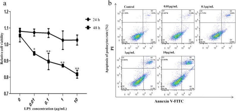
Fig. 3.
LPS inhibited podocyte viability and induced apoptosis in a dose-dependent manner. Cells were incubated with indicated concentrations (0.01, 0.1, 1, and 10 μg/mL) of LPS for 24 and 48 h. Cell growth inhibition activity of LPS was assessed by the CCK-8 assay and apoptosis was measured by Annexin V-FITC/PI staining and flow cytometry. a The viability of podocytes at different concentrations of LPS at 24 and 48 h and b ratio of apoptotic cells to the total number of cells induced by LPS at 48 h. The number of apoptotic cells equals the sum of the cells in the Q2 (early-stage cell apoptosis rate) and Q3 (late-stage cell apoptosis rate) regions. *p < 0.05; **p < 0.01 when compared with control group.

