



Abstract
Background
Chest pain decision unit (CDU) evaluation of patients with acute chest pain (ACP) and nondiagnostic electrocardiogram (ECG) usually includes noninvasive testing for coronary artery disease (CAD).
Hypothesis
CAD evaluation will not improve clinical outcome in low‐risk ACP patients.
Methods
We studied 459 adults admitted to CDU with ACP and no troponin release who underwent noninvasive CAD testing (stress testing in 396 and coronary computed tomographic angiography in 63). Multivariate logistic regression was used to determine predictors of adverse outcome over a 3‐year follow‐up period.
Results
Initial noninvasive test was normal in 367 (80%) and abnormal (positive or indeterminate) in 92 (20%). A total of 42 (9%) patients underwent invasive coronary angiography, and 16 (3.5%) underwent revascularization. During follow‐up, 33 patients had a total of 36 major clinical events: 12 revascularizations, 9 myocardial infarctions, and 15 deaths. Multivariate logistic regression analysis identified abnormal ECG (odds ratio [OR]: 2.7, P = 0.03), typical chest pain (OR: 3.8, P = 0.002), diabetes (OR: 4.1, P = 0.001), and known CAD (OR: 2.3, P = 0.03) as independent predictors for adverse outcome, but not noninvasive test result. Thus, in 187 patients with no high‐risk features (41% of the cohort), the annualized event rate was 0.5%. In 272 patients with at least 1 high‐risk feature, annualized event rates were 2.8% and 5.7% when noninvasive test was normal or abnormal, respectively (P = 0.04).
Conclusions
Clinical risk stratification allows identification of patients at low risk of adverse outcome over an intermediate period of follow‐up. Noninvasive testing is not warranted in such patients.
Introduction
Chest pain is the chief complaint of nearly 5% of all patients seen in emergency departments (EDs). In the United States, it accounts for ∼ 6 million visits annually, with an estimated cost of ∼ $6 billion.1 One‐third of patients presenting to EDs with chest pain have a cardiac cause such as myocardial infarction (MI) or unstable angina, with the remaining two‐thirds having noncardiac causes for their symptoms.1, 2 Consequently, the annual cost for cardiac evaluation of patients with noncardiac chest pain is ∼ $4 billion.1 Nevertheless, ED evaluation of patients with chest pain and nondiagnostic electrocardiogram (ECG) remains a frequent and difficult problem because of the challenge of identifying and treating patients with truly life‐threatening acute coronary syndromes, while avoiding unnecessary and costly evaluation of the others.3
Chest pain decision units (CDUs) have been developed to streamline the evaluation of low‐ to intermediate‐risk patients with chest pain and have been demonstrated to be cost‐effective when compared to the “admit or discharge” approach.4, 5, 6, 7 A typical CDU protocol consists of 2 sets of cardiac enzymes over a 6‐hour observation period followed by noninvasive testing for exclusion of obstructive coronary artery disease (CAD). However, in most hospitals, neither stress testing nor coronary computed tomographic angiography (CCTA) is available 24 hours per day. This often results in delays in evaluation, prolonged length of stay, patient inconvenience, and increased healthcare costs.
The objective of the study was to assess the role of clinical predictors and noninvasive CAD evaluation in predicting major adverse clinical events (MACEs) in patients admitted to the CDU.
Methods
Study Patients
We retrospectively reviewed the medical records of 612 patients who were admitted to our CDU and underwent formal cardiology evaluation over a 1‐year period. A total of 459 patients met the following inclusion criteria: 2 negative sets of cardiac enzymes ∼ 6 hours apart (total and MB isoenzyme of creatine phosphokinase and troponin T) and noninvasive evaluation for CAD by stress testing (exercise or pharmacologic) or CCTA (Figure 1). Patients with evidence of acute coronary syndrome (ischemic ECG changes or elevated cardiac enzymes) or clear non‐CAD etiology of chest pain were excluded (n = 153, 25%). Information regarding MI, repeat stress testing or catheterization, revascularization, and death was collected 3 years following initial CDU evaluation. Patients who had MACEs defined as death, MI, or revascularization were censored at the time of the event.
Figure 1.
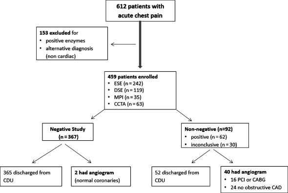
Flowchart demonstrating the clinical and diagnostic algorithm followed in 612 patients with acute chest pain evaluated over a 1‐year period. Abbreviations: CABG, coronary artery bypass graft surgery; CAD, coronary artery disease; CCTA, coronary computed tomographic angiography; CDU, chest pain decision unit; DSE, dobutamine stress echocardiography; ESE, exercise stress echocardiography; MPI, myocardial perfusion imaging; PCI, percutaneous coronary intervention.
Noninvasive Evaluation for CAD (n = 459)
Exercise (n = 242, 52%) or dobutamine (n = 119, 26%) stress echocardiography, myocardial perfusion imaging (MPI; n = 35, 8%), or CCTA (n = 63, 14%) were performed for noninvasive CAD evaluation. The type of stress testing was at the discretion of the consulting cardiologist. At our institution, exercise stress echocardiography is the preferred initial test unless contraindicated. Standard protocols were used for exercise (Bruce protocol) or pharmacologic stress (adenosine for MPI and dobutamine ± atropine for dobutamine stress echocardiography) and for CCTA. All images obtained during noninvasive testing were captured and stored digitally and were available for off‐line review.
Definitions
Typical chest pain (or chest pain equivalent) was defined as symptoms triggered by exertion or emotional stress that lasted <30 minutes and were relieved by rest or sublingual nitroglycerine or as documented by the consulting cardiologist. Atypical chest pain thus had none of these features. Normal ECG was defined as normal sinus rhythm with normal QRS duration and QT interval and no ischemic ST segment or T‐wave changes (T waves concordant with the QRS axis and isoelectric ST segments). Incomplete right bundle branch block and mild first‐degree atrioventricular block (PR interval = 200–230 msec) were not considered abnormal.
Statistical Analysis
Data are presented as range (mean ± standard deviation) or number (%). Between‐group comparisons were made using Student t test for continuous variables and χ2 test for categorical variables. A nonparametric test was used for continuous variables with skewed (non‐normal) distribution. Multivariate logistic regression was used to identify predictors for MI, revascularization, and death.
Investigation was done according to the Declaration of Helsinki. The study was reviewed and approved by the institutional review board.
Results
Clinical and demographic characteristics of the 459 patients enrolled in the study are shown in Table 1. Overall, 367 (80%) of the 459 patients had a normal noninvasive CAD evaluation, whereas 92 (20%) had an abnormal or inconclusive study. Invasive coronary angiography was performed in 40 (9%), and significant CAD requiring revascularization (percutaneous or surgical) was found in 16 (3.5%) patients (Figure 1).
Table 1.
Clinical and Demographic Characteristics of 459 Patients With Suspected Acute Coronary Syndrome
| Characteristic | Value |
|---|---|
| Age, y (mean ± SD) | 26–90 (56 ± 13) |
| Women, No. [%] | 231 [50] |
| Risk factors | |
| Hypertension, No. [%] | 255 [56] |
| Systolic blood pressure, mm Hg (mean ± SD) | 89–236 (127 ± 18) |
| Diabetes mellitus, No. [%] | 100 [22] |
| Hypercholesterolemia, No. [%] | 238 [53] |
| Low‐density lipoprotein, mg/dL (mean ± SD) | 29–210 (103 ± 36) |
| High‐density lipoprotein, mg/dL (mean ± SD) | 9–120 (51 ± 15) |
| Smoking, No. [%] | |
| Ever | 234 [51] |
| Current | 100 [22] |
| Family history of premature CAD, No. [%] | 188/369 [51] |
| Known CAD, No. [%] | 116 [25] |
| Framingham risk score (mean ± SD) | 1–40 (8.3 ± 6%) |
| Medications, No. [%] | |
| Aspirin | 192 [42] |
| Clopidogrel | 36 [19] |
| Statin | 161[35] |
| β‐Blocker | 171 [37] |
| Angiotensin‐converting enzyme inhibitors | 128 [28] |
| Calcium channel blockers | 39 [8.5] |
Abbreviations: CAD, coronary artery disease; SD, standard deviation.
Predictors for MI, Revascularization, and Death During Follow‐up
Overall, 15 of 459 (3.2%) patients died, 9 (2%) had an MI, and 12 (2.6%) underwent a revascularization procedure during follow‐up. A total of 28 revascularizations (16 at index visit and 12 during follow‐up) were performed in 25 patients. During follow‐up, 3 patients had percutaneous coronary intervention at the time of MI. Therefore, 33 (7.2%) patients had MACEs (death, MI, or revascularization) during a mean follow‐up of 3.3 years (range, 0.3–4.1 years).
By univariate analysis, predictors for MI, revascularization, and death were older age, typical chest pain, abnormal ECG, presence of diabetes, hypercholesterolemia and previous CAD, higher Framingham score (FS), and abnormal/indeterminate stress test (Table 2). Baseline revascularization rate was not statistically different between those with and without MACEs during follow‐up (3.1% vs 9.1%, P = 0.1, Table 2). Family history data were excluded from analysis because data were incomplete (available in 369, 80%) and in many cases the age at which first‐degree relatives had CAD events was not documented. Multivariate logistic regression analysis identified abnormal ECG (odds ratio [OR]: = 2.7, P = 0.03), typical chest pain (relative risk [RR] = 3.8, P = 0.002), known history of CAD (OR: 2.3, P = 0.03), and diabetes (OR: 4.1, P = 0.001) as independent predictors for MACEs, whereas stress test was not found to be an independent predictor (model 1, Table 3). Initial revascularization rate was introduced in the multivariate logistic regression model to correct for any baseline differences. FS emerged as an independent predictor for bad outcomes, but it was highly correlated with diabetes, which is part of the FS calculation. When FS with a cutoff of 10% was introduced in the multivariate analysis (model 2), the prediction model was not significantly different and the influence of diabetes was less as reflected by lower OR in model 2 compared to model 1 (Table 2). During follow‐up, 3 (1.6%) patients had MACEs in the low‐risk group compared to 30 (11%) in the group with at least 1 high‐risk factor (P = 0.001). When the low‐risk group (n = 187) was further stratified with an FS < 10% (n = 148), there were no deaths, MIs, or revascularizations during follow‐up.
Table 2.
Univariate Analysis to Predict Death, MI, or Revascularization During Follow‐up
| Clinical Predictor | Univariate Analysis to Predict Death, MI, or Revascularization | ||
|---|---|---|---|
| No Events, n = 426 | Events, n = 33 | P | |
| Age, y | 55 ± 13 | 60 ± 11 | 0.05 |
| Normal ECG | 306 (72%) | 17 (51%) | 0.01 |
| Atypical chest pain | 376 (88%) | 23 (70%) | 0.002 |
| Known CAD | 98 (23%) | 17 (51%) | 0.001 |
| Diabetes mellitus | 83 (19%) | 17 (51%) | 0.001 |
| Hypertension | 231 (54%) | 23 (70%) | 0.08 |
| Hypercholesterolemia | 216 (51%) | 23 (70%) | 0.05 |
| Framingham risk score | 8 ± 6 | 13 ± 8 | 0.001 |
| Abnormal CAD evaluation | 78 (18%) | 14 (42%) | 0.001 |
| Initial coronary revascularization | 13 (3.1%) | 3 (9.1%) | 0.1 |
Abbreviations: CAD, coronary artery disease; ECG, electrocardiogram; MI, myocardial infarction.
Table 3.
Multivariate Analysis to Predict Death, MI, or Revascularization During Follow‐up (Adjusted for Baseline Revascularization Rate)
| Clinical Predictor | Model 1 | Model 2 | ||||
|---|---|---|---|---|---|---|
| OR | P | CI | OR | P | CI | |
| ECG | 2.7 | 0.03 | 1.05‐4.9 | 2.2 | 0.04 | 1.03‐4.8 |
| CP | 3.8 | 0.002 | 1.6‐9.5 | 3.9 | 0.004 | 1.6‐9.6 |
| CAD | 2.3 | 0.03 | 1.07‐4.9 | 2.3 | 0.03 | 1.06‐4.8 |
| DM | 4.1 | <0.001 | 1.9‐9.1 | 2.9 | 0.01 | 1.3‐6.9 |
| Baseline revascularization | 1.3 | 0.7 | 0.3‐5.4 | 1.1 | 0.9 | 0.3‐4.8 |
| FS < 10% | 2.3 | 0.04 | 1.02‐5.3 | |||
Abbreviations: CAD, coronary artery disease; CI, confidence interval; CP, chest pain; DM, diabetes mellitus; ECG, electrocardiogram; FS, Framingham risk score; MI, myocardial infarction; OR, odds ratio.
CAD Evaluation and Follow‐up of Low‐Risk Patients
Overall, 187 (41%) patients had none of the 4 high‐risk clinical predictors (Figure 2). At the initial CDU visit, 11 (6%) patients had an abnormal or indeterminate CAD evaluation (3 abnormal, 8 indeterminate) and 1 required invasive coronary angiography (normal coronaries). There was no initial revascularization performed in the low‐risk group. During follow‐up, incidence of death, MI, or revascularization was significantly lower among 187 patients without high‐risk clinical predictors than among 366 patients with a normal noninvasive study (3 of 187 [1.6%] vs 19 of 366 [5.2%], P = 0.02). The 3 events (1 MI, 1 revascularization, and 1 noncardiac death) in the low‐risk group occurred despite a normal noninvasive test. Noninvasive CAD testing helped stratify those with at least 1 high‐risk predictor (n = 272). In this group, there were fewer MACEs during follow‐up in those with a normal than among those with an abnormal study (8.4% vs 17.1%, P = 0.04).
Figure 2.
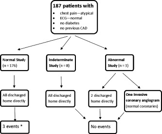
Flowchart demonstrating the results of noninvasive testing, chest pain decision unit disposition, and follow‐up of 187 patients with acute chest pain and low likelihood of coronary etiology. *One myocardial infraction, 1 revascularization, and 1 death. Abbreviations: CAD, coronary artery disease; ECG, electrocardiogram.
Discussion
Summary of the Findings
The results of this study show that noninvasive CAD evaluation (CCTA or stress testing) is not necessary in many patients evaluated in a chest pain decision unit. Clinical predictors alone and biomarkers can identify a subgroup of low‐risk patients who can be discharged from CDU without further CAD evaluation. This low‐risk group had no diabetes or prior history of CAD and presented with atypical chest pain and no ECG abnormalities. Noninvasive testing for CAD in such individuals did not improve their outcome but resulted in unnecessary prolongation of hospital stay, invasive coronary angiography, and increased cost due to false‐positive results. If noninvasive testing had been avoided or deferred, then the CDU length of stay in these low‐risk individuals would have been reduced by two‐thirds from an average of 20 hours.
Chest Pain Decision Units
Chest pain observation units have played a major role in streamlining the management of patients suspected to have acute coronary syndrome. However, due to the medical and legal implications of missing the diagnosis of acute coronary syndrome,8 and particularly in view of the poor prognosis of the erroneously discharged patients,8 attention has been focused primarily on the identification of those with the disease,9, 10 and those with high likelihood of adverse outcomes who may benefit from the highest levels of observation.11, 12 This has been complicated by the concerns raised regarding the potential for missing the diagnosis of acute coronary syndrome even when a conservative strategy is adopted.13 Thus, despite recent guidelines14 and various diagnostic and triage protocols,15, 16, 17, 18 low‐risk individuals with acute chest pain are frequently subjected to noninvasive cardiac testing and an unnecessarily lengthy ED stay.
Low‐Risk Chest Pain Patients
History, physical examination, ECG, and biomarkers of myocardial necrosis constitute the initial evaluation of patients with acute chest pain related to suspected CAD. In the absence of a typical history or ischemic ECG changes, normal troponin levels indicate an excellent short‐term prognosis. Thus, the rate of cardiovascular events at 30 days among such patients is generally low if the truly low‐risk individuals are identified.19, 20, 21, 22 For instance, Sanchis et al21 found a group of low‐risk patients (n = 111) with atypical chest pain, no insulin‐requiring diabetes, and no prior coronary intervention among 646 consecutive patients who presented to the ED with suspected acute coronary syndrome and had no ECG ST‐segment deviation or troponin elevation. At 1 year of follow‐up, no death or MI occurred in these low‐risk individuals compared to 6.7% in the whole cohort.
A few studies have focused on the identification of low‐risk individuals who may be safely discharged home for outpatient follow‐up without undergoing noninvasive testing for CAD. Marsan et al23 reported no 30‐day major adverse cardiac event (revascularization, MI, or death) in 299 acute chest pain patients ≤40 years of age who had normal ECG and no CAD risk factor. Christenson et al24 were also able to identify a group of very low‐risk individuals (age <40 years, normal ECG, no prior ischemic chest pain, and normal initial troponin level) who could be discharged within 2 hours of CDU admission. In a large prospective study, Hess et al developed the North America chest pain rule15 to identify patients with acute chest pain at low risk for death, MI, and revascularization within 30 days following ED discharge. The decision rule used predictors such as history of CAD, atypical chest pain, ischemia on ECG, cardiac biomarkers, and age to identify low‐risk patients. Patient with none of the risk factors who were younger than 40 years could be discharged if the initial troponin was normal, whereas 2 sets of negative troponin tests 6 hours apart were necessary for patients 41 to 50 years old. Although our observations are in general agreement with those outlined above, it does extend the scope of the low‐risk individuals to those >50 years of age and to patients with some risk factors other than diabetes mellitus and known CAD.
Independent Predictors of Risk
The study identified atypical chest pain and absence of ECG abnormalities, diabetes, and known CAD as independent predictors of low risk for death, MI, and revascularization. These parameters also predicted a normal noninvasive evaluation at the initial visit. Thus, it is not surprising that the noninvasive cardiac testing was not an independent predictor for bad outcome.
Certain chest pain characteristics are reported to increase the likelihood of CAD in patients presenting to the emergency room.25 Although a normal ECG does not exclude acute coronary syndrome,8, 26 the presence of ischemic changes markedly increases the likelihood of adverse outcome in patients with chest pain.27 Diabetes is a major risk factor for CAD, and atherosclerosis tends to be more severe in diabetics compared to nondiabetics.28 Even when asymptomatic, diabetic individuals have a high prevalence of coronary atherosclerosis.29, 30 It is, thus, not surprising that among our symptomatic patients, we found diabetes to be a strong predictor for future cardiac events and the absence of diabetes was a strong predictor for a normal cardiac study at the initial CDU evaluation. Symptomatic diabetics have been shown to have higher likelihood of abnormal CAD studies.31 Finally, previous studies have indicated that in patients with chest pain, a history of CAD highly increases the likelihood of acute coronary syndrome as the etiology.10
Deferred Stress Testing
Immediate CAD evaluation is usually performed because of justified concern that a true cardiac etiology for chest pain may be missed. However, outpatient stress testing within 72 hours from ER discharge of low‐risk patients with chest pain has been reported to be feasible, safe, and associated with decreased hospital admission rates.32 Hermann et al33 have also demonstrated that routine provocative CAD testing is of no benefit in young (<40 years old) individuals with acute chest pain, no cardiac enzyme elevation, and no ischemic ECG changes.
Evaluation for CAD in CDUs has a diagnostic and prognostic goal. However, the application of indiscriminate noninvasive CAD testing to very low‐risk subsets of patients with acute chest pain may lead to unnecessary invasive angiography due to a higher chance of false‐positive results. It may also lead to further ED crowding, an issue that has been shown to result in delivery of suboptimal care, with a potential for increasing adverse cardiovascular outcomes.34
Study Limitations
The main limitation of the study is the retrospective design. Our institution serves a large and very stable population and is the only provider in the area for coronary revascularization. Therefore, we believe that information regarding death and revascularization is complete. However, it is possible that some patients might have left the area during follow‐up or had clinical events while traveling outside the home area.
Conclusions
A substantial percentage of patients seen in a chest pain decision unit can be safely discharged after 2 sets of negative cardiac enzyme tests (6–8 hours apart) if they have a normal or near normal ECG, atypical chest pain, no diabetes, and no history of CAD.
The incidence of MACEs (death, MI, or revascularization) was very low (0.5%/y) and was predicted by clinical variables alone. Noninvasive cardiac testing did not predict bad outcome in this low‐risk group and led to false‐positive results.
Supporting information
Figure S1. Kaplan‐Meier MACE Free Survival of low risk patients (bolded) and those with high risk factors (adjusted for baseline revascularization rate)
Table S1. Positive and Negative predictive Values
The authors have no funding, financial relationships, or conflicts of interest to disclose.
References
- 1. Nawar EW, Niska RW, Xu J. National Hospital Ambulatory Medical Care Survey: 2005 emergency department summary. Adv Data. 2007;(386):1–32. [PubMed] [Google Scholar]
- 2. Durairaj L, Reilly B, Das K, et al. Emergency department admissions to inpatient cardiac telemetry beds: a prospective cohort study of risk stratification and outcomes. Am J Med. 2001;110:7–11. [DOI] [PubMed] [Google Scholar]
- 3. Forberg JL, Henriksen LS, Edenbrandt L, et al. Direct hospital costs of chest pain patients attending the emergency department: a retrospective study. BMC Emerg Med. 2006;6:6. [DOI] [PMC free article] [PubMed] [Google Scholar]
- 4. Farkouh ME, Smars PA, Reeder GS, et al. A clinical trial of a chest‐pain observation unit for patients with unstable angina. Chest Pain Evaluation in the Emergency Room (CHEER) Investigators. N Engl J Med. 1998;339:1882–1888. [DOI] [PubMed] [Google Scholar]
- 5. Goodacre S, Nicholl J. A randomised controlled trial to measure the effect of chest pain unit care upon anxiety, depression, and health‐related quality of life [ISRCTN85078221]. Health Qual Life Outcomes. 2004;2:39. [DOI] [PMC free article] [PubMed] [Google Scholar]
- 6. Jagminas L, Partridge R. A comparison of emergency department versus inhospital chest pain observation units. Am J Emerg Med. 2005;23:111–113. [DOI] [PubMed] [Google Scholar]
- 7. Cullen MW, Reeder GS, Farkouh ME, et al. Outcomes in patients with chest pain evaluated in a chest pain unit: the chest pain evaluation in the emergency room study cohort. Am Heart J. 2011;161:871–877. [DOI] [PubMed] [Google Scholar]
- 8. Pope JH, Aufderheide TP, Ruthazer R, et al. Missed diagnoses of acute cardiac ischemia in the emergency department. N Engl J Med. 2000;342:1163–1170. [DOI] [PubMed] [Google Scholar]
- 9. Selker HP, Beshansky JR, Griffith JL, et al. Use of the acute cardiac ischemia time‐insensitive predictive instrument (ACI‐TIPI) to assist with triage of patients with chest pain or other symptoms suggestive of acute cardiac ischemia. A multicenter, controlled clinical trial. Ann Intern Med. 1998;129:845–855. [DOI] [PubMed] [Google Scholar]
- 10. Bjork J, Forberg JL, Ohlsson M, et al. A simple statistical model for prediction of acute coronary syndrome in chest pain patients in the emergency department. BMC Med Inform Decis Mak. 2006;6:28. [DOI] [PMC free article] [PubMed] [Google Scholar]
- 11. Goldman L, Cook EF, Johnson PA, et al. Prediction of the need for intensive care in patients who come to the emergency departments with acute chest pain. N Engl J Med. 1996;334:1498–1504. [DOI] [PubMed] [Google Scholar]
- 12. Reilly BM, Evans AT, Schaider JJ, et al. Triage of patients with chest pain in the emergency department: a comparative study of physicians' decisions. Am J Med. 2002;112:95–103. [DOI] [PubMed] [Google Scholar]
- 13. Madsen T, Mallin M, Bledsoe J, et al. Utility of the emergency department observation unit in ensuring stress testing in low‐risk chest pain patients. Crit Pathw Cardiol. 2009;8:122–124. [DOI] [PubMed] [Google Scholar]
- 14. Amsterdam EA, Kirk JD, Bluemke DA, et al. Testing of low‐risk patients presenting to the emergency department with chest pain: a scientific statement from the American Heart Association. Circulation. 2010;122:1756–1776. [DOI] [PMC free article] [PubMed] [Google Scholar]
- 15. Hess EP, Brison RJ, Perry JJ, et al. Development of a clinical prediction rule for 30‐day cardiac events in emergency department patients with chest pain and possible acute coronary syndrome. Ann Emerg Med. 2012;59:115–125. [DOI] [PubMed] [Google Scholar]
- 16. Hess EP, Knoedler MA, Shah ND, et al. The chest pain choice decision aid: a randomized trial. Circ Cardiovasc Qual Outcomes. 2012;5:251–259. [DOI] [PubMed] [Google Scholar]
- 17. Than M, Cullen L, Aldous S, et al. 2‐Hour accelerated diagnostic protocol to assess patients with chest pain symptoms using contemporary troponins as the only biomarker: the ADAPT Trial. J Am Coll Cardiol. 2012;59:2091–2098. [DOI] [PubMed] [Google Scholar]
- 18. Aldous SJ, Richards MA, Cullen L, et al. A new improved accelerated diagnostic protocol safely identifies low‐risk patients with chest pain in the emergency department. Acad Emerg Med. 2012;19:510–516. [DOI] [PubMed] [Google Scholar]
- 19. Hamm CW, Goldmann BU, Heeschen C, et al. Emergency room triage of patients with acute chest pain by means of rapid testing for cardiac troponin T or troponin I. N Engl J Med. 1997;337:1648–1653. [DOI] [PubMed] [Google Scholar]
- 20. Smith SW, Tibbles CD, Apple FS, et al. Outcome of low‐risk patients discharged home after a normal cardiac troponin I. J Emerg Med. 2004;26:401–406. [DOI] [PubMed] [Google Scholar]
- 21. Sanchis J, Bodi V, Nunez J, et al. New risk score for patients with acute chest pain, non‐ST‐segment deviation, and normal troponin concentrations: a comparison with the TIMI risk score. J Am Coll Cardiol. 2005;46:443–449. [DOI] [PubMed] [Google Scholar]
- 22. Koukkunen H, Pyorala K, O Halinen M. Low‐risk patients with chest pain and without evidence of myocardial infarction may be safely discharged from emergency department. Eur Heart J. 2004;25:329–334. [DOI] [PubMed] [Google Scholar]
- 23. Marsan RJ Jr, Shaver KJ, Sease KL, et al. Evaluation of a clinical decision rule for young adult patients with chest pain. Acad Emerg Med. 2005;12:26–31. [DOI] [PubMed] [Google Scholar]
- 24. Christenson J, Innes G, McKnight D, et al. A clinical prediction rule for early discharge of patients with chest pain. Ann Emerg Med. 2006;47:1–10. [DOI] [PubMed] [Google Scholar]
- 25. Swap CJ, Nagurney JT. Value and limitations of chest pain history in the evaluation of patients with suspected acute coronary syndromes. JAMA. 2005;294:2623–2629. [DOI] [PubMed] [Google Scholar]
- 26. Welch RD, Zalenski RJ, Frederick PD, et al. Prognostic value of a normal or nonspecific initial electrocardiogram in acute myocardial infarction. JAMA. 2001;286:1977–1984. [DOI] [PubMed] [Google Scholar]
- 27. Challa PK, Smith KM, Conti CR. Initial presenting electrocardiogram as determinant for hospital admission in patients presenting to the emergency department with chest pain: a pilot investigation. Clin Cardiol. 2007;30:558–561. [DOI] [PMC free article] [PubMed] [Google Scholar]
- 28. Goraya TY, Leibson CL, Palumbo PJ, et al. Coronary atherosclerosis in diabetes mellitus: a population‐based autopsy study. J Am Coll Cardiol. 2002;40:946–953. [DOI] [PubMed] [Google Scholar]
- 29. De Lorenzo A, Lima RS, Siqueira‐Filho AG, et al. Prevalence and prognostic value of perfusion defects detected by stress technetium‐99m sestamibi myocardial perfusion single‐photon emission computed tomography in asymptomatic patients with diabetes mellitus and no known coronary artery disease. Am J Cardiol. 2002;90:827–832. [DOI] [PubMed] [Google Scholar]
- 30. Scholte AJ, Schuijf JD, Kharagjitsingh AV, et al. Prevalence of coronary artery disease and plaque morphology assessed by multi‐slice computed tomography coronary angiography and calcium scoring in asymptomatic patients with type 2 diabetes. Heart. 2008;94:290–295. [DOI] [PubMed] [Google Scholar]
- 31. Zellweger MJ, Hachamovitch R, Kang X, et al. Prognostic relevance of symptoms versus objective evidence of coronary artery disease in diabetic patients. Eur Heart J. 2004;25:543–550. [DOI] [PubMed] [Google Scholar]
- 32. Meyer MC, Mooney RP, Sekera AK. A critical pathway for patients with acute chest pain and low risk for short‐term adverse cardiac events: role of outpatient stress testing. Ann Emerg Med. 2006;47:427–435. [DOI] [PubMed] [Google Scholar]
- 33. Hermann LK, Weingart SD, Duvall WL, et al. The limited utility of routine cardiac stress testing in emergency department chest pain patients younger than 40 years. Ann Emerg Med. 2009;54:12–16. [DOI] [PubMed] [Google Scholar]
- 34. Pines JM, Pollack CV Jr, Diercks DB, et al. The association between emergency department crowding and adverse cardiovascular outcomes in patients with chest pain. Acad Emerg Med. 2009;16:617–625. [DOI] [PubMed] [Google Scholar]
Associated Data
This section collects any data citations, data availability statements, or supplementary materials included in this article.
Supplementary Materials
Figure S1. Kaplan‐Meier MACE Free Survival of low risk patients (bolded) and those with high risk factors (adjusted for baseline revascularization rate)
Table S1. Positive and Negative predictive Values