Key Points
Abnormal levels of IgA at diagnosis predict time to treatment, survival, and need for immunoglobulin replacement in CLL.
Abnormal levels of IgG at diagnosis predict the need for immunoglobulin replacement in CLL.
Abstract
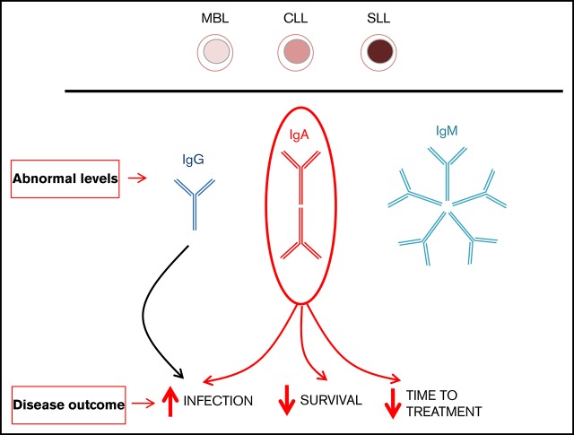
To better understand the relationship between baseline immunoglobulin measurements and subsequent clinical outcomes in chronic lymphocytic leukemia (CLL), we performed a retrospective analysis on 660 patients with CLL (72%), monoclonal B-cell lymphocytosis (MBL) (13%), and small lymphocytic lymphoma (SLL) (14%), diagnosed between 2005 and 2014 at CancerCare Manitoba. Of 511 patients who had their first immunoglobulin level determined within 3 months of diagnosis, abnormal (either increased or decreased) immunoglobulin M (IgM), IgG, and IgA values were observed in 58% of patients with CLL, 27% of patients with MBL, and 20% of patients with SLL. Immunoglobulin deviances were similar for MBL and CLL Rai stage 0 and for SLL and Rai stages I and II; for CLL, IgG and IgA abnormalities occurred with increasing frequency with advancing Rai stage. In contrast, the frequency of IgM abnormalities was similar in all patient groups. IgA abnormalities significantly correlated with high β2-microglobulin (B2M) expression, whereas abnormal IgG and IgA levels were associated with the use of IGHV1-69, 3-21, and 3-49 subtypes. Increases in IgG or IgM were commonly associated with the presence of a CLL-type M-band, whereas oligoclonal bands were frequently observed with increased IgA levels. Although abnormal levels of IgG and IgA at diagnosis were independent predictors for future immunoglobulin replacement, only abnormal IgA levels were associated with shorter time to first treatment and overall survival. These findings indicate that both reduced and elevated levels of IgG and IgA at diagnosis are important and independent prognostic markers for infection in CLL, with IgA being more relevant as a marker of disease progression and survival.
Visual Abstract
Introduction
Immune suppression is a fundamental feature of chronic lymphocytic leukemia (CLL) and small lymphocytic lymphoma (SLL), leading to an increased incidence of infections and second malignancies.1-3 Interestingly, whereas immune dysfunction is apparent at diagnosis, altered serum light chain ratios, serum paraproteins, and low immunoglobulin G (IgG) levels have been observed years before the diagnosis of CLL.4 Moreover, patients with monoclonal B-cell lymphocytosis (MBL), the precursor to CLL or SLL, also have an increased incidence of infections and second malignancies.5,6 Hypogammaglobulinemia (a reduction in the immunoglobulins) is a commonly seen immune defect and a major cause of infections in patients with CLL.7 In addition, a reduction in more than 1 immunoglobulin or a decrease in a specific IgG subclass may be associated with the development of infections.8,9 The use of immunoglobulin replacement therapy, either intravenous or subcutaneous, reduces the incidence and severity of infections in patients with CLL.7
Several studies have evaluated whether baseline immune function reflects the underlying biology of CLL and is of prognostic value.10-17 Decreased IgG levels have been most widely reported and are also the most variable, occurring in 9.9% to 76% of patients, whereas reductions in IgA occur in 12% to 68% of patients and reductions in IgM occur in 4% to 56% of patients.10-19 The clinical significance of decreased immunoglobulins at diagnosis is uncertain, because some authors have reported that low IgG or IgA levels may be independent predictors for time to first treatment (TTFT),14,17 findings that have not been observed by others.10,12,13,16,19 In addition, an older study by Rozman et al11 demonstrated that reduced levels of IgA were an independent prognostic marker for overall survival (OS), although this study was performed in an era before fludarabine and chemoimmunotherapy. Moreover, low IgM has been reported to be an independent predictor of OS, but others have found it to have no prognostic significance.11,18 Decreased IgG at the time of CLL diagnosis has been shown to be associated with later development of severe infections, but this significance was lost in multivariable analysis.20 The variability of these findings is likely a result of small sample sizes and variations in the patient populations. Furthermore, little is known about the incidence and significance of immunoglobulin abnormalities in patients with SLL and MBL.
Serum paraproteins have been observed in <10% of patients at diagnosis, and usually they are IgM.10,12,13,21 Previous studies have shown that these paraproteins have no prognostic significance, but a recent study demonstrated that the presence of a CLL-derived IgM paraprotein is associated with more aggressive disease and shorter OS.21
Materials and methods
Patients
CancerCare Manitoba (CCMB) is the primary referral center for patients with cancer who are seen in the province of Manitoba, Canada, with a catchment population of 1.2 million. In this retrospective review, all newly diagnosed patients referred to the CLL clinic at CCMB from 1 January 2005 to 31 December 2014 were evaluated. Patient follow-up continued until 30 September 2017. Patients were diagnosed on the basis of flow cytometry results and were classified as having MBL, CLL, or SLL, according to 2008 International Workshop on Chronic Lymphocytic Leukemia criteria.22 The majority of MBL patients were high-count patients, with a median lymphocyte count of 5.5 × 10⁹/L (range, 1.2 to 9.4 × 10⁹/L). Clinical characteristics and prognostic markers were obtained from our clinical database, and all baseline values reflect laboratory values at the time of diagnosis or within 3 months of diagnosis. Relevant clinical characteristics included age, sex, Rai stage, and Cumulative Illness Rating Scale (CIRS) score.23 Prognostic markers included β-2 microglobulin (B2M), CD38, and ZAP-70 expression (measured by flow cytometry, with positivity defined as greater than 20% of cells staining positively), and IGHV mutational status/gene family use.24,25 Normal immunoglobulin levels in our clinical laboratory are as follows: IgG, 6.9 to 16.2 g/L; IgA, 0.7 to 3.8 g/L; and IgM, 0.6 to 2.6 g/L. The normal B2M range was 1.1 to 2.4 mg/L or 708 to 1315 μg/L. Fluorescence in situ hybridization analysis was not available at diagnosis in these patients.
Study approval was obtained from the Research Ethics Board at the University of Manitoba. Study end points were TTFT, time to immunoglobulin replacement (TTIR), and OS. Immunoglobulin replacement was considered for patients with CLL who demonstrated hypogammaglobulinemia (documented IgG <4 g/L or a reduction in a specific IgG subtype) requiring at least 2 courses of oral antibiotics in the previous 12-month period or the occurrence of 1 severe infection requiring hospital admission or intravenous antibiotics. IgG subclass was not routinely measured but was assessed in the few patients with IgG >4 g/L who had an excessive number of infections.8,9
Statistical analysis
Immunoglobulin distributions were plotted using density plots and Spearman correlations to measure association between immunoglobulin types. Cox regression was used to predict OS, and competing risk regression models (with death as a competing risk) were used to predict TTFT and TTIR. Schoenfeld plots were used to test the proportional hazard assumption, linearity was tested with fractional polynomials, and influence plots were used to detect influential outliers. Immunoglobulin variables were analyzed as continuous variables and included a polynomial function if the assumption of linearity was not satisfied. Dichotomizing continuous predictors is often a poor approach in which there is a loss in statistical power and a loss of variation between groups (eg, individuals on opposite sides of a cut point are seen as being very different); it does not demonstrate the possible nonlinear relationship (eg, those found in this study).26-28 When not being analyzed as outcomes, TTFT and TTIR were included in models as time-varying predictors. Likelihood ratio testing was used for model building.
Because there were missing data, results were also produced with imputed values, which can potentially correct coefficients and increase power in the analyses. We attempted to correct the impact of missing data through multiple imputation. Imputations were created by using a substantive model-compatible modification of fully conditional specifications,29 which has demonstrated better performance in imputing values when the outcome is nonlinear (eg, survival analysis) or when a model includes interactions or nonlinear effects compared with the traditional fully conditional specification. Fifty imputations were run with 20 iterations. Convergence based on the number of iterations was also assessed. Time-varying covariates were entered by including an event indicator and the Nelson-Aalen estimator.30 One exception was the time-varying predictor of IgG replacement therapy in the competing risk regression model that predicted treatment, which did not reach convergence. Results from both unimputed and imputed analyses are presented to demonstrate the impact of imputations on the results, because unspecified imputations typically provide results biased toward a null effect.31 Significant immunoglobulin relationships found in the analysis with imputed data were plotted with covariates held at their mean; plots of coefficients were produced, as well as Kaplan-Meier curves and cumulative incidence plots but with predicted values at the 10th, 50th, and 90th percentiles of immunoglobulin values. STATA 14.2 was used for the analyses and included the mfp (fractional polynomials), smcfcs (multiple imputation), stcox (Cox regression), and stcrreg (competing risk) procedures.
Results
Clinical features and description of cohort
We retrospectively analyzed 660 CLL, SLL, and MBL patients newly diagnosed between 2005 and 2014 who had been referred to the CLL clinic at CCMB (Table 1). Seventy-two percent of the patients had CLL, 14% had SLL, and 13% had MBL. The median age of patients was 68 years at diagnosis for CLL and SLL but 70 years for MBL. The CLL patients were slightly younger than patients seen in the general population, but were similar in age to patients at other CLL clinics.32 The median follow-up was 66 months (range, 0.6-137 months). MBL patients were slightly older and had more favorable prognostic indicators (CD38–, ZAP-70–, and IGHV mutated) than did CLL and SLL patients. Approximately half the patients with CLL (48%) and SLL (58%) required treatment of their CLL during follow-up, whereas 8% of MBL patients required treatment for disease progression. Only 1% of all patients required chemotherapy for a solid tumor. Twenty-seven percent of patients died during follow-up and 7.4% of patients required immunoglobulin replacement (46 patients for infectious complications, 2 for immune thrombocytopenia secondary to CLL, and 1 for an associated dermatitis).
Table 1.
Clinical features of patients newly diagnosed between 2005 and 2014
| Variable | CLL (n = 479; 72%) | SLL (n = 95; 14%) | MBL (n = 86; 13%) | All patients (N = 660) |
|---|---|---|---|---|
| Ratio of males to females | 299:179 (1.6:1) | 57:38 (1.5:1) | 53:34 (1.6:1) | 409:251 (1.6:1) |
| Median age (range), y | 68 (37-99) | 68 (34-91) | 70 (45-93) | 68 (34-99) |
| CLL Rai stage | ||||
| 0 | 263 (55) | |||
| I/II | 167 (35) | |||
| III/IV | 48 (48) | |||
| Lymphocyte doubling time, mo | ||||
| <6 | 38 (8) | 3 (3) | 1 (1) | 42 (6) |
| 6-12 | 67 (14) | 10 (11) | 3 (3) | 80 (12) |
| >12 | 361 (76) | 82 (86) | 80 (92) | 523 (79) |
| Ratio of mutated IGHV to unmutated IGHV | 189/150 (1.3:1) | 25/24 (1:1) | 42/11 (3.8:1) | 256/185 (1.4:1) |
| Ratio of ZAP-70 positive/ZAP-70 negative | 122/275 (1:2.3) | 20/40 (1:2) | 14/54 (1:3.9) | 156/369 (1:2.4) |
| Ratio of CD38 positive/CD38 negative | 118/299 (1:2.5) | 37/34 (1.1:1) | 10/63 (1:6.3) | 165/396 (1:2.5) |
| Ratio of B2M high/B2M normal | 152/90 (1.7:1) | 37/29 (1.3:1) | 14/30 (1:2.1) | 203/149 (1.4:1) |
| Chemotherapy | ||||
| For CLL | 232 (48) | 55 (58) | 7 (8) | 294 (44) |
| For solid tumors | 6 (1) | 2 (1) | 1 (1) | 9 (1) |
| IgG replacement | 41 (9) | 4 (4) | 4 (5) | 49 (7) |
| Deaths | 144 (30) | 25 (26) | 11 (13) | 180 (27) |
| Mean CIRS | 5 | 5 | 4 | 5 |
All data are n (%) unless otherwise indicated. All values are rounded up to the nearest whole number.
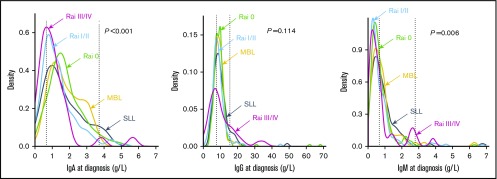
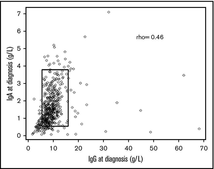
In all, 511 patients (77%) had baseline immunoglobulins measured at diagnosis (or within the first 3 months) and 23% of patients had reduced IgG, 16% reduced IgA, and 54% reduced IgM values (Table 2). There was a progressive increase in the incidence of reduced IgG or IgA with advancing Rai stage, but this was not observed for IgM. IgM levels were reduced by 43% to 63% in patients with MBL, CLL, or SLL. In contrast, the decreases in IgG and IgA were similar for MBL and Rai 0 CLL, whereas patients with SLL had a reduction in IgA and IgG similar to that in patients with Rai 0 to II CLL. To visualize the immunoglobulin distribution within each disease group, the continuous variation in immunoglobulin levels was examined by density plots (Figure 1). The majority of patients had IgM values clustering below the normal range. In contrast, the majority of the IgG and IgA values were within the low-normal range. IgA and IgG levels tended to congregate together (ρ, 0.46), with most cases congregating at the lower levels of normal or below the normal range (Figure 2).
Table 2.
Immunoglobulin levels in MBL, CLL, and SLL patients at diagnosis
| Immunoglobulin | CLL, n (%) | SLL, n (%) | MBL, n (%) | All , N (%) | ||
|---|---|---|---|---|---|---|
| Rai 0 | Rai I/II | Rai III/IV | ||||
| IgG | ||||||
| Low | 36 (18) | 36 (29) | 13 (39) | 17 (21) | 13 (19) | 115 (23) |
| Normal | 162 (80) | 87 (67) | 14 (42) | 58 (71) | 51 (78) | 372 (73) |
| High | 4 (2) | 6 (4) | 6 (18) | 6 (8) | 2 (3) | 24 (5) |
| Total patients | 202 | 129 | 33 | 81 | 66 | 511 |
| IgA | ||||||
| Low | 19 (9) | 29 (23) | 12 (37) | 16 (20) | 6 (9) | 82 (16) |
| Normal | 174 (87) | 95 (73) | 17 (53) | 60 (74) | 60 (91) | 406 (80) |
| High | 8 (4) | 5 (4) | 3 (9) | 5 (6) | 0 (0) | 21 (4) |
| Total patients | 201 | 129 | 32 | 81 | 66 | 509 |
| IgM | ||||||
| Low | 112 (55) | 82 (63) | 20 (61) | 35 (43) | 29 (45) | 278 (54) |
| Normal | 85 (42) | 42 (33) | 9 (27) | 43 (53) | 33 (50) | 212 (42) |
| High | 5 (3) | 5 (4) | 4 (12) | 3 (4) | 3 (5) | 20 (4) |
| Total patients | 202 | 129 | 33 | 81 | 65 | 510 |
Figure 1.
Distribution of IgG, IgA, and IgM baseline levels in the cohort, with hatched vertical lines showing normal ranges. IgM levels were most frequently reduced (P = .006), followed by IgA (P = .001) whereas the reductions in IgG were least significant (0.114). Most patients had levels in the low-normal range. About 5% of patients had increased levels.
Figure 2.
Relationship between baseline IgG and IgA levels and by Spearman’s ρ calculation (r = 0.46). The boxed area shows the normal ranges, and a small percentage of patients had abnormal levels of 1 immunoglobulin and normal levels of the other.
Elevated levels of immunoglobulin occurred in ∼5% of patients, affecting all 3 classes of immunoglobulin (Table 2). In CLL, high immunoglobulin levels (IgG, IgA, and IgM) occurred more frequently in patients with advanced Rai stage. In MBL, high IgG and IgM occurred at rates similar to those in patients with Rai stage 0 CLL, and no patients had elevated IgA. Increased immunoglobulin levels occurred in 4% to 8% of SLL patients (Table 2). To determine the potential cause for the increased levels of Ig, serum electrophoresis was assessed in each patient who had known immunoglobulin levels (Table 3). For IgG and IgM, about half the patients had a paraprotein, which usually had light chain restriction similar to that of the CLL cells (CLL-like). In contrast, for IgA, half the patients had oligoclonal bands, and a single paraprotein was less common. Thirty-six patients with elevated immunoglobulins required treatment: 34 patients received chemotherapy for progressive CLL, 1 for Richter transformation, and 1 for concomitant multiple myeloma.
Table 3.
Causes of increased immunoglobulinsootnote:
| Increased IgM, n (%) | Increased IgA, n (%) | Increased IgG, n (%) | |
|---|---|---|---|
| Total, n /N (%) | 20/510 (4) | 21/509 (4) | 24/511 (5) |
| M-bands | |||
| CLL-type | 9 (45) | 2 (9.5) | 7 (29) |
| Non-CLL type | 3 (15) | 1 (4.5) | 3 (12.5) |
| Oligoclonal bands | 3 (15) | 8 (38) | 6 (25) |
| Polyclonal | 2 (10) | 5 (24) | 3 (12.5) |
| Unknown | 3 (15) | 5 (24) | 5 (21) |
Abnormal immunoglobulin levels and prognostic markers
A significant correlation was observed between abnormal (reduced or increased) levels of all immunoglobulins (IgA, IgG, IgM) and the diagnosis of MBL, SLL, or CLL (Table 4). Abnormal IgA levels showed a highly significant positive correlation with elevated B2M levels (P = .005) and CD38 positivity (P < .001), both of which are markers of aggressive disease (Table 4). Abnormal IgM levels correlated with increased CD38 positivity (P = .034) and mutated IGHV (P = .005). In contrast, abnormal IgG levels did not correlate with any of these prognostic markers apart from advancing Rai stage. Unfortunately, fluorescence in situ hybridization analysis was not carried out at diagnosis, so correlations with genomic abnormalities could not be determined.
Table 4.
Differences between baseline immunoglobulin levels and prognostic markers
| Variables | IgA, n (%) | IgG | IgM, n (%) | ||||||
|---|---|---|---|---|---|---|---|---|---|
| Abnormal (n = 103) | Normal (n = 406) | P | Abnormal (n = 139) | Normal (n = 372) | P | Abnormal (n = 298) | Normal (n = 212) | P | |
| Ratio of males to females | 1.5:1 | 1.8:1 | .457 | 1.5:1 | 1.8:1 | .562 | 1.8:1 | 1.6:1 | .585 |
| Median age (range), y | 70 (40-94) | 68 (34-99) | .081 | 68 (40-90) | 68 (34-99) | .58 | 68 (34-99) | 68 (44-93) | .646 |
| CLL Rai stage | <.001 | <.001 | .008 | ||||||
| 0 | 27 (26) | 174 (43) | 40 (29) | 162 (44) | 117 (39) | 85 (40) | |||
| I/II | 34 (33) | 95 (23) | 42 (30) | 87 (23) | 87 (29) | 42 (20) | |||
| III/IV | 15 (15) | 17 (4) | 19 (14) | 14 (4) | 24 (8) | 9 (4) | |||
| MBL | 6 (6) | 60 (15) | 15 (11) | 51 (14) | 32 (11) | 33 (16) | |||
| SLL | 21 (20) | 60 (15) | 23 (17) | 58 (16) | 38 (13) | 43 (20) | |||
| LDT, mo | .071 | .269 | .899 | ||||||
| >12 | 77 (75) | 331 (82) | 106 (76) | 303 (81) | 237 (80) | 33 (16) | |||
| 6-12 | 12 (12) | 44 (11) | 17 (12) | 40 (11) | 32 (11) | 44 (21) | |||
| <6 | 12 (12) | 22 (5) | 13 (9) | 21 (6) | 21 (7) | 22 (10) | |||
| ZAP-70 positive/ZAP-70 negative | 31/49 (30/48) | 92/245 (23/60) | .06 | 35/78 (25/56) | 88/218 (24/59) | .748 | 74/177 (25/59) | 49/118 (23/56) | 1 |
| CD38 positive/CD38 negative | 39/47 (38/46) | 84/260 (21/64) | <.001 | 41/76 (30/55) | 82/233 (22/63) | .085 | 83/174 (28/58) | 39/135 (18/64) | .034 |
| B2M | .005 | .169 | .723 | ||||||
| High | 55 (53) | 141 (35) | 65 (47) | 131 (35) | 115 (39) | 81 (38) | |||
| Normal | 21 (20) | 124 (31) | 37 (27) | 108 (29) | 89 (30) | 56 (26) | |||
| IGHV mutated/unmutated | 38/36 (37/35) | 168/112 (41/28) | .227 | 56/47 (40/34) | 150/103 (40/28) | .463 | 138/78 (46/26) | 67/72 (32/34) | .005 |
All data are n (%) of cases where data available, unless otherwise indicated. Abnormal immunoglobulins include patients with both high and low levels.
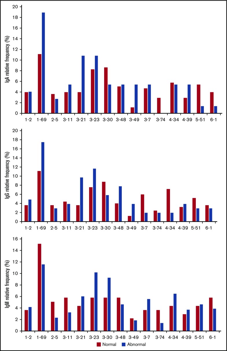
Because it has recently been demonstrated that serum IgM and IgG paraproteins in CLL are associated with increased use of IGHV3-74 and IGHV4-39, respectively, we evaluated IGHV gene family use in patients with abnormal immunoglobulins.21 Abnormal levels of IgG and IgA were associated with overrepresentation of IGHV1-69, IGHV3-21, and IGHV3-49, whereas this pattern was not seen for IgM (Figure 3). Reduced levels of IgG have previously been associated with IGHV3-21 and 3-23.21 In contrast, patients with abnormal IgA seemed less likely to use IGHV3-7 and IGHV4-34 than patients with abnormal IgG. Statistical analysis was not feasible because of the small sample size, but these results suggest that abnormal levels of IgA or IgG may be associated with specific IGHV gene subtypes, which in turn may be associated with more aggressive and advanced disease.
Figure 3.
Normal and abnormal expression of IgG, IgA, and IgM within the different IGHV family subfamilies. Relative frequency is the percentage of patients within the patient population.
Predictive value of abnormal immunoglobulins for TTFT, OS, and immunoglobulin replacement
Results from both imputed and unimputed data were compared in these analyses to demonstrate the impact of missing data and imputations (supplemental Tables 1 and 2). The ratios produced from the OS analyses were similar between unimputed and imputed data, suggesting that the imputations could have been properly specified. Ratios produced for the TTFT and TTIR analyses were closer to the null effect for the immunoglobulin variables in the imputed data than in the unimputed data. This could indicate that the imputations were partially misspecified. However, the differences between unimputed and imputed data for immunoglobulin variables were much greater when using alternative imputation approaches (passive imputation and “just another variable”).33
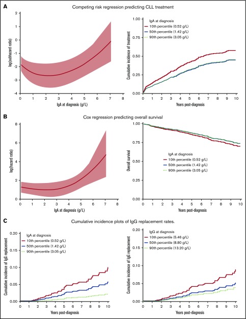
By using multivariable analysis and the imputed model, we showed that several baseline variables (supplemental Table 1) correlated with shorter TTFT. Variables associated with a shorter TTFT were low IgA levels (subdistribution hazard ratio [SHR], 0.34/1.09; 95% CI, 0.18-0.64/1.04-1.14; P = .001), unmutated IGHV (SHR, 3.01; 95% CI, 2.19-4.15; P < .001), and high B2M level (SHR, 1.57; 95% CI, 1.17-2.10; P = .002) (see supplemental Table 1 for details). Additional significant variables, compared with MBL, included SLL (SHR, 7.32; 95% CI, 3.58-14.99; P ≤ .001), Rai stages III to IV CLL (SHR, 12.24; 95% CI, 5.50-27.25; P ≤ .001), and Rai stages I to II CLL (SHR, 8.25; 95% CI, 4.17-16.30; P ≤ .001). Interestingly, a profound U-shaped relationship was observed between baseline IgA and TTFT (Figure 4A, left graph), suggesting that patients with low and high or very high IgA levels at diagnosis have more aggressive or advanced disease, which necessitates earlier treatment. Patients with low IgA values (10th percentile) had substantially more need for treatment than patients with values at the 50th and 90th percentiles who had similar risk (Figure 4A, right graph). Thus, independent prognostic markers for TTFT included either very high IgA (>4 g/L) or low IgA (<1 g/L) (Figure 4A, left graph), unmutated IGHV, high B2M level, and advanced disease.
Figure 4.
Influence of immunoglobulin levels on clinical outcome. The relationship between altered levels of IgA and TTFT (A), OS (B), and requirement for immunoglobulin replacement (C) for severe infections.
By using multivariable imputed models, prognostic factors at diagnosis that significantly influenced survival included abnormal IgA level (SHR, 0.81/1.01; 95% CI, 0.61-1.08/1.00-1.03; P = .026), sex (SHR, 0.52; 95% CI, 0.38-0.73; P < .001), advancing age (SHR, 1.72; 95% CI, 1.52-1.95; P < .001), high B2M level (SHR, 3.01; 95% CI, 1.84-4.92; P < .001), advancing Rai stage (P = .045), and treatment for CLL (SHR, 1.83; 95% CI, 1.29-2.59; P = .001) (supplemental Table 2). Interestingly, the plot based on imputed data showed a U-shaped nonlinear relationship between the continuum of IgA level at diagnosis and OS, indicating that decreased survival was associated with both low and very high levels of the immunoglobulin (Figure 4B, left graph). However, the Kaplan-Meier plot showing OS demonstrated that survival rates differed slightly between patients with IgA values at the 10th, 50th, and 90th percentiles (Figure 4B, right graph). Thus, an abnormal IgA level, high B2M level, male sex, older age, advanced disease, and need for CLL or SLL treatment were independently associated with decreased survival.
Abnormal low levels of IgG (P = .047) or IgA (P = .031) at diagnosis were marginally significant for predicting the need for immunoglobulin replacement (supplemental Table 3). However, the number of patients with elevated IgG and IgA at diagnosis was low. Future need for IgG replacement also significantly correlated with increasing CIRS score (P < .001), advancing age (P = .001), and need for CLL treatment (P = .002) (supplemental Table 3). The cumulative incidence plot for IgA (Figure 4C, left graph) demonstrates the rates of immunoglobulin replacement for the 10th, 50th, and 90th percentile values showing the close relationship between baseline IgA and subsequent need for immunoglobulin replacement. In contrast, the cumulative incidence plot for IgG (Figure 4C, right graph) shows that patients at the 10th percentile value of IgG had an increased rate of immunoglobulin replacement compared with those who had either 50th or 90th percentile values, which were similar. Thus, there is a closer relationship between absolute level of IgA at diagnosis and need for replacement therapy.
Discussion
Findings in this study indicate the importance of measuring immunoglobulin levels in CLL and its variants at diagnosis. Previous studies have focused on the clinical and prognostic relevance of low immunoglobulin levels and have been limited by sample size and composition, typically excluding patients with MBL and SLL. Our retrospective review included these patient populations and examined the significance of both low and high immunoglobulin levels at the time of diagnosis. Highly significant differences were observed between abnormal IgG or IgA (either increased or decreased) in MBL, SLL, and in the different Rai stages of CLL at diagnosis, whereas the difference was less marked with IgM.
We have confirmed that the immunoglobulins are decreased in a significant number of patients with CLL, SLL, and MBL at diagnosis, with half the patients reporting low IgM (54%), 23% low IgG, and IgA (the least likely to be reduced) at 16%. Interestingly, significant reductions in immunoglobulins (IgG, 19%; IgA, 9%; IgM, 45%) were even seen in the 66 MBL patients. Glancy et al34 previously noted low IgG in one-third of patients with MBL but did not report on IgA or IgM levels. In this study, the degree of suppression correlated with tumor burden; low IgG and IgA levels were more likely to be present in patients with CLL than in patients with MBL, and there was a greater decline in those patients with advanced Rai stage. The decreases in IgG and IgA levels in SLL were similar to those in Rai stage 0 to II CLL. In contrast, IgM reduction occurred at equal frequency in all patients. The reason for this is unclear, but it is unlikely to be related to aging, because IgM levels remain stable throughout life, whereas IgG and IgA levels actually increase with age.35,36
Elevated immunoglobulin levels were observed in about 5% of patients, with the incidence depending on tumor burden, being most frequent in patients with advanced Rai stage CLL. Serum protein electrophoresis reported a monoclonal band (M-band) in half the patients with elevated IgG and IgM, with most bands being CLL-like. However, although IgA paraproteins were uncommon, oligoclonal bands were frequently seen with increased IgA.21,37 By using a highly sensitive immunoblotting technique, Beaume et al37 showed that multiple monoclonal proteins can be detected in 80% of patients with early-stage CLL, with a mean of 3.3 bands in affected patients. These oligoclonal bands are also present in the elderly and in individuals with immune abnormalities, suggesting that oligoclonal banding in CLL is related to the presence of multiple B-cell clones as a result of T-cell dysfunction. Thus, the presence of hypogammaglobulinemia and increased immunoglobulin levels with oligoclonal bands likely reflect underlying immune deficiency.
Recent studies have demonstrated that low levels of IgG, IgA, or IgM may predict for TTFT or OS by univariable analysis, but the correlations were usually lost with multivariable analysis.18,19 Parikh et al14 did note, however, that reduced IgG at diagnosis predicted for TTFT by multivariable analysis, but not OS. An important observation in our study was the association of either low or high IgA levels at diagnosis with shortened TTFT and OS, with abnormal levels being an independent prognostic factor in multivariable analysis. These findings support previous observations that reduced levels of IgA at diagnosis are an independent predictor of TTFT in CLL but also demonstrate the additional prognostic relevance of increased levels of IgA.11,17 As with IgG and IgM, abnormal levels of IgA correlated with disease burden, but IgA was unique in that abnormal levels also significantly correlated with increased B2M levels and CD38 positivity. B2M is noncovalently linked to the α-chain of the class I major histocompatibility complex on all nucleated cells, including CLL cells, increasing with tumor burden, renal dysfunction, and elevated plasma inflammatory cytokines (interleukin-6 [IL-6], IL-8, and tumor necrosis factor).38 In contrast, CD38 is involved in cell proliferation and survival.39 In addition, although abnormal IgM levels highly correlated with mutated IGHV, this was not observed with IgG or IgA. However, in contradistinction to IgM, altered levels of IgG and IgA were associated with IGHV1-69 and IGHV3-21, subgroups that are associated with aggressive disease. These results suggest that alterations in the immunoglobulins are likely a function of the CLL cell of origin and tumor burden. The patient numbers in the other IGHV subgroups were too small to make a meaningful comparison between IgG and IgA. IgA differs from the other immunoglobulins in that recombination occurs in Peyer’s patches in the distal ileum, other gut lymphoid tissue, and nasopharyngeal lymphoid tissue, with subsequent passage of the memory B cells to mucosal tissue where they may become secreting plasma cells.40 Thus, alterations in plasma IgA levels may reflect involvement of these sites by CLL.
In our study, 7.4% of patients required immunoglobulin replacement, almost always for frequent or severe infections, and independent prognostic factors for replacement therapy were advancing age, high CIRS score, chemotherapy, or abnormal levels of IgA or IgG at diagnosis. Moreover, when stratified according to the level of immunoglobulin, patients with the lowest levels of IgG or IgA had the greatest likelihood of requiring replacement. Similarly, Anderson et al41 and Crassini et al42have demonstrated that low levels of IgA at diagnosis is a better predictor of serious infections than low levels of IgG or IgM. Infections in CLL usually involve the respiratory and urinary tracts, areas that are protected by locally secreted dimeric IgA and IgG from the systemic circulation.40,43 Despite the fact that IgA is produced and secreted by the mucosa, plasma IgA levels do predict for the need for immunoglobulin replacement, which suggests that they correlate with secreted levels. This is an important finding because although patients may not be experiencing infectious complications at the time of diagnosis, those patients with low levels of IgG and IgA should be counseled more thoroughly regarding their future risk of infections and the importance of early assessment and intervention. As previously reported,11,14 immunoglobulin levels also tend to decrease during the course of CLL, and patients have a high incidence of infections early in their disease, even before treatment. These infections are a significant problem, and they predict for reduced survival.41,42 The fact that the Bruton tyrosine kinase inhibitor ibrutinib can increase plasma IgA levels in CLL suggests that early used of this agent in patients presenting with low or decreasing levels of IgA may prevent these early infections.44,45
The mechanisms responsible for decreased immunoglobulins in CLL at diagnosis is still unclear, but the CLL cells themselves or factors secreted by the CLL cells may suppress normal B and plasma cells.2 CLL cells may have a direct effect on plasma cells through the surface expression of Fas ligand, CD27, and PD-L1.2 In addition, transforming growth factor-β is secreted by CLL and marrow stromal cells in CLL and is a potent inhibitor of normal B and T cells.46,47 Finally, CLL cells have features similar to those of regulatory B cells, producing IL-10 which has immunosuppressive properties.48,49 Interestingly, IL-10 production has been shown to be greater in IGHV-mutated cells than in unmutated cells as a result of reduced DNA methylation.49 Plasma levels of IL-10 are similar in IGHV-mutated and unmutated disease, possibly reflecting the greater tumor burden with unmutated disease.49 However, although chemoimmunotherapy could further reduce immunoglobulin levels, presumably through effects on normal B cells, agents acting through the microenvironment may actually improve these levels.44,50
In summary, this study demonstrates that alterations in IgG and IgA at diagnosis reflect both CLL biology and tumor burden. Although abnormalities in both immunoglobulins predict for subsequent infections, IgA is unique because alterations reflect aggressive disease with shortened TTFT and OS.
Supplementary Material
The full-text version of this article contains a data supplement.
Acknowledgment
The authors thank Mandy Squires, Jayce Bi, Yun Li, and Laurie Lange from the Manitoba Tumor Bank for compiling clinical information and processing clinical samples and Donna Hewitt for obtaining informed consent.
This work was supported by grants from the Research Manitoba and CancerCare Manitoba Foundation. Data collection and the database were supported by grants from Gilead and Janssen pharmaceuticals.
Authorship
Contribution: J.B.J., G.I., and E.S. contributed to study design, literature review, data interpretation, and preparation of the manuscript; P.L. performed statistical analyses; and S.B.G., V.B., H.S.D., S.M.M., and A.J.M. provided conceptual advice and editing.
Conflict-of-interest disclosure: The authors declare no competing financial interests.
Correspondence: James B. Johnston, Section of Hematology/Oncology, Department of Internal Medicine, University of Manitoba, CancerCare Manitoba, 675 McDermot Ave, Winnipeg, MB R3E 0V9, Canada; e-mail: james.johnston@cancercare.mb.ca.
References
- 1.Morrison VA. Infectious complications of chronic lymphocytic leukaemia: pathogenesis, spectrum of infection, preventive approaches. Best Pract Res Clin Haematol. 2010;23(1):145-153. [DOI] [PubMed] [Google Scholar]
- 2.Forconi F, Moss P. Perturbation of the normal immune system in patients with CLL. Blood. 2015;126(5):573-581. [DOI] [PubMed] [Google Scholar]
- 3.Riches JC, Gribben JG; Potential Clinical Implications . Understanding the immunodeficiency in chronic lymphocytic leukemia: potential clinical implications. Hematol Oncol Clin North Am. 2013;27(2):207-235. [DOI] [PubMed] [Google Scholar]
- 4.Tsai HT, Caporaso NE, Kyle RA, et al. . Evidence of serum immunoglobulin abnormalities up to 9.8 years before diagnosis of chronic lymphocytic leukemia: a prospective study. Blood. 2009;114(24):4928-4932. [DOI] [PMC free article] [PubMed] [Google Scholar]
- 5.Moreira J, Rabe KG, Cerhan JR, et al. . Infectious complications among individuals with clinical monoclonal B-cell lymphocytosis (MBL): a cohort study of newly diagnosed cases compared to controls. Leukemia. 2013;27(1):136-141. [DOI] [PubMed] [Google Scholar]
- 6.Solomon BM, Chaffee KG, Moreira J, et al. . Risk of non-hematologic cancer in individuals with high-count monoclonal B-cell lymphocytosis. Leukemia. 2016;30(2):331-336. [DOI] [PMC free article] [PubMed] [Google Scholar]
- 7.Compagno N, Malipiero G, Cinetto F, Agostini C. Immunoglobulin replacement therapy in secondary hypogammaglobulinemia. Front Immunol. 2014;5:626. [DOI] [PMC free article] [PubMed] [Google Scholar]
- 8.Visentin A, Compagno N, Cinetto F, et al. . Clinical profile associated with infections in patients with chronic lymphocytic leukemia. Protective role of immunoglobulin replacement therapy. Haematologica. 2015;100(12):e515-e518. [DOI] [PMC free article] [PubMed] [Google Scholar]
- 9.Freeman JA, Crassini KR, Best OG, et al. . Immunoglobulin G subclass deficiency and infection risk in 150 patients with chronic lymphocytic leukemia. Leuk Lymphoma. 2013;54(1):99-104. [DOI] [PubMed] [Google Scholar]
- 10.Ben-Bassat I, Many A, Modan M, Peretz C, Ramot B. Serum immunoglobulins in chronic lymphocytic leukemia. Am J Med Sci. 1979;278(1):4-9. [DOI] [PubMed] [Google Scholar]
- 11.Rozman C, Montserrat E, Viñolas N. Serum immunoglobulins in B-chronic lymphocytic leukemia. Natural history and prognostic significance. Cancer. 1988;61(2):279-283. [DOI] [PubMed] [Google Scholar]
- 12.Shvidel L, Tadmor T, Braester A, et al. ; Israeli CLL Study Group . Serum immunoglobulin levels at diagnosis have no prognostic significance in stage A chronic lymphocytic leukemia: a study of 1113 cases from the Israeli CLL Study Group. Eur J Haematol. 2014;93(1):29-33. [DOI] [PubMed] [Google Scholar]
- 13.Lee JS, Dixon DO, Kantarjian HM, Keating MJ, Talpaz M. Prognosis of chronic lymphocytic leukemia: a multivariate regression analysis of 325 untreated patients. Blood. 1987;69(3):929-936. [PubMed] [Google Scholar]
- 14.Parikh SA, Leis JF, Chaffee KG, et al. . Hypogammaglobulinemia in newly diagnosed chronic lymphocytic leukemia: Natural history, clinical correlates, and outcomes. Cancer. 2015;121(17):2883-2891. [DOI] [PMC free article] [PubMed] [Google Scholar]
- 15.Hansen DA, Robbins BA, Bylund DJ, Piro LD, Saven A, Ellison DJ. Identification of monoclonal immunoglobulins and quantitative immunoglobulin abnormalities in hairy cell leukemia and chronic lymphocytic leukemia. Am J Clin Pathol. 1994;102(5):580-585. [DOI] [PubMed] [Google Scholar]
- 16.Baliakas P, Xochelli A, Minga E, et al. . Revisiting hypogammaglobulinemia in chronic lymphocytic leukemia: A combined clinicobiological approach [abstract]. Blood. 2014;124(21):Abstract 5633. [Google Scholar]
- 17.Reda G, Cassin R, Levati G, et al. . Hypogammaglobulinemia in chronic lymphocytic leukemia: A predictor of outcome? [abstract]. Blood. 2017;130(suppl 1):Abstract 2997. [Google Scholar]
- 18.Andersen MA, Vojdeman FJ, Andersen MK, et al. . Hypogammaglobulinemia in newly diagnosed chronic lymphocytic leukemia is a predictor of early death. Leuk Lymphoma. 2016;57(7):1592-1599. [DOI] [PubMed] [Google Scholar]
- 19.Crassini KR, Zhang E, Balendran S, et al. . Humoral immune failure defined by immunoglobulin class and immunoglobulin G subclass deficiency is associated with shorter treatment-free and overall survival in Chronic Lymphocytic Leukaemia. Br J Haematol. 2018;181(1):97. [DOI] [PubMed] [Google Scholar]
- 20.Hensel M, Kornacker M, Yammeni S, Egerer G, Ho AD. Disease activity and pretreatment, rather than hypogammaglobulinaemia, are major risk factors for infectious complications in patients with chronic lymphocytic leukaemia. Br J Haematol. 2003;122(4):600-606. [DOI] [PubMed] [Google Scholar]
- 21.Rizzo D, Chauzeix J, Trimoreau F, et al. . IgM peak independently predicts treatment-free survival in chronic lymphocytic leukemia and correlates with accumulation of adverse oncogenetic events. Leukemia. 2015;29(2):337-345. [DOI] [PubMed] [Google Scholar]
- 22.Hallek M, Cheson BD, Catovsky D, et al. ; International Workshop on Chronic Lymphocytic Leukemia . Guidelines for the diagnosis and treatment of chronic lymphocytic leukemia: a report from the International Workshop on Chronic Lymphocytic Leukemia updating the National Cancer Institute-Working Group 1996 guidelines. Blood. 2008;111(12):5446-5456. [DOI] [PMC free article] [PubMed] [Google Scholar]
- 23.Salvi F, Miller MD, Grilli A, et al. . A manual of guidelines to score the modified cumulative illness rating scale and its validation in acute hospitalized elderly patients. J Am Geriatr Soc. 2008;56(10):1926-1931. [DOI] [PubMed] [Google Scholar]
- 24.Kost SE, Bouchard ED, LaBossière É, et al. . Cross-resistance and synergy with bendamustine in chronic lymphocytic leukemia. Leuk Res. 2016;50:63-71. [DOI] [PubMed] [Google Scholar]
- 25.Rosenquist R, Ghia P, Hadzidimitriou A, et al. . Immunoglobulin gene sequence analysis in chronic lymphocytic leukemia: updated ERIC recommendations. Leukemia. 2017;31(7):1477-1481. [DOI] [PMC free article] [PubMed] [Google Scholar]
- 26.Altman DG, Royston P. The cost of dichotomising continuous variables. BMJ. 2006;332(7549):1080. [DOI] [PMC free article] [PubMed] [Google Scholar]
- 27.Royston P, Altman DG, Sauerbrei W. Dichotomizing continuous predictors in multiple regression: a bad idea. Stat Med. 2006;25(1):127-141. [DOI] [PubMed] [Google Scholar]
- 28.Senn SJ. Dichotomania: an obsessive compulsive disorder that is badly affecting the quality of analysis of pharmaceutical trials. In: Proceedings of The 55th Session of the International Statistical Institute Sydney; 6-12 April 2005; Sydney, Australia. [Google Scholar]
- 29.Bartlett JW, Seaman SR, White IR, Carpenter JR; Alzheimer’s Disease Neuroimaging Initiative . Multiple imputation of covariates by fully conditional specification: Accommodating the substantive model. Stat Methods Med Res. 2015;24(4):462-487. [DOI] [PMC free article] [PubMed] [Google Scholar]
- 30.White IR, Royston P. Imputing missing covariate values for the Cox model. Stat Med. 2009;28(15):1982-1998. [DOI] [PMC free article] [PubMed] [Google Scholar]
- 31.White IR, Royston P, Wood AM. Multiple imputation using chained equations: Issues and guidance for practice. Stat Med. 2011;30(4):377-399. [DOI] [PubMed] [Google Scholar]
- 32.Beiggi S, Banerji V, Deneka A, Griffith J, Gibson SB, Johnston JB. Comparison of outcome of patients with CLL who are referred or nonreferred to a specialized CLL clinic: a Canadian population-based study. Cancer Med. 2016;5(6):971-079. [DOI] [PMC free article] [PubMed] [Google Scholar]
- 33.Von Hippel PT. How to impute interactions, squares, and other transformed variables. Sociol Methodol. 2009;39(1):265-291. [Google Scholar]
- 34.Glancy E, Siles R. Monoclonal B-cell lymphocytosis and hypogammaglobulinaemia. Br J Haematol. 2016;173(2):316-317. [DOI] [PubMed] [Google Scholar]
- 35.Buckley CE III, Dorsey FC. Serum immunoglobulin levels throughout the life-span of healthy man. Ann Intern Med. 1971;75(5):673-682. [DOI] [PubMed] [Google Scholar]
- 36.Cassidy JT, Nordby GL, Dodge HJ. Biologic variation of human serum immunoglobulin concentrations: sex-age specific effects. J Chronic Dis. 1974;27(11-12):507-516. [DOI] [PubMed] [Google Scholar]
- 37.Beaume A, Brizard A, Dreyfus B, Preud’homme JL. High incidence of serum monoclonal Igs detected by a sensitive immunoblotting technique in B-cell chronic lymphocytic leukemia. Blood. 1994;84(4):1216-1219. [PubMed] [Google Scholar]
- 38.Yoon JY, Lafarge S, Dawe D, et al. . Association of interleukin-6 and interleukin-8 with poor prognosis in elderly patients with chronic lymphocytic leukemia. Leuk Lymphoma. 2012;53(9):1735-1742. [DOI] [PubMed] [Google Scholar]
- 39.Malavasi F, Deaglio S, Damle R, Cutrona G, Ferrarini M, Chiorazzi N. CD38 and chronic lymphocytic leukemia: a decade later. Blood. 2011;118(13):3470-3478. [DOI] [PMC free article] [PubMed] [Google Scholar]
- 40.Macpherson AJ, McCoy KD, Johansen FE, Brandtzaeg P. The immune geography of IgA induction and function. Mucosal Immunol. 2008;1(1):11-22. [DOI] [PubMed] [Google Scholar]
- 41.Andersen MA, Eriksen CT, Brieghel C, et al. . Incidence and predictors of infection among patients prior to treatment of chronic lymphocytic leukemia: a Danish nationwide cohort study. Haematologica. 2018;103(7):e300-e303. [DOI] [PMC free article] [PubMed] [Google Scholar]
- 42.Crassini KR, Best OG, Mulligan SP. Immune failure, infection and survival in chronic lymphocytic leukemia. Haematologica. 2018;103(7):e329. [DOI] [PMC free article] [PubMed] [Google Scholar]
- 43.Brandtzaeg P. Secretory immunity with special reference to the oral cavity. J Oral Microbiol. 2013;5:20401. [DOI] [PMC free article] [PubMed] [Google Scholar]
- 44.Sun C, Tian X, Lee YS, et al. . Partial reconstitution of humoral immunity and fewer infections in patients with chronic lymphocytic leukemia treated with ibrutinib. Blood. 2015;126(19):2213-2219. [DOI] [PMC free article] [PubMed] [Google Scholar]
- 45.Andersen MA, Niemann CU. Immune failure, infection and survival in chronic lymphocytic leukemia in Denmark. Haematologica. 2018;103(7):e330. [DOI] [PMC free article] [PubMed] [Google Scholar]
- 46.Kremer JP, Reisbach G, Nerl C, Dörmer P. B-cell chronic lymphocytic leukaemia cells express and release transforming growth factor-beta. Br J Haematol. 1992;80(4):480-487. [DOI] [PubMed] [Google Scholar]
- 47.Lagneaux L, Delforge A, Dorval C, Bron D, Stryckmans P. Excessive production of transforming growth factor-beta by bone marrow stromal cells in B-cell chronic lymphocytic leukemia inhibits growth of hematopoietic precursors and interleukin-6 production. Blood. 1993;82(8):2379-2385. [PubMed] [Google Scholar]
- 48.Rossi M, Gentile M, Toscano R, et al. . Enumeration of interleukin-10-positive B cells from peripheral blood of patients with chronic lymphocytic leukemia. Leuk Lymphoma. 2014;55(6):1394-1396. [DOI] [PubMed] [Google Scholar]
- 49.Drennan S, D’Avola A, Gao Y, et al. . IL-10 production by CLL cells is enhanced in the anergic IGHV mutated subset and associates with reduced DNA methylation of the IL10 locus. Leukemia. 2017;31(8):1686-1694. [DOI] [PubMed] [Google Scholar]
- 50.Strati P, Keating MJ, Wierda WG, et al. . Lenalidomide induces long-lasting responses in elderly patients with chronic lymphocytic leukemia. Blood. 2013;122(5):734-737. [DOI] [PMC free article] [PubMed] [Google Scholar]
Associated Data
This section collects any data citations, data availability statements, or supplementary materials included in this article.


