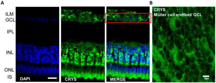
Figure 1.
Immunolocalization of αA-crystallin in the caiman retina. (A) αA-crystallin (CRYS, green) is expressed in the inner nuclear layer (INL) surrounding the nuclei of neurons (DAPI nuclear staining, blue). Crystallin is also localized surrounding ganglion cell nuclei (red box) in the ganglion cell layer and in the inner segment area of photoreceptors. (B) αA-crystallin observed from the top of whole-retina tissue. Crystallin is confined to the endfeet of Müller cells in the ganglion cell layer (white arrow). IPL, inner plexiform layer; ILM, inner limiting membrane; GCL, ganglion cell layer; INL, inner nuclear layer; ONL, outer nuclear layer; IS, inner segments of photoreceptors. Scale bar in A, 20 μm and in B, 10 μm.

