Abstract
Urethral leiomyoma is an infrequent benign tumor. Much more infrequent is recurrence. It has been described in exceptional cases. We report a rare case of a 46 year old woman who had a surgery for a urethral leiomyoma eight years ago. Now, she presents with nodulation in her vagina with no other symptoms. The patient underwent surgical excision of the tumor, and pathological examination revealed an recurrence of urethral leiomyoma.
Introduction
Leiomyoma is a benign tumor that arises from the smooth muscle. Uterus is the most common location among women during reproductive age. In the urethra it is extremely rare. Few more than 100 cases are reported to date. Recurrence has been described only in counted cases.1, 2, 3
Case presentation
A 46 year old woman was admitted to the Department of Urology (University Hospital Nuestra Señora de la Candelaria, Tenerife, Spain) presenting a protruding mass at the urethral opening. Her medical history revealed no chronic diseases. Eight years before she was operated by a excision of a mass in the same place. At that time, pathological examination revealed an urethral leiomyoma.
At the present time she presented a new nodulation in her vagina which was enlargering gradually for the last year, without any other symptoms. The physical examination showed a round and firm mass between the external urethral meatus and the vagina from 3 to 9 o'clock, measuring approximately 2 × 2 x 1,5 cm.
In order to complete the diagnosis, several complementary tests were performed. Cystourethroscopy was normal. Magnetic resonance imaging (MRI) showed a solid polylobulated lesion, well defined, measuring 29× 24 × 29 mm, arising from the posterior wall of the distal urethra. It appeared isointense to muscle on T1-weighted images and hypointense predominance on T2-weighted images (Fig. 1 (A,B)). After contrast administration it enhances heterogeneously, with no signs of diffusion restriction.
Fig. 1.
Magnetic resonance imaging showing solid polilobulated lesion in continuity with posterior wall of the distal urethra, which is hypointense predominance to muscle on T2-weighted image (A: axial image; B: coronal image).
Based on physical examination and MRI findings, a preoperative diagnosis of urethral leiomyoma recurrence was made.
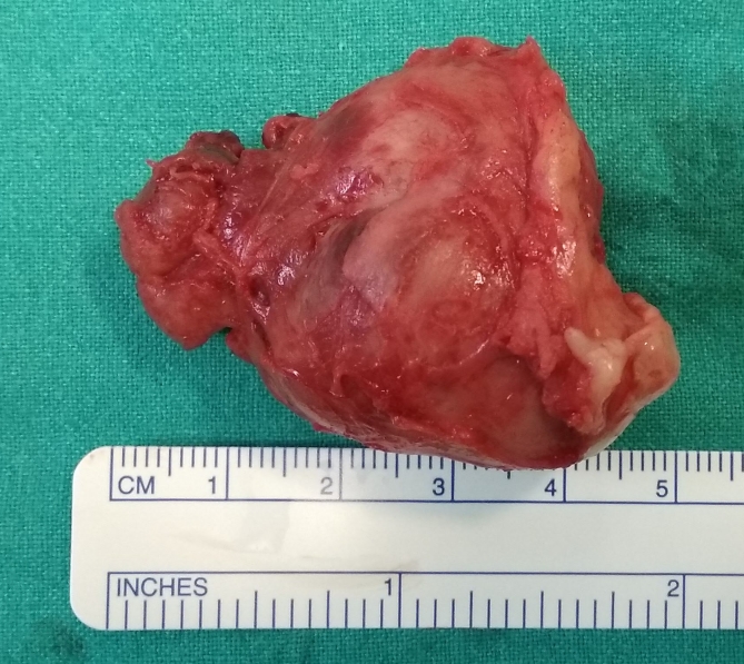
Surgical excision of the mass (Fig. 2) was performed under spinal anesthesia, in lithotomy position. A 16 French-Foley catheter was placed. Haemostatic solution with bupivacaine 0,25% and adrenaline diluted 1:200000 was injected over the vaginal mucosa. The lesion was excised through an U-shaped inverted suburethral incision. It was very attached to the posterior wall of the distal urethra, probably due to the previous surgery in the same place. At this point the urethra was injured, so it was repaired with a vicryl rapid 3.0 suture. Incision was closed using vicryl rapid 3.0 suture. She was discharged from the hospital on first postoperative day with Foley catheter.
Fig. 2.
Surgical specimen.
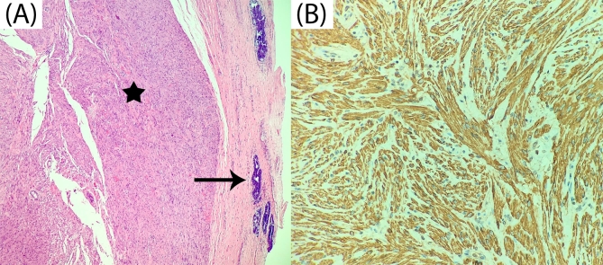
Histopathological examination (Fig. 3 (A,B)) revealed a round mass of 3,7 × 3,5 × 2,2 cm. It was composed of interlacing bundles of smooth muscle fibres. There was no mitotic activity or necrosis. Inmunohistochemical test demonstrated that the tumor were positive for actin and desmin. The proliferation index (Ki67) was around <2–3%. The features confirmed diagnosis of urethral leiomyoma.
Fig. 3.
(A) Microscopic histological image showing interlacing bundles of smooth muscle fibres without atypia or mitosis (star) with urothelial ducts of posterior wall of the distal urethra (arrow). Hematoxylin-eosin staining x100. (B) Positive staining for desmin, specific marker of smooth muscle, x400.
Discussion
The first case of urethral leiomyoma was described by Buttner in 1894. To date, around 120 cases have been reported in the literature.
The differential diagnosis of urethral masses includes bening tumors as urethral prolapse, urethral caruncle, diverticulum, paraurethral cyst, polyps, papillomas, hemangiomas, leiomyomas, fibromas, neurinomas and adenomas. Malignant causes as leiomyosarcoma, lymphoma, transitional cell carcinoma, squamous cell carcinoma, clear cell adenocarcinoma and metastasis have been also described.4
Urethral leiomyoma is most common in women during reproductive age. The most common location is proximal urethra. However, it may affect the distal segment, as in the case we report.
Despite around 20% of patients remain asymptomatic, the clinical picture includes a diversity of symptoms depending on mass location, as urinary tract infection, palpable mass, dyspareunia, acute urinary retention, irritative lower urinary tract symptoms, haematuria or vaginal bleeding.
The diagnosis is based on anamnesis and physical examination. Due to the anathomical proximity to anterior vaginal wall, it is important to perform a preoperative evaluation with imaging studies. MRI is the gold standar. It shows delineation anathomy of the mass and the relationship with surrounding organs. Typical findings on MRI are a hypointense lesion or isointense to muscle on T1-weighted and hyperintense or isointense to muscle on T2-weighted images. In this patient, as a particularity, it showed hypointense on T2 weighted. Final diagnosis is made by histopathological examination and no malignant transformation had being reported.
There is clear no protocol for management and treatment. Nonetheless, complete surgical excision has been performed as the first choice of treatment in all published cases. To date, only three patients have developed recurrence, and it has been treated with a repeated excision. In this case, there was hardly a margin with urethra and it was injured to get a complete resection. Probably the cause of recurrence was an incomplete first excision in order to avoid damage in the urethra.
Ozel et al.5 described clinical findings to differentiate urethral from paraurethral leiomyomas. They support that if urethra is damaged during excision, it is an urethral leiomyoma.
Conclusion
Recurrence of urethral leiomyoma is an exceptional picture. Location in distal urethra is unusual. Surgical excision is the treatment of choice and the objective is to achieve a complete excision, even with urethral damage, in order to avoid new recurrence.
Contributor Information
Marta Jiménez Navarro, Email: marta_jn12@hotmail.com.
Begoña Ballesta Martínez, Email: bballestamartinez@gmail.com.
Jonathan Rodríguez Talavera, Email: jonathantala@hotmail.com.
Adrián Amador Robayna, Email: adriamadorobayna@gmail.com.
References
- 1.Shen Y.H., Yang K. Recurrent huge leiomyoma of the urethra in a female patient: a case report. Oncol Lett. 2014;7:1933–1935. doi: 10.3892/ol.2014.1991. [DOI] [PMC free article] [PubMed] [Google Scholar]
- 2.Merrell R.W., Brown H.E. Recurrent urethral leiomyoma presenting as stress incontinence. Urology. 1981;17:588–589. doi: 10.1016/0090-4295(81)90082-0. [DOI] [PubMed] [Google Scholar]
- 3.Hoshii T., Hasegawa G., Ikeda Y. Angiomyomatous leiomyoma of a female urethral meatus recurrence after seven years of the resection: a Case Report. Urol Case Rep. 2016;9:24–26. doi: 10.1016/j.eucr.2016.08.006. [DOI] [PMC free article] [PubMed] [Google Scholar]
- 4.Harada K., Ishikawa Y., Fujiwara H. Female paraurethral leiomyoma successfully excised through a vaginal approach: a case report. J Obstet Gynaecol Res. 2018;44:1174–1176. doi: 10.1111/jog.13641. [DOI] [PubMed] [Google Scholar]
- 5.Ozel B., Ballard C. Urethral and paraurethral leiomyomas in the female patient. Int Urogynecol J Pelvic Floor Dysfunct. 2006;17:93–95. doi: 10.1007/s00192-005-1316-3. [DOI] [PubMed] [Google Scholar]


