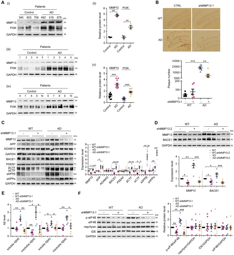
Figure 4.
Mmp13 knockdown ameliorates Alzheimer’s disease-associated pathology in APP/PS1 mice. (A) Representative western blots and bar plot summary of MMP13 and PI3K expression in the prefrontal cortex of control (n = 13) and Alzheimer’s disease patients (n = 13). Data in (i and ii) and in (iii–v) were from samples provided by Sydney Brain Bank and NIH NeuroBiobank, respectively. (B) Immunohistochemical images and bar plot summary of neuritic plaques in the hippocampi of wild-type (WT) and APP/PS1 mice (AD) treated with vehicle (CTRL) and Mmp13 shRNA-1 (shMMP13-1). Neuritic plaques were probed with the amyloid-β-specific monoclonal antibody 6E10 (n = 6). (C) Representative western blots and bar plot summary of MMP13, APP, ADAM10, BACE1, PSEN1, βCTF, αCTF, sAPPβ and sAPPα expression in the hippocampi of wild-type (n = 8), wild-type with Mmp13 shRNA-1 (WT-shMMP13-1, n = 8), Alzheimer’s disease (n = 8) and Alzheimer’s disease with Mmp13 shRNA-1 mice (AD-shMMP13-1, n = 8). The soluble fractions were used to detect sAPPβ and sAPPα with sAPPβ and 6E10 antibodies. (D) Representative western blots and bar plot summary of MMP13 and BACE1 expression in the hippocampi of wild-type (n = 6), wild-type with Mmp13 shRNA-2 (WT-shMMP13-2, n = 6), Alzheimer’s disease (n = 6) and Alzheimer’s disease with Mmp13 shRNA-2 mice (AD-shMMP13-2, n = 6). (E) ELISAs were used to measure soluble and insoluble amyloid-β40/42 levels in the brain homogenates of wild-type (n = 6), WT-shMMP13-1 (n = 6), Alzheimer’s disease (n = 6) and AD-shMMP13-1 (n = 6) mice. (F) Representative western blots and bar plot summary of p-eIF4B, eIF4B, neprilysin and insulin-degrading enzyme (IDE) levels in the hippocampi of wild-type (n = 8), WT-shMMP13-1 (n = 8), Alzheimer’s disease (n = 8) and AD-shMMP13-1 (n = 8) mice. All values were normalized to CTRL or wild-type (1.0) within each experiment. The error bars are the SEM. n.s. = no significant difference; *P < 0.05, **P < 0.01, ***P < 0.001 (ANOVA).

