Abstract
Resistance of pathogenic bacteria to standard antibiotics is an issue of great concern, and new treatments for bacterial infections are needed. Antimicrobial peptides (AMPs) are small, cationic, and amphipathic molecules expressed by metazoans that kill pathogens. They are a key part of the innate immune system in both vertebrates and invertebrates. Due to their low toxicity and broad antimicrobial activities, there has been increasing attention to their therapeutic usage. Our previous research demonstrated that four peptides—DAN1, DAN2, HOLO1 and LOUDEF1—derived from recently sequenced arthropod genomes exhibited potent antimicrobial effects in-vitro. In this study, we show that DAN2 protected 100% of mice when it was administered at a concentration of 20 mg/kg thirty minutes after the inoculation of a lethal dose of E. coli intraperitoneally. Lower concentrations of DAN2—10mg/kg and 5mg/kg protected more than 2/3s of the mice. All three dose levels reduced bacterial loads in blood and peritoneal fluid by 10-fold or more when counted six hours after bacterial challenge. We determined that DAN2 acts by compromising the integrity of the E. coli membrane. This study supports the potential of DAN2 peptide as a therapeutic agent for treating antibiotic resistant Gram-negative bacterial infections.
Introduction
Prior to the discovery of antibiotics, the majority of deaths in all age groups were associated with infection by pathogenic bacteria. Over the past 80 years, conventional antibiotics have been routinely used to treat bacterial infections, and this has markedly reduced morbidity and mortality from bacterial infections. The widespread feeding of antibiotics to farm animals for non-therapeutic purposes and an excessive use of antibiotics to treat people who often have no bacterial infection has selected for bacterial strains that resist multiple antibiotics. The threats from multidrug resistant bacteria such as Methicillin-resistant Staphylococcus aureus (MRSA), extended-spectrum beta-lactamase (ESBLs) multi-drug resistant tuberculosis (MDR), and all-antibiotic resistant superbug gonorrhea are increasing at an alarming rate worldwide [1,2]. According to the WHO report, there were 490,000 cases of multi-drug resistant TB globally in 2016 [2]. Increasing the available treatments for antibiotic resistant bacteria is necessary to prevent mortality and morbidity due to these infectious agents. In this context, the potential of antimicrobial peptides (AMPs) as promising candidates for novel antimicrobial agents deserves attention.
The biological world abounds in antimicrobial peptides since, for many organisms, they are the primary antimicrobial defense against bacteria, fungi and viruses. Over millions of years, natural selection of organisms that survive infections has driven the diversity of antimicrobial peptides, and metazoans have all evolved unique cohorts of antimicrobial peptides. The immense number, structural diversity, and multiple modes of action of these AMPs confers advantages when dealing with antibiotic resistance [3,4]. These peptides are usually 12–100 amino acids long, positively charged and amphipathic [5]. The positive charge is a consistent feature of these peptides and likely supports electrostatic interaction with negatively charged bacterial membranes. These highly positively charged amino acids rich in arginine and lysine have been shown to be important in AMP-mediated killing of various food borne pathogens [6]. More than 2,500 AMPs have been identified in various organisms [7]. Several peptides have been used clinically or are in clinical trials to treat bacterial infection, chronic wound healing, cystic fibrosis and other pathologies characterized by difficult-to-treat infections. Polymyxins, lipopeptides discovered in 1947, and colistin are cytotoxic peptides that are clinically used as last resort drugs for patients with multi-drug resistant bacterial infection [8]. The LTX series of synthetic antimicrobial peptides have promising antibacterial activity against Staphylococcus aureus infections [9]. Daptomycin, a lipopeptide, is active against Gram-positive bacteria only [10]. P-113, a histidine rich antimicrobial peptide that is originally derived from human saliva, has potent activity against fungal infections in HIV patients with oral candidiasis [11]. These peptides all display rapid action of killing and low minimum inhibitory concentrations (MIC) [8].
Every organism has a defense system against pathogenic infections [12]. The highly sophisticated vertebrate defense system utilizes both an innate and an adaptive immune system whereas invertebrates, for the most part, lack the latter [13]. AMPs are integral components of immunity in multicellular animals and play a major role in protecting these organisms from pathogens. Individual AMPs vary in efficacy against different classes of pathogens, but AMPs as a group have activity against all classes of single-cell pathogenic microorganisms, including Gram-positive and Gram-negative bacteria, protozoa, yeast, fungi, and viruses. Antimicrobial peptides are grouped structurally and by sequence and organism source as cecropins, insect-defensins, glycine-rich proteins, proline-rich proteins.
A variety of cecropins and insect defensins have been studied and reported in the literature [14,15]. Cecropins are a family of cationic antimicrobial peptides of 31–39 residues with a broad spectrum of activity against Gram-negative and Gram-positive bacteria, as well as fungi. They were first isolated from the hemolymph of the giant silk moth, Hyalophora cecropia [16]. They lack cysteine, so they cannot form disulfide bridges [17]. Another large group of AMPs, defensins are 28–42 (~4 kDa) cationic AMPs with six conserved cysteine residues that can form three disulfide bridges [15,18]. They typically affect Gram-positive bacteria [15,18].
In our previous research, we have identified putative insect AMPs by searching newly sequenced arthropod genomes using known AMPs as homology templates [19]. We screened six cecropin and defensin derived peptides-DAN1, DAN2, HOLO1, LOUDEF1, INVICT1, and IXI for antimicrobial activity in vitro against several microbes including Gram-positive bacteria, Gram-negative bacteria and a single fungus. The results from radial diffusion assays and broth microdilution assays demonstrated potent antimicrobial activities of four peptides—DAN1, DAN2, LOUDEF1, and HOLO1. Cecropin family members, DAN1 and DAN2, were most effective against Gram-negative bacteria including E. coli and P. aeruginosa. In addition, these peptides were not toxic to mammalian cells even at concentrations ten and twenty times higher than the minimum inhibitory concentration (MIC) to inhibit the growth of bacteria as demonstrated by minimal hemolysis of sheep erythrocytes [1].
In vitro studies for efficacy against Gram-negative bacteria and lack of toxicity against erythrocytes encouraged us to perform more definitive in vivo preclinical studies to determine whether DAN2 could protect against lethal infection without overt toxicity. In this paper, we present the results of DAN2 treatment of acute lethal infection with E. coli and its mechanism of microbicidal activity.
Materials and methods
2.1. Synthesis and application of peptide
DAN2 was identified in the inferred translation of genomic sequence data from the monarch butterfly (Danaus plexippus). The peptide was commercially synthesized by GenScript (Piscataway, NJ), which utilized solid phase peptide synthesis, and purified the peptide using HPLC to >85% purity. We dissolved the peptide in 0.01% acetic acid solution and stored it at -70°C in working stock solutions (5 mg/mL) for antimicrobial assays. An aliquot of stock solution was diluted in sterile PBS solution before administering it into mice.
2.2. Bacterial strains
E.coli strain ATCC 25922 (Serotype 06, Biotype 1) was purchased from American Type Culture Collection. This is a standard laboratory and non-multi drug resistant strain that does not express a toxin.
2.3. Mouse strains
Female C57BL/6 mice (6–8 weeks of age, approximately 20 g) were obtained from Jackson Laboratory. Animal studies were specifically approved by the IACUC of Dartmouth (Protocol number 00002014). They were kept in a temperature-controlled room under a 12 h light 12 h dark cycle with free access to commercial solid food and water. The mice were anesthetized using isoflurane prior to drawing blood and euthanized by an approved method while under anesthesia.
2.4. Preparation of bacteria
Bacteria were cultured in Mueller Hinton Broth (MH broth) with aeration at 37°C for 12 hours to obtain a stationary growth phase. On the day of infection, a fresh culture was made by inoculating bacteria into MH broth to make a final 100-fold dilution. The number of viable bacteria in the fresh culture was estimated based on the optical density at 600 nm. After doing manual colony counts, we calculated that an O.D.600 of 0.4 contains approximately 2.0 × 10⁸ CFU/ml E. coli (ATCC 25922). The bacteria were washed with sterile PBS two times (8700 rpm for 5 mins) and re-suspended in PBS before administering to mice.
2.5. Mouse infection model
Mice were inoculated intraperitoneally (i.p.) with 2.2*107CFU, (130 μl) of E. coli ATCC 25922. 30 minutes after bacterial challenges, the control mice received PBS by i.p. injection whereas the treatment mice received peptide by i.p. injection. Mice were monitored at least every hour for the first 24 hours. We observed that the infected mice started showing symptoms of illness such as hunched posture and ruffled fur 6–8 hours after bacterial challenge. Some of these mice appearing ill would recover and the endpoint criteria used was lack of responsiveness, at which point the animals were euthanized. Animals that had symptoms of illness were monitored every 30 minutes until they either appeared normal or were euthanized. Mice that survived for the first 24 hours were monitored daily for 5 days after infection. None of the mice that survived the 24 hours after bacterial challenge had observable symptoms of illness during subsequent days.
2.6. Bacterial counts in blood and peritoneal lavage
Bacterial counts were determined from blood and peritoneal fluid after 6 hours of bacterial challenge using a protocol described previously [20]. After injecting mice with 2.2 × 10⁷ CFU of E. coli and different doses of DAN2, the mice were anesthetized using isoflurane prior to drawing blood. The blood was collected retro-orbitally using a heparinized capillary and placed in a heparinized Eppendorf. Mice were then euthanized by cervical dislocation while still under anesthesia. 3 mL of PBS was then injected into mice intraperitoneally, and the abdomen was gently massaged. Approximately 1 ml of fluid was drawn using a syringe and collected in a tube. Blood and peritoneal fluids were then diluted to an appropriate dilution, from which 100 μl was plated on MH agar plate. The plates were incubated overnight at 37° C and colonies were counted manually after 12–18 hours of incubation. A total of 20 mice were used to determine the bacterial loads in blood and peritoneal fluid in mice treated with different concentration of DAN2.
2.7. Flow cytometric analysis
The integrity of the bacterial membrane after the treatment with DAN2 was determined via staining with Propidium Iodide (PI)[21,22]. If there are holes in the membrane, PI enters the bacteria, binds to DNA and fluoresces. The bacterial strain was grown to the exponential phase (O.D.600 = 0.4) and re-suspended in PBS to a final concentration of 106 CFU/ml after washing twice with PBS. Bacteria were mixed with DAN2 at a concentration of 24 μg/ml (twice the MIC) and were incubated for 30 min at 37° C. The bacteria were stained with PI solution (1 mg/ml) for 10 minutes at room temperature in the dark. Flow cytometric measurements were performed and the data were evaluated using Flojo software. Heat killed bacteria was used as a positive control. The bacterial solution was incubated in flow cytometry tube at 85° C water bath for five minutes before adding PI.
Results
3.1. DAN2 protects mice from lethal bacterial challenge
The antimicrobial effect of DAN2, confirmed by several in-vitro tests such as the radial diffusion test and a broth micro-dilution assay, was assessed for the ability to protect mice from infection using an acute mouse model of infection. Bacterial concentration of 2. 2 × 10⁷ CFU was determined to be a lethal concentration in all mice (LC100). We challenged mice with this concentration of bacteria by i.p injection and treated i.p. with different concentrations of DAN2 30 minutes after infection. Infected untreated mice started showing symptoms 6–8 hours after bacterial challenge and reached the endpoint within 12 hours.
A preliminary experiment involved six mice in a treatment group that received 20 mg/kg of peptide and six mice in a control group that received PBS only after 30 minutes of bacterial challenge. All the mice treated with 20 mg/kg survived for five days, while all the control mice reached the endpoint within 12 hours (S1 Fig). In order to determine the least effective concentration that could protect all the mice from lethality, we performed another set of experiments with four different concentrations of DAN2 (0 mg/kg, 5 mg/kg, 10 mg/kg, and 20 mg/kg).
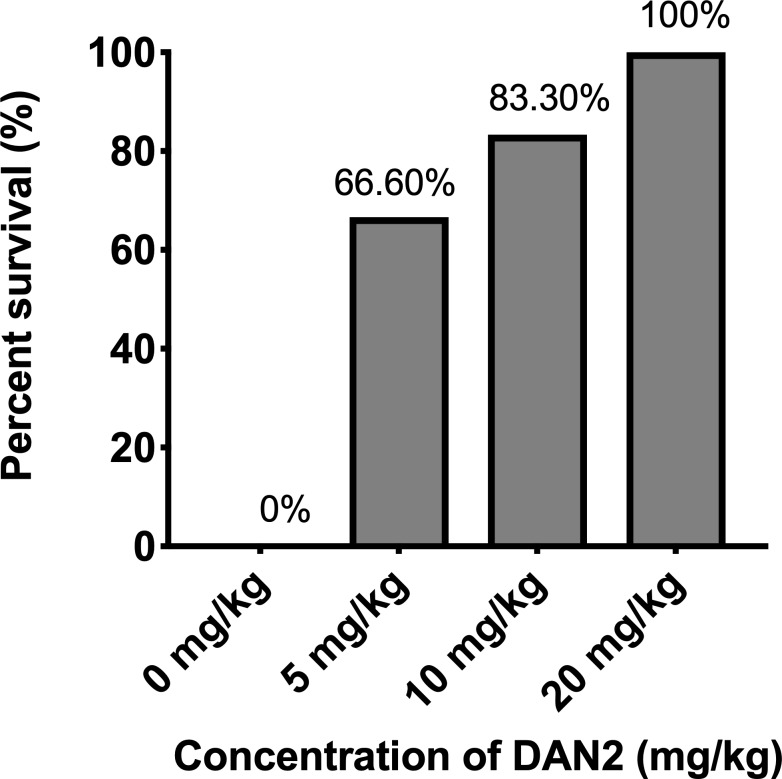
As shown in the Fig 1, when the mice were treated with 5 mg/kg and 10 mg/kg of DAN2, we observed survival rates of 67% and 83% respectively. Out of six mice that received 5 mg/kg, two mice reached the endpoint in 20 hours, while one mouse that received 10 mg/kg reached the endpoint at around 22 hours (S2 Fig). This indicates that 5 mg/kg and 10 mg/kg of DAN2 prolonged the survival of those mice. However, all the infected mice that were treated with 20 mg/kg of DAN2 survived, demonstrating that the effective dose 100 (EC100) of DAN2 is 20mg/kg.
Fig 1. The infected mice exhibited dose-dependent response to survival.
All the mice of four groups were injected intraperitoneally (i. p.) with a lethal dose, 2.2 × 10⁷ CFU of E. coli and treated with different concentration of DAN2 after 30 minutes of bacterial challenge. The control group only received bacterial suspension and PBS. All the treated mice were monitored for five days. There were 6 mice/group.
3.2. DAN2 reduces bacterial counts in blood and peritoneal fluid after 6 hours of challenge
In order to document the impact of DAN2 on bacterial counts in vivo, we assayed bacterial loads in blood and peritoneal fluid in mice that received varying doses of peptide. This approach assesses the microbicidal or microbiostatic activity of DAN2. 4 mice were used in each treatment and were infected with 2. 2 × 10⁷ CFU. All mice in a group received a given concentration of DAN2 (0 mg/kg, 5 mg/kg, 10 mg/kg and 20 mg/kg) after 30 minutes of bacterial infection. Peritoneal fluid and blood were collected after 6 hours of bacteria challenge and the blood or peritoneal wash was plated in dilutions to provide bacterial counts.
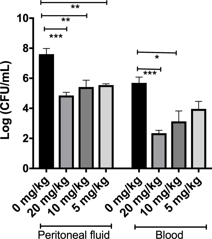
As shown in Fig 2, bacterial loads in peritoneal fluid of the treated mice were significantly lower than that of the control mice and similar to each other regardless of the DAN2 dose. There was an approximately 150-fold decrease in bacteria in the treated mice compared to control mice after 6 hours of infection. One-way ANOVA analysis was performed to compare the effect of DAN2 dose on bacterial loads. A significant difference was observed in the bacterial loads between the experimental treatment groups (F 3, 11 = 17. 8, p = 0.0002). Post-hoc comparisons using Tukey HSD tests (α = 0.05) indicated that there was a significant reduction in bacterial growth in mice treated with 20 mg/kg when compared with the control mice (p < 0.0005). Similarly, the difference between 10 mg/kg and control or 5 mg/kg and control was also statistically significant (p < 0.005).
Fig 2. DAN2 treated mice have reduced bacterial loads both in peritoneal fluid and blood.
Four mice were infected and treated with 0 mg/kg, 20 mg/kg, 10 mg/kg and 5 mg/kg of DAN2 individually and bacterial count was determined after 6 hours of infections. The error bars represent the standard deviation of the mean. The number of asterisks was used to denote the extent of statistical significance amongst groups (* denotes p < 0.05, ** denotes p < 0.005, *** denotes p < 0.0005).
Similarly, the bacterial load in blood was found to have decreased approximately 100-fold in mice treated with 20 mg/kg and 10-fold in mice treated with 10 mg/kg in comparison to the control mice. There was no significant decrease in bacteria in the mice treated with 5 mg/kg compared to PBS-treated mice. An analysis of variance (ANOVA) yielded significant difference in bacterial loads between the control and each treatment group (F 3, 12 = 12.57, p = 0.0005). Post-hoc comparisons using Tukey HSD tests (α = 0.05) indicated that there was a significant reduction in bacterial growth in blood in mice treated with 20 mg/kg as compared to the control mice (p < 0.0005). The difference between 10 mg/kg and control was also statistically significant (p < 0.05). However, a statistical significance was not observed between control and 5 mg/kg (p > 0.05).
3.3. DAN2 permeabilizes E.coli membrane
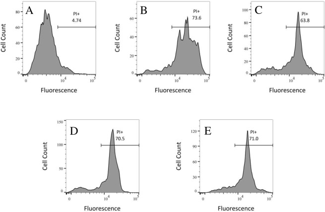
One identified mechanism of antimicrobial action of AMPs is forming pores in cytoplasmic membranes which if not repaired quickly is lethal. We evaluated the integrity of the cell membrane of E. coli using propidium iodide (PI), a DNA interacting dye that intercalates into DNA of permeabilized membrane and fluoresces brightly. The fluorescence of PI in a bacterial culture demonstrates that the membrane integrity is compromised. Fig 3 shows the fluorescence intensity of bacteria under different conditions. The bacterial cells treated with any of the tested concentrations of peptide or heated to 85° C have large fractions of the population that have admitted PI and fluoresce intensely. The area under the peak of higher fluorescence quantitates the cell population. Most of the heat shocked bacterial cells (74%, Fig 3B) were fluorescently labeled compared to the cells treated with peptide. The percentage of fluorescently labeled cells were 64%, 70% and 71% for the cells treated with 48 μg/ml, 36 μg/ml and 24 μg/ml of DAN2 respectively. Surprisingly, the intensity of fluorescing bacteria did not increase in proportion to the increasing concentration of DAN2, which indicates that many cells had compromised membranes at all of these treatment concentrations (Fig 3C, 3D and 3E). None of the samples, either heat shocked or peptide treated, showed any colony growth when 100 μl of the samples were plated for colony count showing that virtually all the bacteria were unable to divide, although not all fluoresced at the time of the assay.
Fig 3. DAN2 disrupts the integrity of E.coli cell membrane.
The DNA binding dye propidium iodide (PI) was used to evaluate cell membrane permeability of E. coli ATCC 25922 via flow cytometry. 2.0 × 10⁶ CFU/ml was incubated with varying concentrations of peptide for an hour and PI added subsequently. Flow cytometry was performed using a FACScan instrument. (A) Bacteria; (B) Heat treated (positive control); (C) DAN2 (48 μg/ml); (D) DAN2 (36 μg/ml); (E) DAN2 (24 μg/ml). Bacterial cells treated with either peptide or heat shocked have increased cellular fluorescence intensity of PI.
Discussion
In the presence of the global threat from antibiotic resistance, antimicrobial peptides are promising anti-infective agents. AMPs are particularly attractive because microbes are less likely to develop resistance [23,24]. AMPs primarily target bacterial cell membranes, and it is challenging for bacteria to preserve cell membrane structure and function while avoiding membrane disrupting activity of peptide. In addition, unlike conventional antibiotics that target a specific biochemical process or a cell component, AMPs as a group have multiple modes of action, making them resilient against bacterial resistance [23,24].
Our current study provides an insight into the antimicrobial activity of DAN2 in-vivo. We found that DAN2 prolongs the survival of the mice in a dose dependent manner and more importantly, a single bolus dose protects infected mice from lethal bacterial challenge. The effective dose of DAN2 is comparable to other AMPs studied in the literature. A synthetic AMP named M33 (9 amino acid residues long) protected 100% of mice infected with lethal doses of E. coli and P. aeruginosa when administered at 12.5 and 25 mg/kg respectively [25]. Another study conducted in a rat model of septic shock demonstrated that a cecropin B reduced the lethality when given i. p. immediately after E.coli challenge at 1 mg/kg [26]. This suggests that the peptides belonging to the cecropin family can be effective in vivo. However, only a few in vivo studies of cecropins have been reported.
Several studies suggest that the cationic AMPs interact with the negatively charged membrane and form either ion channels or pores [23,24]. These cationic AMPs can also block intracellular processes by inhibiting protein folding or activity of enzymes, [14,27,28]. However, the cell membrane is reported as the primary target of cecropins [14]. We have found that DAN2 compromises the integrity of the bacterial membrane. Our in vitro study demonstrated that DAN2 does not lyse mammalian RBC which supports its potential use clinically [19]. Unlike bacterial cells which have 25% more anionic lipids that favor stronger electrostatic interaction, the mammalian cell membrane has large amounts of charge-neutral components, such as phosphatidylethanolamine, phosphatidylcholine, and sphingomyelin [29]. In addition, the presence of cholesterol in mammalian cell membranes stabilizes the phospholipid bilayer and hence reduces the pore-forming activity of AMPs.
While in vitro studies support lack of mammalian cell cytolysis by DAN2, it is possible that in vivo utilization causes toxicity through a mechanism other than cytolysis. To further evaluate potential toxicity of DAN2, we IP-injected C57BL/6 mice with 40 mg/kg (twice the highest concentration tested in the protection assay). The mice were observed over 5 days for behavioral changes associated with illness including weight loss, hunched posture, ruffled fur, and slow response to handling. However, there were no symptoms of illness (data not shown). After euthanasia the organs were observed by gross dissection and had no discernible abnormalities. Additionally, during treatment the infected mice that got sufficient DAN2 to protect them from the infection did not demonstrate murine illness behaviors and following euthanasia the organs appeared normal upon dissection. Our in vivo studies show that DAN2 is not toxic to mice when used to treat acute infection.
Several studies have suggested that AMPs work not only by disrupting the cell membrane, but also by exerting immune-modulatory activity[30–36]. To date, a class of cecropin family has been shown to stimulate the migration of leukocytes to the site of infection, reduce plasma levels of tumor necrosis factor, endotoxins, and cytokines responsible for septic shock [26,37–42] There has also been a report of increased anti-inflammatory cytokines (IL-4, IL-10) and/or reduced pro-inflammatory molecules (IL-6, IL-8, TNF-alpha) following an administration of cecropins [39,43]. These multiple modes of action of cecropin peptide awaits further investigation in the field.
As science has revealed the importance of normal flora in the host, the problem with disruption of normal flora from antibiotic use has been recognized. The standard antibiotics that are administered orally are disruptive of the normal flora [44]. However, there is considerably less information on the influence of AMPs on commensals. A report suggested that some commensal bacteria defend themselves against AMPs the host secretes by modifying the negatively charged phosphate group in their LPS [45]. Overall, there has been minimal investigation of the impact of IV or IP administration of AMP on the normal flora, and our study of intraperitoneal administration of DAN2 did not monitor normal flora changes. Our expectation is that while many commensals are likely susceptible to DAN2, IP administration would limit access of a charged peptide to the normal flora and impact on normal flora would be minimal.
An argument against the potential use of AMPs as anti-infective agent is the expected development of neutralizing antibodies when a given AMP is repeatedly applied to the same patient [46]. This is an important issue since antibodies against a given peptide would be likely to reduce the antimicrobial activity and since the peptide administration is temporally and perhaps physically associated with an infection, the immune system response against it is more likely to occur. One approach to avoid this problem is to not use a given peptide more than a few times in the same patient. The number of potentially clinically useful AMPs in nature is almost without limit. There could be hundreds of available peptides to treat any given infection and the repeated use of the same peptide could be minimized to avoid neutralizing antibodies.
To date, most of the characterized peptides have been identified in arthropods and vertebrates, particularly amphibian [47]. However, in virtually any organism in which they have been sought, novel AMPs have been found. Recently AMPs have also been identified in a broader range of organisms such as plants, Feijoa sellowiana Berg fruit [48] and shellfish Mytilus galloprovincialis mussel [49]. This argues that there is an inexhaustible supply of natural AMPs and we have barely begun to identify, catalog and test them experimentally and clinically. The developed technology to generate peptide in large quantities inexpensively further supports the potential for AMPs to contribute to new treatments for bacteria resistant to standard antibiotics. The availability of such a pharmacy of studied AMPs would provide a crucial tool to treat microbes that are resistant to standard small molecule antibiotics.
Our findings indicate that a cecropin type peptide, DAN2, has potential for clinical use. The present research is a further step in examining the antimicrobial activity of DAN2 in the process of developing this peptide as a therapeutic drug.
Funding information: Research was supported by New Hampshire-INBRE through an Institutional Development Award (IDeA), P20GM103506, from the National Institute of General Medical Sciences of the NIH.
Supporting information
All twelve mice were infected with a lethal dose of E. coli ATCC 25922 intraperitoneally (i.p.). The control group received 300μl of PBS after 30 minutes of bacterial challenge. Control mice showed 0% survivability, whereas 20 mg/kg peptide ensured 100% survivability in E. coli infected mice.
(TIFF)
The control group did not receive any peptide. Six mice were used per group. 5 mg/kg, 10 mg/kg and 20 mg/kg of peptide prolonged the survival of mice, but all control mice reached the endpoint within 12 hours of bacterial infections.
(TIFF)
Acknowledgments
We thank the Department of Natural Science at Colby-Sawyer College for their continuous support, Jennifer Fields and the Dartmouth Mouse Modeling Shared Resource for mice related assistance, and Gary A. Ward and Mikayla Kravetz Hernandez and the DartLab Immune Monitoring and Flow Cytometry Shared Resource for flow cytometry assay. Shared resources at Dartmouth are supported by the Norris Cotton Cancer Center and the supporting grant 5 P30 CA023108-38.
Data Availability
All relevant data are within the manuscript and its Supporting Information files.
Funding Statement
This work was supported by the National Institutes of Health, National Institute of General Medical Sciences, IDeA Program, NH-INBRE, P20GM103506 to SF and JJ. The funder had no role in study design, data collection and analysis, decision to publish, or preparation of the manuscript.
References
- 1.Organización Mundial de la Salud. High levels of antibiotic resistance found worldwide, new data shows [Internet]. Media centre. 2018. p. 1–7. Available from: https://www.who.int/news-room/detail/29-01-2018-high-levels-of-antibiotic-resistance-found-worldwide-new-data-shows#.XSFbetAnzs8.email
- 2.WHO. Antimicrobial Resistance Fact sheet [Internet]. WHO, Antimicrobial resistance. 2018. Available from: https://www.who.int/news-room/fact-sheets/detail/antimicrobial-resistance?fbclid=IwAR2jOtjXMuuFgyDOjh-dvo2guPPvF72yo5oRBJDMYR3vYPBJKPt3fuDfq70
- 3.da Costa JP, Cova M, Ferreira R, Vitorino R. Antimicrobial peptides: an alternative for innovative medicines? Appl Microbiol Biotechnol. 2015;99(5):2023–40. 10.1007/s00253-015-6375-x [DOI] [PubMed] [Google Scholar]
- 4.Lyu Y, Yang Y, Lyu X, Dong N, Shan A. Antimicrobial activity, improved cell selectivity and mode of action of short PMAP-36-derived peptides against bacteria and Candida. Sci Rep. 2016;6(May):1–12. [DOI] [PMC free article] [PubMed] [Google Scholar]
- 5.Martin E, Ganz T, Lehrer RI. Defensins and other endogenous peptide antibiotics of vertebrates. J Leukoc Biol. 1995;58(2):128–36. 10.1002/jlb.58.2.128 [DOI] [PubMed] [Google Scholar]
- 6.Conte M, Aliberti F, Fucci L, Piscopo M. Antimicrobial activity of various cationic molecules on foodborne pathogens. World J Microbiol Biotechnol. 2007;23(12):1679–83. 10.1007/s11274-007-9415-6 [DOI] [PubMed] [Google Scholar]
- 7.Zhang LJ, Gallo RL. Antimicrobial peptides. Curr Biol. 2016;26(1):R14–9. 10.1016/j.cub.2015.11.017 [DOI] [PubMed] [Google Scholar]
- 8.Giuliani A, Pirri G, Nicoletto SF. Antimicrobial peptides: An overview of a promising class of therapeutics. Vol. 2, Central European Journal of Biology. 2007. 1–33 p. [Google Scholar]
- 9.Saravolatz LD, Pawlak J, Martin H, Saravolatz S, Johnson L, Wold H, et al. Postantibiotic effect and postantibiotic sub-MIC effect of LTX-109 and mupirocin on Staphylococcus aureus blood isolates. Lett Appl Microbiol. 2017;65(5):410–3. 10.1111/lam.12792 [DOI] [PubMed] [Google Scholar]
- 10.Carpenter CF, Chambers HF. Daptomycin: Another Novel Agent for Treating Infections Due to Drug‐Resistant Gram‐Positive Pathogens. Clin Infect Dis. 2004;38(7):994–1000. 10.1086/383472 [DOI] [PubMed] [Google Scholar]
- 11.Cheng KT, Wu CL, Yip BS, Yu HY, Cheng HT, Chih YH, et al. High level expression and purification of the clinically active antimicrobial peptide P-113 in Escherichia coli. Molecules. 2018;23(4):800. [DOI] [PMC free article] [PubMed] [Google Scholar]
- 12.Bulet P, Stöcklin R, Menin L. _Anti-microbial peptides- from invertebrates to vertebrates.pdf. Immunol Rev. 2004;198:169–84. [DOI] [PubMed] [Google Scholar]
- 13.Buchmann K. Evolution of innate immunity: Clues from invertebrates via fish to mammals. Front Immunol. 2014;5(September). [DOI] [PMC free article] [PubMed] [Google Scholar]
- 14.Wu Q, Patočka J, Kuča K. Insect Antimicrobial Peptides, a Mini Review. Toxins (Basel). 2018;10(11):461. [DOI] [PMC free article] [PubMed] [Google Scholar]
- 15.Yi HY, Chowdhury M, Huang YD, Yu XQ. Insect antimicrobial peptides and their applications. Appl Microbiol Biotechnol. 2014;98(13):5807–22. 10.1007/s00253-014-5792-6 [DOI] [PMC free article] [PubMed] [Google Scholar]
- 16.Reddy KVR, Yedery RD, Aranha C. Antimicrobial peptides: Premises and promises. Vol. 24, International Journal of Antimicrobial Agents. 2004. p. 536–47. 10.1016/j.ijantimicag.2004.09.005 [DOI] [PubMed] [Google Scholar]
- 17.Hancock REW, Sahl HG. Antimicrobial and host-defense peptides as new anti-infective therapeutic strategies. Nat Biotechnol [Internet]. 2006;24(12):1551–7. Available from: http://www.ncbi.nlm.nih.gov/pubmed/17160061 10.1038/nbt1267 [DOI] [PubMed] [Google Scholar]
- 18.Ganz T, Lehrer RI. Defensins. Pharmacol Ther. 1995;66(2):191–205. [DOI] [PubMed] [Google Scholar]
- 19.Duwadi D, Shrestha A, Yilma B, Kozlovski I, Sa-eed M, Dahal N, et al. Identification and screening of potent antimicrobial peptides in arthropod genomes. Peptides [Internet]. 2018;103:26–30. Available from: http://www.sciencedirect.com/science/article/pii/S0196978118300251 10.1016/j.peptides.2018.01.017 [DOI] [PMC free article] [PubMed] [Google Scholar]
- 20.Erlendsdottir H, Knudsen JD, Odenholt I, Cars O, Espersen F, Frimodt-Møller N, et al. Penicillin pharmacodynamics in four experimental pneumococcal infection models. Antimicrob Agents Chemother. 2001;45(4):1078–85. 10.1128/AAC.45.4.1078-1085.2001 [DOI] [PMC free article] [PubMed] [Google Scholar]
- 21.Jang WS, Kim HK, Lee KY, Kim SA, Han YS, Lee IH. Antifungal activity of synthetic peptide derived from halocidin, antimicrobial peptide from the tunicate, Halocynthia aurantium. FEBS Lett. 2006;580(5):1490–6. 10.1016/j.febslet.2006.01.041 [DOI] [PubMed] [Google Scholar]
- 22.Yeaman MR, Bayer AS, Koo SP, Foss W, Sullam PM. Platelet microbicidal proteins and neutrophil defensin disrupt the Staphylococcus aureus cytoplasmic membrane by distinct mechanisms of action. J Clin Invest. 1998;101(1):178–87. 10.1172/JCI562 [DOI] [PMC free article] [PubMed] [Google Scholar]
- 23.Park AJ, Okhovat JP, Kim J. Antimicrobial peptides. Clin Basic Immunodermatology Second Ed. 2017;26(1):81–95. [Google Scholar]
- 24.Mahlapuu M, Håkansson J, Ringstad L, Björn C. Antimicrobial Peptides: An Emerging Category of Therapeutic Agents. Front Cell Infect Microbiol. 2016;6(December):1–12. [DOI] [PMC free article] [PubMed] [Google Scholar]
- 25.Teixeira LD, Silva ON, Migliolo L, Fensterseifer ICM, Franco OL. In vivo antimicrobial evaluation of an alanine-rich peptide derived from Pleuronectes americanus. Peptides. 2013;42:144–8. 10.1016/j.peptides.2013.02.001 [DOI] [PubMed] [Google Scholar]
- 26.Giacometti A, Cirioni O, Ghiselli R, Viticchi C, Mocchegiani F, Riva A, et al. Effect of mono-dose intraperitoneal cecropins in experimental septic shock. Crit Care Med. 2001;29(9):1666–9. 10.1097/00003246-200109000-00002 [DOI] [PubMed] [Google Scholar]
- 27.Bechinger B, Gorr SU. Antimicrobial Peptides: Mechanisms of Action and Resistance. J Dent Res. 2017;96(3):254–60. 10.1177/0022034516679973 [DOI] [PMC free article] [PubMed] [Google Scholar]
- 28.Le C-F, Fang C-M, Sekaran SD. Intracellular Targeting Mechanisms by Antimicrobial Peptides. Antimicrob Agents Chemother. 2017;61(4):e02340–16. 10.1128/AAC.02340-16 [DOI] [PMC free article] [PubMed] [Google Scholar]
- 29.Van Meer G, Voelker DR, Feigenson GW. Membrane lipids: Where they are and how they behave. Nat Rev Mol Cell Biol. 2008;9(2):112–24. 10.1038/nrm2330 [DOI] [PMC free article] [PubMed] [Google Scholar]
- 30.Silva ON, De La Fuente-Núñez C, Haney EF, Fensterseifer ICM, Ribeiro SM, Porto WF, et al. An anti-infective synthetic peptide with dual antimicrobial and immunomodulatory activities. Sci Rep. 2016;6:1–11. 10.1038/s41598-016-0001-8 [DOI] [PMC free article] [PubMed] [Google Scholar]
- 31.Choi KYG, Mookherjee N. Multiple immune-modulatory functions of cathelicidin host defense peptides. Front Immunol. 2012;3(June):1–4. [DOI] [PMC free article] [PubMed] [Google Scholar]
- 32.Haney EF, Mansour SC, Hilchie AL, de la Fuente-Núñez C, Hancock REW. High throughput screening methods for assessing antibiofilm and immunomodulatory activities of synthetic peptides. Peptides. 2015;71:276–85. 10.1016/j.peptides.2015.03.015 [DOI] [PMC free article] [PubMed] [Google Scholar]
- 33.Nijnik A, Hancock REW. The roles of cathelicidin LL-37 in immune defences and novel clinical applications. Curr Opin Hematol. 2009;16(1):41–7. [DOI] [PubMed] [Google Scholar]
- 34.Nijnik A, Madera L, Ma S, Waldbrook M, Elliott MR, Easton DM, et al. Synthetic Cationic Peptide IDR-1002 Provides Protection against Bacterial Infections through Chemokine Induction and Enhanced Leukocyte Recruitment. J Immunol. 2010;184(5):2539–50. 10.4049/jimmunol.0901813 [DOI] [PubMed] [Google Scholar]
- 35.Lee E, Jeong KW, Lee J, Shin A, Kim JK, Lee J, et al. Structure-activity relationships of cecropin-like peptides and their interactions with phospholipid membrane. BMB Rep. 2013;46(5):282–7. 10.5483/BMBRep.2013.46.5.252 [DOI] [PMC free article] [PubMed] [Google Scholar]
- 36.Kamysz W, Okrój M, Łukasiak J. Novel properties of antimicrobial peptides. Vol. 50, Acta Biochimica Polonica. 2003. p. 461–9. doi: 035002461 [PubMed] [Google Scholar]
- 37.Fink MP. Animal models of sepsis. Virulence. 2014;5(1):143–53. 10.4161/viru.26083 [DOI] [PMC free article] [PubMed] [Google Scholar]
- 38.Mogensen TH. Pathogen recognition and inflammatory signaling in innate immune defenses. Clin Microbiol Rev. 2009;22(2):240–73. 10.1128/CMR.00046-08 [DOI] [PMC free article] [PubMed] [Google Scholar]
- 39.Wu J, Mu L, Zhuang L, Han Y, Liu T, Li J, et al. A cecropin-like antimicrobial peptide with anti-inflammatory activity from the black fly salivary glands. Parasites and Vectors. 2015;8(1):1–13. [DOI] [PMC free article] [PubMed] [Google Scholar]
- 40.Wei L, Huang C, Yang H, Li M, Yang J, Qiao X, et al. A potent anti-inflammatory peptide from the salivary glands of horsefly. Parasites and Vectors. 2015;8:556 10.1186/s13071-015-1149-y [DOI] [PMC free article] [PubMed] [Google Scholar]
- 41.Ruan M, Fu Y V., Song Y, Wang J, Ma K, Li Y, et al. A novel cecropin B-derived peptide with antibacterial and potential anti-inflammatory properties. PeerJ. 2018;6:e5369 10.7717/peerj.5369 [DOI] [PMC free article] [PubMed] [Google Scholar]
- 42.Wei L, Yang Y, Zhou Y, Li M, Yang H, Mu L, et al. Anti-inflammatory activities of Aedes aegypti cecropins and their protection against murine endotoxin shock. Parasites and Vectors. 2018;11(1):470 10.1186/s13071-018-3000-8 [DOI] [PMC free article] [PubMed] [Google Scholar]
- 43.Sun Y, Shang D. Inhibitory Effects of Antimicrobial Peptides on Lipopolysaccharide-Induced Inflammation. Mediators Inflamm. 2015;2015:167572 10.1155/2015/167572 [DOI] [PMC free article] [PubMed] [Google Scholar]
- 44.Langdon A, Crook N, Dantas G. The effects of antibiotics on the microbiome throughout development and alternative approaches for therapeutic modulation. Genome Med. 2016;8:39 10.1186/s13073-016-0294-z [DOI] [PMC free article] [PubMed] [Google Scholar]
- 45.Cullen TW, Schofield WB, Barry NA, Putnam EE, Rundell EA, Trent MS, et al. Antimicrobial peptide resistance mediates resilience of prominent gut commensals during inflammation. Science (80-). 2015;347(6218):170–5. [DOI] [PMC free article] [PubMed] [Google Scholar]
- 46.Deptuła M, Wardowska A, Dzierzy´nska M, Rodziewicz-Motowidło S, Pikuła M. Antibacterial peptides in dermatology–strategies for evaluation of allergic potential. Molecules. 2018;23(2):414. [DOI] [PMC free article] [PubMed] [Google Scholar]
- 47.Kumar P, Kizhakkedathu JN, Straus SK. Antimicrobial peptides: Diversity, mechanism of action and strategies to improve the activity and biocompatibility in vivo. Biomolecules. 2018;8(1):4. [DOI] [PMC free article] [PubMed] [Google Scholar]
- 48.Piscopo M, Tenore GC, Notariale R, Maresca V, Maisto M, de Ruberto F, et al. Antimicrobial and antioxidant activity of proteins from Feijoa sellowiana Berg. fruit before and after in vitro gastrointestinal digestion. Nat Prod Res. 2019;1–5. [DOI] [PubMed] [Google Scholar]
- 49.Notariale R, Basile A, Montana E, Romano NC, Cacciapuoti MG, Aliberti F, et al. Protamine-like proteins have bactericidal activity. The first evidence in Mytilus galloprovincialis. Acta Biochim Pol. 2018;65(4):585–94. 10.18388/abp.2018_2638 [DOI] [PubMed] [Google Scholar]
Associated Data
This section collects any data citations, data availability statements, or supplementary materials included in this article.
Supplementary Materials
All twelve mice were infected with a lethal dose of E. coli ATCC 25922 intraperitoneally (i.p.). The control group received 300μl of PBS after 30 minutes of bacterial challenge. Control mice showed 0% survivability, whereas 20 mg/kg peptide ensured 100% survivability in E. coli infected mice.
(TIFF)
The control group did not receive any peptide. Six mice were used per group. 5 mg/kg, 10 mg/kg and 20 mg/kg of peptide prolonged the survival of mice, but all control mice reached the endpoint within 12 hours of bacterial infections.
(TIFF)
Data Availability Statement
All relevant data are within the manuscript and its Supporting Information files.


