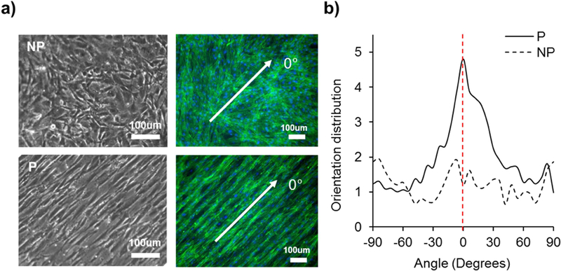
Figure 2.

Patterning of hydrogel substrate regulated cellular alignment. (a) Microscopic (left) and F-actin stained images (right) of cells grown on nonpatterned and patterned hydrogel. Green indicates actin filament and blue indicates nuclei. (b) F-actin stained images were further analyzed using 2D FFT alignment analysis. Cells grown on patterned hydrogel showed guided cellular alignment in the direction parallel to the pattern while cells grown on nonpatterned hydrogel showed random direction. White arrow in (a) indicates the direction of substrate patterning, where its angle is 0°, shown as the red dotted line in (b).
