

Adv. Sci. 2019, 6, 1801555
Several images in Figure 3, Figure 4, and Figure S7, Supporting Information, accidentally presented duplicate samples in the original article. The correct figures are presented below. The authors apologize for any inconvenience this may have caused.
Figure 3.
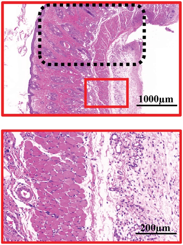
e, third row) H&E staining of the necrosis and survival junction area of the skin flaps in the 40 μM MF‐Lip treated sample. e, last row) Magnified view of the survival area in the 40 μM MF‐Lip treated sample. *p < 0.05.
Figure 4.
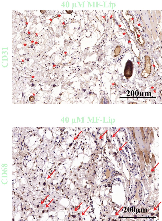
Effect of MF‐Lip@PEG on random‐pattern skin flap neovascularization, inflammation, and infection. Immunohistochemical images of flaps highlighting a) blood vessel CD31‐positive endothelial cells (red dots mark the microvessels) and CD68‐positive macrophage/monocytes (red arrows mark the cells).
Figure S7.
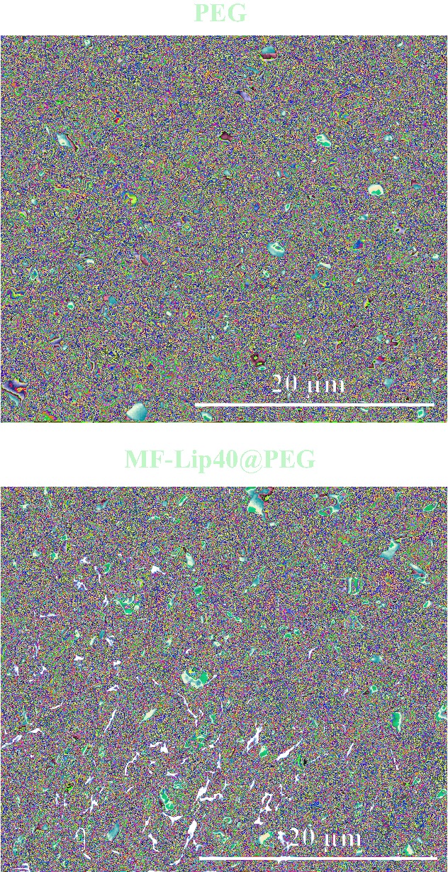
The Morphological examination of hydrogels. SEM results at low magnification.