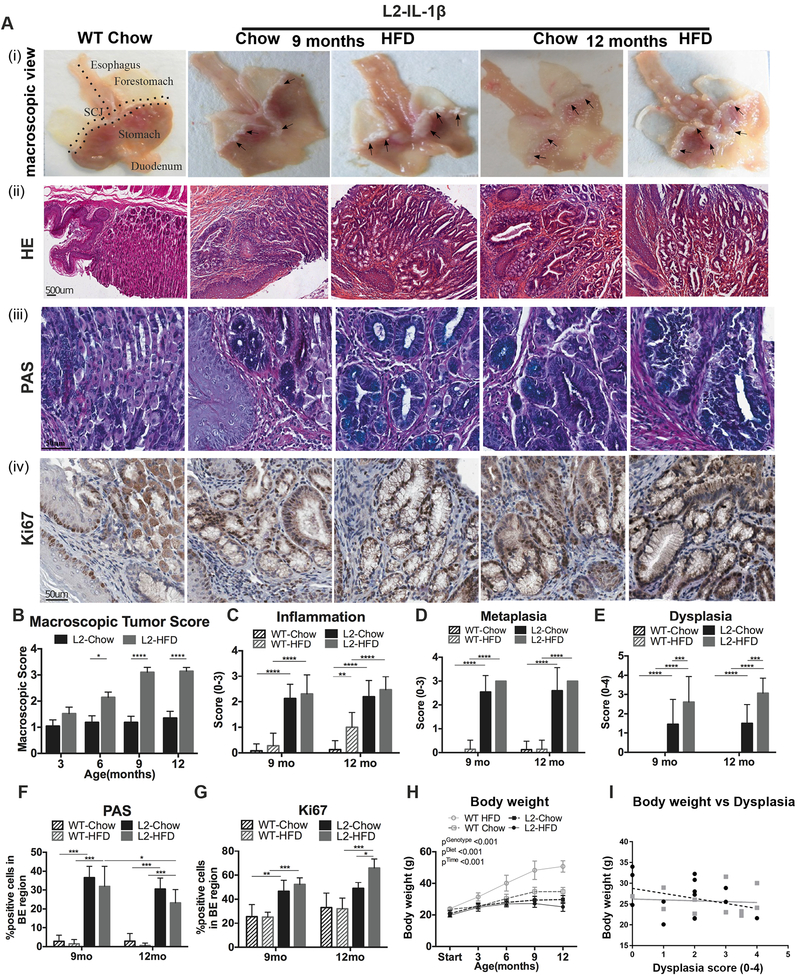
Figure 1. HFD accelerates esophageal dysplasia in the L2-IL1B mouse model.
(A) (i) macroscopic image and epresentative pictures of (ii) Haematoxylin & Eosin (H&E), (iii) Periodic acid–Schiff (PAS) and (iv) Ki67 of the SCJ. A WT Chow mouse was used as a representative for all WT mice. Macroscopic tumor score at 3–12 months (mean ±SEM) (B), inflammation (C) metaplasia (D), and dysplasia scores (E) (n=8–12, mean ± SD). Quantification of (F) PAS and (G) Ki67 staining (n=8–12). (H) Body weight of L2-IL1B mice. (I) Correlations between body mass and dysplasia score. Quantification of 4 high-magnification-field (20X) of esophagus and SCJ tissue (****p<0.0001, ***p<0.001, **p<0.01, *p<0.05) For (B-E) 2-way ANOVA with Sidak-Holm post-hoc was used, for (F,G) a 1 way ANOVA with Tukey post-hoc and for (H,I) 2 way ANOVA with Tukey Post-hoc. BE region was defined as the region between squamous epithelium and oxyntic mucosa of the stomach. L2=L2-IL-1β, WT=wildtype, HFD=High fat diet

