Summary
Pancreatitis is an increasingly common disease that carries a significant mortality and which lacks specific therapy. Pathological calcium signalling is an important contributor to the initiating cell injury, caused by or acting through mitochondrial inhibition. A principal effect of disordered cell signalling and impaired mitochondrial function is cell death, either by apoptosis that is primarily protective, or by necrosis that is deleterious, both locally and systemically. Mitochondrial calcium overload is particularly important in necrotic injury, which may include damage mediated by the mitochondrial permeability transition pore. The role of reactive oxygen species remains controversial. Present understanding of the part played by disordered pancreatic acinar calcium signalling and mitochondrial inhibition offers several new potential therapeutic targets.
Keywords: Pancreatitis, Mitochondria, Calcium signaling, Apoptosis, Necrosis
Pancreatitis is a disease of increasing incidence caused by gallstones, ethanol excess, hyperlipidaemia and other, rarer precipitating factors that in severe cases induces pancreatic necrosis, multiple organ failure and death [1]. Pancreatic necrosis is a critical contributing event, in part through the local and systemic release of activated digestive enzymes, accounting for extreme effects despite the relatively small volume of the gland (<200 ml). Dysregulation of Ca2+ homeostasis was proposed over a decade ago [2] and since confirmed by a number of groups to be the key trigger of pancreatic acinar cell injury induced by hyperstimulation [3,4], bile salts [5,6], ethanol metabolites (fatty acid ethyl esters, FAEEs) [7,8] and fatty acids (FAs) [7,8]. These agents are implicated in the pathogenesis of pancreatitis [1], as bile salts may reflux into the pancreatic duct during passage of gallstones through the sphincter of Oddi [9,10], FAEEs are produced in the pancreas through non-oxidative metabolism of ethanol [11-14], and FAs accumulate in the pancreas after FAEE hydrolysis [11,12] or in hyperlipidaemia [1]. The central role of mitochondria in the transduction of intracellular Ca2+ signals [15], the supply of ATP and determination of cell fate [16,17] places mitochondrial injury centre stage in this scenario [18]. This review outlines current understanding of the place of mitochondrial injury in the pathogenesis of pancreatitis.
1. Pancreatitis and the lack of specific therapies
Acute pancreatitis was first described comprehensively by Fitz, a pathologist at Harvard who identified severe forms of the disease as haemorrhagic, suppurative or gangrenous [19] now recognised as various stages of acute necrotising pancreatitis [1]. During the first half of the last century, Moynihan described acute pancreatitis as ‘most terrible of all calamities that occur in connection with the abdominal viscera’ [20]. The disease occurs acutely in up to 100 per 100,000 people per year [21] and causes severe disease in 20% [22]. Although gallstones have accounted for almost half of all cases for several decades, alcohol abuse is the cause of an increasing number, in some countries the majority [21]. Rarer causes include hypercalcaemia, viral infections, drugs, endoscopic retrograde cholangiopancreatography, surgery, ductal tumours and autoimmune causes [1]. Whatever the cause, acute inflammation of the pancreas features necrosis (>30% of the gland) in up to 20% of patients, which may extend into peri-pancreatic tissues and be accompanied by injury of remote organ systems as well; up to 50% of deaths occur in the first week from multiple organ failure [1,22]. In subsequent weeks, secondary infection is the most significant factor-determining outcome [22]. In general, the more severe the pancreatic injury and the greater the extent of necrosis, the greater the accompanying systemic inflammatory response, subsequent organ failure and risk from superimposed infection [22]. Thus, in patients with necrosis compared to those without, organ failure is far more common (50% vs. 5—10%, respectively), as is mortality (17% vs. 3%) [1]. Although the mechanism by which necrosis leads to organ failure is unclear, experimental studies demonstrate that the severity and extent of pancreatic injury is an instrumental factor.
While digestive enzyme secretion is the primary function of the exocrine pancreas, premature intracellular digestive enzyme activation is the hallmark of, and a key component of tissue damage in, pancreatitis [23]. This is illustrated by hereditary pancreatitis, as is the continuum between acute and chronic pancreatitis, the result of cationic trypsinogen gene mutations that render trypsinogen more readily activated or once activated, less readily broken down [24]. Cationic trypsinogen gene mutations result in an autosomal dominant disease with 80% penetrance, beginning with recurrent attacks of acute pancreatitis in childhood and/or adolescence, developing into chronic pancreatitis during early adult life, and a 40% lifetime risk of pancreatic carcinoma [25]. In a similar manner continued alcohol excess and uncontrolled hyperlipidaemia can induce recurrent acute pancreatitis that is likely to become chronic pancreatitis, usually but not always at a later stage in adult life, the sporadic form of which increases the lifetime risk of pancreatic carcinoma by an order of ×15 [26].
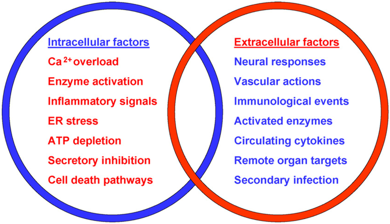
Despite the major burden of disease resulting from both acute and chronic pancreatitis, little specific treatment is available to halt their progression, let alone prevent their occurrence [1,22]. Primarily for this reason research has been undertaken to understand the pathogenesis of pancreatitis, in the hope of identifying new prophylactic and therapeutic targets that may be altered to improve outcome. Many data now indicate that the initial site of injury is within pancreatic acinar cells, which make up the bulk of the organ [3-8,13,14,18,23]. Subsequently both intra- and extracellular factors contribute to the inflammation, oedema and cell death responses observed in pancreatitis as well as the progression to the systemic inflammatory response syndrome and multiple organ failure characteristic of severe disease [1], as summarised in Fig. 1. To understand what goes wrong in pancreatic acinar cells, however, normal mechanisms must first be outlined.
Figure 1.
Intra- and extracellular factors which contribute to the pathogenesis of pancreatitis. Acinar cell injury is the principal driving event, initially mediated via Ca2+ overload, leading to inflammatory signals that occur following cell stress. The loss of normal enzyme secretion and ATP production induce cell death pathways, which with severe stress are most likely to result in necrosis of cells. These factors contribute to and interact with extracellular factors, mediated initially via neural and vascular—endothelial mechanisms that accelerate inflammation. Activated resident and infiltrating monocyte-macrophages and/or neutrophils exacerbate the tissue injury, adding to the effects of activated interstitial and circulating digestive enzymes. Circulating cytokine responses increase the likelihood of remote organ damage, compromising recovery, while secondary infection adds to the likelihood of poor outcome.
2. Pancreatic acinar cell Ca2+ signalling
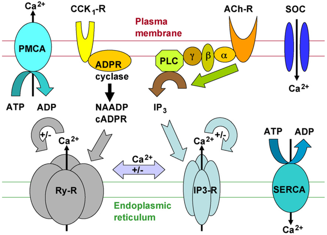
The pancreatic acinar cell is the classical, polarised, secretory cell, with Ca2+ centre stage in the control of its activities (see Fig. 2) [23,27]. The copiously productive endoplasmic reticulum (ER) is densely packed around the nucleus in the basolateral region, and extends into the apical region, where the Golgi extends into a zymogen granule- (ZG-) rich and endo-lysosome- (EL-) rich secretory pole containing a panoply of inactive digestive enzymes [23,27-31]. The enzymes within ZGs that have a low pH (5.5—5.8) are held in a condensed matrix, tightly bound by high concentrations of Ca2+ [32,33]. Mitochondria surround the apical pole and nucleus, also lying beneath the basolateral plasma membrane, providing energy for the essential functions of these regions [28-30]. Pancreatic acinar cells normally synthesise inactive digestive enzymes, stored in apical ZGs which are subsequently secreted into the duodenum in response to stimulation by acetylcholine or cholecystokinin. These enzymes are triggered into an activation cascade through conversion of trypsinogen into active trypsin by duodenal enterokinase.
Figure 2.
Principal Ca2+ signalling machinery of the pancreatic acinar cell. Binding of cholecystokinin (CCK) and acetylcholine (ACh) to their respective receptors activates ADP ribose cyclase and phospholipase C, respectively to form the second messengers nicotinic acid adenine dinucleotide phosphate (NAADP), cyclic ADP ribose (cADPr) and inositol trisphosphate (IP3). Although not shown, CCK- and ACh-elicited Ca2+ signals also depend on the generation of IP3 and cADPr, respectively, notably for Ca2+ signal amplification. The second messengers bind to their endoplasmic reticulum (ER) receptor Ca2+ release channels (RyRs and IP3Rs), to elicit release of Ca2+ signals from this principal Ca2+ store (also from acid stores in the apical region). Ca2+ elicits further Ca2+ release (Ca2+-induced Ca2+ release, CICR) from these receptors (seen as ‘+’ in the figure), which becomes inhibitory at higher concentrations (seen as ‘−’ in the figure); there is also mutual interaction between RyRs and IP3Rs through Ca2+. Ca2+ is then cleared from the cytosol by Ca2+-ATPase pumps (sarcoendoplasmic reticulum Ca2+-ATPase, SERCA; plasma membrane Ca2+-ATPase, PMCA), and any losses from cells stores are made up by Ca2+ entry via store-operated Ca2+ entry channels (SOCs).
In response to physiological stimulation of G-proteinlinked receptors by the neurotransmitter acetylcholine (ACh) or hormone cholecystokinin (CCK), the second messengers inositol trisphosphate (IP3), cyclic ADP ribose (cADPr and nicotinic acid adenine dinucleotide phosphate (NAADP) are generated then rapidly diffuse and bind to their receptors (IP3R, ryanodine receptors, RyR, and probably NAADPR), which are Ca2+ channels on the ER, ZGs and ELs [27,31]. As a result, repetitive local rises (spikes) in the free cytosolic Ca2+ concentration ([Ca2+]i) occur, buffered by peri-apical mitochondria that respond by increasing ATP production to drive secretion, and terminate the signal [28-30]. Ca2+- induced Ca2+ release (CICR), notably via RyR, as well as the combinatorial action of two or more second messengers, gives rise to less frequent Ca2+ signals that spread beyond the peri-apical buffer zone into the basolateral region of the cell, again driving mitochondrial ATP production for transcription, translation and ATPase pump activity [27-31]. The sarcoendoplasmic reticulum Ca2+-ATPase (SERCA) pump refills the ER Ca2+ store while the plasma membrane Ca2+- ATPase (PMCA) pump assists to restore the normal basal [Ca2+]i [34]. These two pumps also clear a continuous ER Ca2+ leak, the origin and function of which remains unclear [27,31,34].
Ca2+ entry into the cell is regulated at the plasma membrane by store-operated Ca2+ channels [35]. In non-excitatory cells such as acinar cells, extracellular Ca2+ entry does not typically initiate signalling, but does sustain continued signalling, and is required to replace PMCA-mediated extrusion and following secretion, to replenish stores that will contribute to new ZGs [27]. In such cells, however, plasma membrane Na+/Ca2+ exchange activity is minimal, so the potential for Ca2+ clearance is restricted to high-affinity, low capacity PMCA pumps should cytosolic Ca2+ overload occur [23,27].
3. Pathological Ca2+ signalling
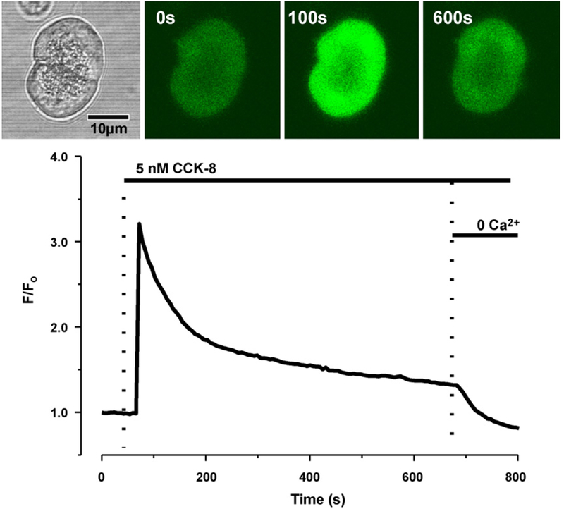
Abnormal, prolonged elevation of [Ca2+]i was proposed to be the crucial trigger of pancreatitic acinar cell injury in pancreatitis over a decade ago [2]. First tested by CCK octapeptide hyperstimulation (see Fig. 3) [3,4], an established means of inducing experimental acute pancreatitis, subsequent work with bile salts [5,6], FAs and FAEEs [7,8] confirmed the essential role of cytosolic Ca2+ overload in acinar cell injury. All these agents induce profound, global Ca2+ release from the ER, also likely from acid stores (ZGs and ELs), although the mechanisms and role of release from acid stores are less well characterised. Without sustained Ca2+ entry, cellular injury does not occur, the early signs of which include premature intracellular digestive enzyme activation, vacuolisation, secretory block, basolateral secretion and disorganisation of the cytoskeleton, all of which lead to cellular necrosis [3-8]. Nevertheless it is the sustained, global elevation of [Ca2+]i and not ER Ca2+ depletion that induces the injury, since abrogation of elevated [Ca2+]i with a Ca2+ chelator prevents these abnormal changes [3,6,8].
Figure 3.
Changes in the free ionised cytosolic Ca2+ concentration ([Ca2+]i) during hyperstimulation of a pancreatic acinar cell by CCK. Supra-maximal stimulation with 5 nM CCK induces a high peak and global, prolonged elevation of cytosolic Ca2+, sustained by Ca2+ entry. Upon withdrawal of external Ca2+, despite continuing hyperstimulation, [Ca2+]i returns to basal levels. Continued hyperstimulation in the presence of external Ca2+ induces premature, intracellular digestive enzyme activation, vacuolisation, disruption of the cytoskeleton, mitochondrial impairment and necrosis.
Three principal sites have been identified that are targets of toxins which induce excessively elevated [Ca2+]i, these being IP3Rs, RyRs and Ca2+-ATPase pumps [5,6,8,39]. Thus, bile salts induce Ca2+ release by increasing the open probability of IP3Rs and RyRs [5,39], whereas FAEEs induce Ca2+ release via IP3Rs [8]. FAs, however, do not induce significant release via ER Ca2+ channels at levels reached in vivo [8]. All these agents inhibit Ca2+-ATPase pumps through inhibition of mitochonrial ATP production, but in the case of bile salts this may also occur via a direct action on SERCA pumps [6].
4. Normal acinar cell mitochondrial function
Peri-apical, peri-nuclear and sub-plasmalemmal mitochondria comprise the principal aerobic powerhouses for all ATP-dependent activities of the acinar cell (see Fig. 4) [28-30]. Much as elsewhere, mitochondria integrate local cytosolic Ca2+ signals that activate Kreb’s cycle enzymes and drive ATP production, while maintaining a mitochondrial membrane potential (ΔψM) of 150—180 mV across the inner mitochondrial membrane, negative inside, by pumping protons into the inter-membrane space [15]. ΔψM concentrates Ca2+ within the matrix to effect Kreb’s cycle metabolism [15]. Some reactive oxygen species (ROS) generation is normal, and their effect on ΔψM is unclear, but there are many mitochondrial targets of injury from excessive ROS generation, including Ca2+ channels, transporters and microdomains, as well as complexes and enzymes that produce ATP [16-18,36,37]. Should this occur, acinar cell mitochondria initiate the intrinsic apoptotic pathway [18], as in most other cell types [36]. Mitochondria within the acinar cell — unlike in some other cell types [40] — play a pivotal role in maintaining Ca2+ homeostasis, directly and indirectly [28-30]. This role may depend, in part, upon their close apposition to ER Ca2+-release terminals [15,40]. Directly, the peri-apical mitochondria take up local cytosolic Ca2+ spikes through the Ca2+ uniporter [38], preventing further spread of the signal, terminated via the mitochondrial Na+/Ca2+ exchanger [15]. Such signal termination prevents further emptying of the Ca2+ stores, prevents large, frequent Ca2+ fluxes, and reduces metabolic demands for ATP production and energy expenditure in the cell.
Figure 4.
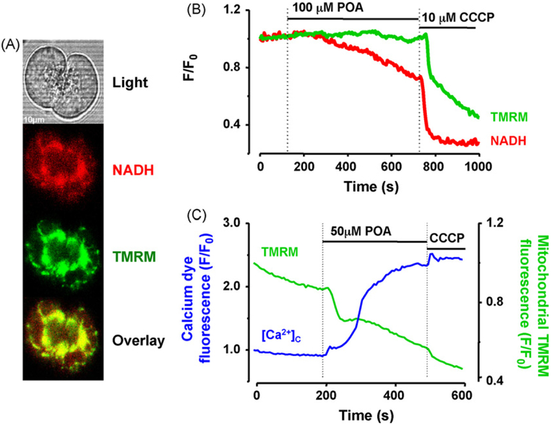
Pancreatic acinar cell mitochondrial impairment induced by POA which is independent of, but exacerbated by, Ca2+ overload. (A) Typical light microscopic appearance of a doublet with NADH (auto-fluorescence) and tetramethyl rhodamine methyl ester (TMRM, ΔψM-dependent) fluorescence shown with overlay in the bottom panel. (B) Fall in NADH autofluorescence after POA administration in a cell pre-treated with a Ca2+ chelator that results in little if any change in [Ca2+]i, or fall in ΔψM, suggesting diminished ATP production without an immediate loss in electrochemical gradient, such as might occur with diminished βoxidation. (C) Large, sustained rise in [Ca2+]i during POA administration to cell without pre-treatment with a Ca2+ chelator, preceding and accompanied by a fall in ΔψM, indicated by a drop in TMRM fluorescence. The drop in ΔψM is to such a level that the protonophore CCCP induces little further fall (reproduced in part with permission from reference [8], copyright Elsevier).
Within pancreatic acinar cells physiological Ca2+ signals always begin within the apical pole, but on occasions the combinatorial effects of multiple second messengers (IP3, cADPr and NAADP) and CICR exceed the immediate buffering capacity of peri-apical mitochondria, such that Ca2+ signals spread into the basolateral region to enter peri-nuclear and sub-plasmalemmal mitochondria and initiate cellular responses in that part of the cell [27,31]. These mitochondrial groups again respond to and assist in termination of the signal, supplying ATP for SERCA and PMCA activity that completes the process.
5. Abnormal mitochondrial function
The most significant mechanism of Ca2+-ATPase inhibition by toxins that induce pancreatitis is likely to be that of direct impairment of mitochondrial ATP production [8,15-18,23,36,41]. FAEEs are formed non-oxidatively from ethanol and fatty acids by synthases that are relatively abundant in pancreatic acinar cells [11]. FAEEs induce experimental pancreatitis and are found in high concentrations within the pancreata of people who have died from alcohol intoxication [11,13,14]. FAEEs bind to and accumulate within the inner mitochondrial membrane, where hydrolases release locally high concentrations of FAs, which uncouple oxidative phosphorylation, deplete ΔψM, and impair ATP production [8,12]. Hyperlipidaemia, another cause of pancreatitis, also results in high concentrations of long-chain FAs within cells, may be transported into mitochondria by carnitine palmitoyltransferase and induce mitochondrial impairment similar to that caused by FAEEs [8]. Failure of ATP production reduces the capacity of the acinar cell to clear Ca2+, whatever the cause, whether from Ca2+ signals, Ca2+ leak [15,18], or an abnormal elevation in [Ca2+]i caused by toxins [39]. Under these circumstances, peri-granular mitochondria cannot buffer apical Ca2+ elevations. The resulting elevation of [Ca2+]i increases the Ca2+ load on mitochondria, induces further collapse of ΔψM and impairment of ATP production, until the cell passes beyond any capacity to sustain sufficient ATP production for apoptosis, such that necrosis results [18]. Protection of cells from this sequence can be provided by excluding Ca2+ from the external medium [3,5,7,8], or providing supplementary intracellular ATP [8]. This has been demonstrated in patch clamp experiments where 4mM ATP has been supplied via the patch pipette, maintaining activity of SERCA and PMCA pumps to avoid global, sustained, toxic elevations of [Ca2+]i [8].
One important effect of FAs on pancreatic acinar cell mitochondria is uncoupling of oxidative phosphorylation, an effect that does not depend on gross depolarisation of ΔψM per se [8]. It is, however, noteworthy that loss of ΔψM is an early event in experimental pancreatitis [48], in large measure attributable to toxic elevation of [Ca2+]i [8]. Thus, in cells loaded with a cytosolic Ca2+ chelator and treated with FAs, diminished ATP production is seen (suggested by a fall in NADH auto-fluorescence, see Fig. 4) without obvious changes in ΔψM. Without Ca2+ chelation, large, global, sustained elevations in [Ca2+]i occur that induce obvious loss of ΔψM and accelerate mitochondrial impairment [8,18,27]. Bile salts, however, may induce a modest loss of ΔψM even in the presence of Ca2+ chelation, although detection of such a loss of ΔψM depends on high concentrations (>100 μM, which may have detergent effects) of bile salts and high ‘dequench’ concentrations of mitochondrial fluorescent dyes, since ‘classic’ concentrations of these dyes fail to demonstrate any loss of ΔψM [37].
6. Apoptosis versus necrosis in pancreatitis
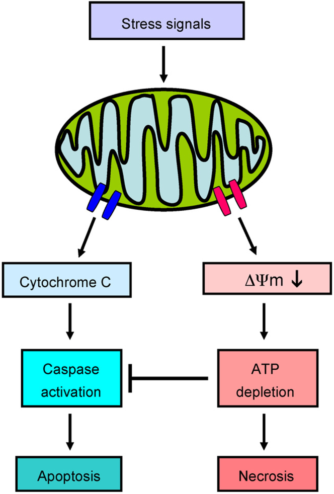
For many years it has been accepted that in pancreatitis the major form of pancreatic acinar cell death is necrosis [42]. Necrosis is characterised by severe pathophysiological changes that include mitochondrial swelling, plasmalemmal disruption and leakage of activated digestive enzymes and other cellular contents, which trigger acute exudative inflammation of the surrounding tissue [1,43-45]. The process is largely uncontrolled but can be initiated by a variety of toxic insults, including bile salts, FAEEs and FAs, inducing cytosolic Ca2+ overload, excessive ROS generation, severe depletion of ΔψM and failure of ATP production, as described [3-8] (see Fig. 5). In contrast, apoptosis is genetically regulated, occurs via both caspase dependent and independent pathways [41] and requires ATP to proceed. Both intrinsic (classical, mitochondrial, see Fig. 5) and extrinsic (receptor-mediated) apoptotic pathways progress via a cascade of events that result in removal of cells from tissue which unlike necrosis occurs with minimal release of intracellular content, inflammation and collateral damage [16-18,41,45].
Figure 5.
Alternative cell death pathways in pancreatic acinar cells associated with release of mitochondrial content. In response to Ca2+ loading of mitochondria and other stresses, cytochrome c is released from the inner mitochondrial membrane, activating caspases and inducing further Ca2+ release via IP3Rs. These events may be controlled by Bax/Bak and Bcl-2 mediated channels (shown in blue). Caspase activation has a protective role in pancreatitis, inhibited by a lack of ATP. Marked loss of mitochondrial membrane potential, notably with induction of the MPTP (shown in red, see Fig. 6), inhibits ATP production, which in turn inhibits apoptosis and leads to necrosis.
In vivo, cell death pathways are less distinct from one another, although apoptotic pathways are more evident in mild pancreatitis, whereas by definition, necrosis is more obvious in severe pancreatitis [18,46-48]. Recent data suggest that promotion of apoptosis is beneficial, whereas inhibition of apoptosis is harmful [46,48]. Thus, promotion of caspase activity, through reduction of endogenous caspase inhibition, has been found to protect from pancreatic necrosis in caerulein pancreatitis, whereas caspase inhibitors were found to increase pancreatic necrosis [48]. A further issue in understanding how cell death occurs in the pancreas is that stressed but surviving acinar cells produce and release cytokines that prime inflammatory responses, which by themselves contribute to further cell injury [1,49]. Promotion of apoptosis may reduce the capacity of acinar cells to produce such inflammatory responses. Infiltrating neutrophils are significant in this regard, since neutrophil depletion or genetic deletion of NADPH oxidase significantly lessens the severity of experimental acute pancreatitis [50], although how — and why — activated neutrophils exacerbate pancreatitis remains unclear. Nonetheless, moderate cell stresses are more likely to induce apoptosis, while severe stresses overcome apoptotic pathways and result in necrosis, especially when the protective functions of mitochondria are overwhelmed, as from massive Ca2+ overload or direct inhibition by FAEEs and/or FAs [8,16-18,36,46-48].
Elevations of [Ca2+]i have been shown to be essential to the induction of apoptosis in isolated pancreatic acinar cells by the ROS-producing oxidant menadione [51,52]. Although global, these elevations are not prolonged (>30s), unlike those induced by supramaximal stimulation with CCK, bile salts, FAEEs or FAs. The combination, however, of a modest, global elevation of [Ca2+]i plus abnormal ROS production induces mitochondrial release of cytochrome c and activation of caspase 9 (~2 min), in keeping with the mitochondrial, apoptotic pathway [51,52]. When ROS production is augmented by inhibition of endogenous anti-oxidant defence mechanisms, pancreatic acinar cell apoptosis is potentiated [52]. Additionally, there is a slower (~30 min), ‘fail-safe’, Ca2+-independent mechanism of caspase 8 activation, dependent on lysosomal release of cathepsins, evident in a minority of cells [53]. Both cytochrome c and caspase 3 have been shown to increase Ca2+ release via the IP3R [54], but the changes in [Ca2+]i that occur in the later stages of pancreatic acinar cell apoptosis have not yet been examined. Nevertheless, should apoptosis fail, necrosis is likely to supervene, as may follow inhibition of caspases or ATP production [18].
Some acinar cells appear more resistant to toxic stresses than others, although the determinants of relative vulnerability have yet to be defined [47]. Initiation of apoptosis prior to the induction of experimental pancreatitis reduces the severity of the disease [55], suggesting that such pretreatment removes cells that would otherwise become necrotic. Unlike acute infarction in other organs, pancreatic necrosis is a slow process, developing over 72 h or more [1,22,23], during which time cells may pass through various phases of apoptosis. Although the determinants, nature and extent of the switch from apoptosis to necrosis in pancreatitis remain to be identified [18,47], it would appear that mitochondria are likely to play a central role in this switch.
7. Role of the mitochondrial permeability transition pore
An evolving hypothesis is that the mitochondrial permeability transition pore (MPTP) determines cell fate from injury, notably in glutamate-induced neurotoxicity as well as ischaemia-reperfusion injury in the brain and heart [56]. Mitochondrial Ca2+ overload and ROS induce a ‘permeability transition’ across the membranes of mitochondria, causing the membrane to become permeable to any molecule of less than 1.5 kDa, including protons, leading to a loss of ΔψM [17,36,40,56]. Induction of the MPTP is a graded phenomenon, in that the fall in ΔψM may be partial and/or transient with continued mitochondrial function, or ΔψM may collapse fully and ATP production may fail [56]. In the former circumstance apoptosis is likely, although this is unlikely to be by the intrinsic apoptotic pathway via cytochrome c release [1,15-17,56-60] (see Fig. 5); without ATP, necrosis follows. Nevertheless, since Ca2+ overload with or without ROS generation and mitochondrial failure are features of pancreatic acinar cell injury from agents that induce pancreatitis [1,3-8], the MPTP could be a key factor in any switch from apoptosis to necrosis in the development of severe, necrotising pancreatitis.
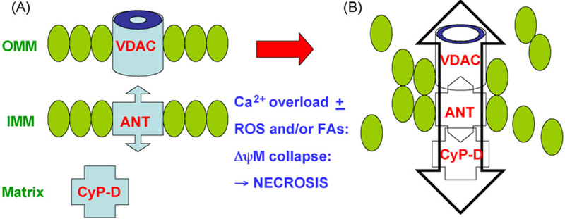
The pore has at least three components, these being a voltage dependent anion channel (VDAC) in the outer membrane, adenine nucleotide translocase (ANT) in the inner mitochondrial membrane and CyP-D within the mitochondrial matrix [17,56]. VDAC is closely associated with ER IP3Rs and localises Ca2+ signals between the ER and mitochondria [15], while ANT exchanges cytosolic ADP for mitochondrial ATP (and can act in reverse) [36,56]; CyP-D is within the mitochondrial matrix and exhibits peptidyl-prolyl cis—trans isomerise (PPIase) activity [56]. High concentrations of Ca2+ induce conformational changes that produce macro-molecular complexes of these proteins to form the MPTP [17,56]. Data implicate each molecule in both apoptotic and necrotic cell death, depending on the effect on ΔψM [15-18,36,41,56] (see Fig. 6). It has been shown in other models that mitochondrial Ca2+ overload together with oxidative stress may lead to sustained opening of the MPTP, mitochondrial swelling and leakage, a profound failure of ATP production and cell necrosis [16,17,56]. In pancreatic acinar cells, blockade of the MPTP with bongkrekic acid inhibits the partial, transient loss of ΔψM and caspase activation typically induced by menadione, and so inhibits menadione-induced apoptosis [51], although any effect on necrotic cell death has not been studied. Recent data, however, bring the role of VDAC [57] and of ANT [58] in MPTP mediated-injury into question. Also, CyP-D knockout mice develop normally and show no protection from a range of stimuli of that induce apoptosis via the intrinsic pathway [59,60], although these mice are protected from Ca2+ overload and ROS in heart and brain ischaemia-reperfusion injury [59,60]. Surprisingly, cyclosporin A, which binds to CyP-D and inhibits the MPTP, does not ameliorate but exacerbates acute pancreatitis by an unclear mechanism [61]. These data are consistent with independent operation of the intrinsic apoptotic pathway by another mechanism, the most likely candidate for which is cytochrome c release via Bax/Bak channels in the outer mitochondrial membrane [1,15-17,58-60]. In the setting of chronic pancreatic injury, cyclosporin A prevents tissue remodelling, and exacerbates fibrosis, perhaps by altering cytokine expression [62]. Clearly, a logical next step would be to determine whether sustained opening of the MPTP has a major role in acute pancreatitis, and to explore relationships between the intrinsic apoptotic pathway, Bcl-2 proteins and the MPTP.
Figure 6.
Key components of the mitochondrial permeability transition pore (MPTP) contributing to partial or complete loss of ΔψM. (A) Normal mitochondrial components of the outer mitochondrial membrane (OMM; voltage-dependent anion transporter, VDAC), inner mitochondrial membrane (IMM; adenine nucleotide transporter, ANT) and mitochondrial matrix (cyclophilin-D, CyP-D) undergo conformational change following moderate or severe stress, resulting in transient or (B) sustained formation of the MPTP, the latter ending in necrosis. Ca2+ overload, FAs and reactive oxygen species (ROS) contribute to induction of the MPTP, which allows passage of all particles up to 1.5 kDa, contributing to mitochondrial swelling and rupture.
8. Therapeutic prospects
Both acute and chronic pancreatitis have no specific pharmacological therapy and a growing mountain of negative randomised control trials [1,63]. Several approaches could be adopted to ameliorate these diseases [23]:
Prevent the disease entirely.
Modify the early course of severe acute pancreatitis.
Halt progression to chronic disease.
The increasing, worldwide abuse of alcohol is likely to be the most stubborn obstacle to the first approach [23], as well as the prevalence of silent gallstones and the diversity of less frequent aetiologies. Thus, a cure is required, most especially for severe, necrotising disease that is the major cause of morbidity and mortality. Acute infarction in the pancreas is very different to that in other organs such as the brain or heart, being a slower process that develops over 72 h or more [1,22,23]. Potentially, this is an important window during which compounds that limit acinar cell injury could abrogate severe disease. Similarly, those agents that limit acute injury could modify continuing injury that drives progression to chronic disease.
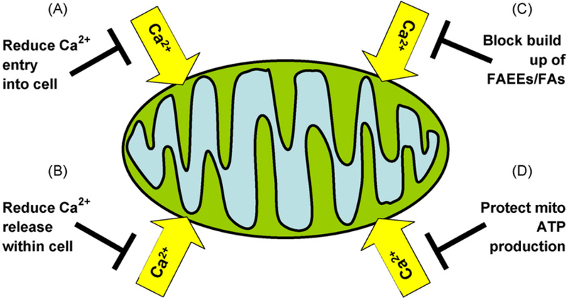
To date, all data indicate that cytosolic Ca2+ overload is common to all forms of pancreatic acinar cell injury from toxins that induce pancreatitis [3-8,23]. Attractive interventions would be to (1) reduce the effect of toxins prior to the induction of Ca2+ release, (2) inhibit Ca2+ entry into or accumulation within the cell, (3) enhance [Ca2+]i clearance (see Fig. 7). Inhibition of FAEE synthesis and/or hydrolysis could lessen the toxicity of ethanol [8]. As yet no agent specifically blocks Ca2+ entry into non-excitable cells, let alone into pancreatic acinar cells [15]; the nature and location of pancreatic acinar cell store-operated Ca2+ entry channels remain controversial [27,35]. Preservation of mitochondrial function to enhance [Ca2+]i clearance may reduce the severity of pancreatitis, using strategies under development for other organs. Thus, enhancement of glucose oxidation in preference to β-oxidation may reduce toxicity from FAs [64], while anti-oxidants and/or chemical preconditioning could be beneficial.
Figure 7.
Strategies to reduce mitochondrial injury and acinar cell damage in pancreatitis. (A) Blockage of Ca2+ entry will probably depend on identification of the SOC channel in the pancreatic acinar cell. (B) Prevention of intracellular Ca2+ release that awaits identification of specific antagonists of pancreatic acinar cell Ca2+ release mechanisms (see also Fig. 2). (C) Inhibition of fatty acid ethyl ester (FAEE) synthases combine ethanol with FAs, after which FAEEs bind to and accumulate within the inner mitochondrial membrane. Inhibition of hydrolases would prevent the release of FAs from FAEEs already accumulated within mitochondria. (D) Anti-oxidant and/or preconditioning strategies could preserve mitochondrial function to deliver adequate ATP for pancreatic acinar cells to survive intact.
Acknowledgements
This work was supported by Programme, Cooperative and Component Grants from the Medical Research Council, UK (OHP, RS) and by National Institutes of Health Grants DK59936 (ASG) and DK59508 (SP); OHP is a Medical Research Council Professor and RM is supported by an Amelie Waring Clinical Research Fellowship from CORE.
Abbreviations:
- ACh
acetylcholine
- ADP
adenosine diphosphate
- ANT
adenine nucleotide transporter
- ATP
adenosine triphosphate
- [Ca2+]i
cytosolic free Ca2+ ion concentration
- cADPr
cyclic ADP ribose
- CCCP
carbonyl cyanide 3-chlorophenylhydrazone
- CCK
cholecystokinin
- CICR
Ca2+-induced Ca2+ release
- CyP-D
cyclophilin D
- Δψm
mitochondrial membrane potential
- EL
endo-lysosome
- ER
endoplasmic reticulum
- FA
fatty acid
- FAEE
fatty acid ethyl ester
- IP3
inositol trisphosphate
- IP3R
inositol trisphosphate receptor
- MPTP
mitochondrial permeability transition pore
- NAADP
nicotinic acid adenine dinucleotide phosphate
- NAADPR
nicotinic acid adenine dinucleotide phosphate receptor
- NADH
reduced nicotinamide adenine dinucleotide
- PMCA
plasma membrane Ca2+ ATPase pump
- POA
palmitoleic acid
- ROS
reactive oxygen species
- RyR
ryanodine receptor
- SERCA
sarcoplasmic/endoplasmic reticulum Ca2+ ATPase pump
- SOC
store-operated Ca2+ entry channel
- TMRM
tetramethyl rhodamine methyl ester
- VDAC
voltage-dependent anion transporter
- ZG
zymogen granule
References
- [1].Pandol SJ, Saluja AK, Imrie CW, Banks PA, Acute pancreatitis: bench to bedside, Gastroenterology 132 (2007) 1127–1151. [DOI] [PubMed] [Google Scholar]
- [2].Ward JB, Petersen OH, Jenkins SA, Sutton R, Is an elevated concentration of acinar cytosolic free ionised calcium the trigger for acute pancreatitis? Lancet 346 (1995) 1016–1019. [DOI] [PubMed] [Google Scholar]
- [3].Raraty M, Ward J, Erdemli G, Vaillant C, Neoptolemos JP, Sutton R, Petersen OH, Calcium-dependent enzyme activation and vacuole formation in the apical granular region of pancreatic acinar cells, Proc. Natl. Acad. Sci. U.S.A. 97 (2000) 13126–13131. [DOI] [PMC free article] [PubMed] [Google Scholar]
- [4].Kruger B, Albrecht E, Lerch MM, The role of intracellular calcium signaling in premature protease activation and the onset of pancreatitis, Am. J. Pathol. 157 (2000) 43–50. [DOI] [PMC free article] [PubMed] [Google Scholar]
- [5].Voronina S, Longbottom R, Sutton R, Petersen OH, Tepikin A, Bile acids induce calcium signals in mouse pancreatic acinar cells. Implications for bile-induced pancreatic pathology, J. Physiol. 540 (2002) 49–55. [DOI] [PMC free article] [PubMed] [Google Scholar]
- [6].Kim JY, Kim KH, Lee JA, Namkung W, Sun AQ, Ananthanarayanan M, Suchy FJ, Shin DM, Muallem S, Lee MG, Transporter-mediated bile acid uptake causes Ca2+-dependent cell death in rat pancreatic acinar cells, Gastroenterology 122 (2002) 1941–1953. [DOI] [PubMed] [Google Scholar]
- [7].Criddle DN, Raraty MG, Neoptolemos JP, Tepikin AV, Petersen OH, Sutton R, Ethanol toxicity in pancreatic acinar cells: mediation by nonoxidative fatty acid metabolites, Proc. Natl. Acad. Sci. U.S.A. 101 (2004) 10738–10743. [DOI] [PMC free article] [PubMed] [Google Scholar]
- [8].Criddle DN, Murphy J, Fistetto G, Barrow S, Tepikin AV, Neoptolemos JP, Sutton R, Petersen OH, Fatty acid ethyl esters cause pancreatic calcium toxicity via inositol trisphosphate receptors and loss of ATP synthesis, Gastroenterology 130 (2006) 781–793. [DOI] [PubMed] [Google Scholar]
- [9].Armstrong CP, Taylor TV, Pancreatic-duct reflux and acute gallstone pancreatitis, Ann. Surg. 204 (1986) 59–64. [DOI] [PMC free article] [PubMed] [Google Scholar]
- [10].Tashiro S, et al. , Pancreatobiliary maljunction: retrospective and nationwide survey in Japan, J. Hepatobiliary Pancreat. Surg. 10 (2003) 345–351. [DOI] [PubMed] [Google Scholar]
- [11].Lange LG, Sobel BE, Mitochondrial dysfunction induced by fatty acid ethyl esters, myocardial metabolites of ethanol, J. Clin. Invest. 72 (1983) 724–731. [DOI] [PMC free article] [PubMed] [Google Scholar]
- [12].Laposata EA, Lange LG, Presence of nonoxidative ethanol metabolism in human organs commonly damaged by ethanol abuse, Science 231 (1986) 497–499. [DOI] [PubMed] [Google Scholar]
- [13].Werner J, Laposata M, Fernandez-del Castillo C, Saghir M, Iozzo RV, Lewandrowski KB, Warshaw AL, Pancreatic injury in rats induced by fatty acid ethyl ester, a nonoxidative metabolite of alcohol, Gastroenterology 113 (1997) 286–294. [DOI] [PubMed] [Google Scholar]
- [14].Werner J, Saghir M, Warshaw AL, Lewandrowski KB, Laposata M, Iozzo RV, Carter EA, Schatz RJ, Fernandez-Del Castillo C, Alcoholic pancreatitis in rats: injury from nonoxidative metabolites of ethanol, Am. J. Physiol. Gastrointest. Liver Physiol. 283 (2002) G65–G73. [DOI] [PubMed] [Google Scholar]
- [15].Rizzuto R, Pozzan T, Microdomains of intracellular calcium: molecular determinants and functional consequences, Physiol. Rev. 86 (2006) 369–408. [DOI] [PubMed] [Google Scholar]
- [16].Orrenius S, Gogvadze V, Zhivotovsky B, Mitochondrial oxidative stress: implications for cell death, Annu. Rev. Pharmacol. Toxicol. 47 (2007) 143–183. [DOI] [PubMed] [Google Scholar]
- [17].Kroemer G, Galluzzi L, Brenner C, Mitochondrial membrane permeabilization in cell death, Physiol. Rev. 87 (2007) 99–163. [DOI] [PubMed] [Google Scholar]
- [18].Criddle DN, Gerasimenko JV, Baumgartner HK, Jaffar M, Voronina S, Sutton R, Petersen OH, Gerasimenko OV, Calcium signalling and pancreatic cell death: apoptosis or necrosis? Cell Death Differ. 14 (2007) 1285–1294. [DOI] [PubMed] [Google Scholar]
- [19].Fitz RH, Acute pancreatitis, a consideration of pancreatic hemorrhage, hemorrhagic, suppurative, and gangrenous pancreatitis, and of disseminated fat necrosis, Boston Med. Surg. J. 70 (1889) 181–235. [Google Scholar]
- [20].Moynihan B, Acute pancreatitis, Ann. Surg. 81 (1925)132–142. [DOI] [PMC free article] [PubMed] [Google Scholar]
- [21].Jaakkola M, Nordback I, Pancreatitis in Finland between 1970 and 1989, Gut 34 (1993) 1255–1260. [DOI] [PMC free article] [PubMed] [Google Scholar]
- [22].Swaroop VS, Chari ST, Clain JE, Severe acute pancreatitis, JAMA 291 (2004) 2865–2868. [DOI] [PubMed] [Google Scholar]
- [23].Petersen OH, Sutton R, Ca2+ signalling and pancreatitis: effects of alcohol, bile and coffee, TIPS 27 (2006) 113–120. [DOI] [PubMed] [Google Scholar]
- [24].Whitcomb DC, Gorry MC, Preston RA, Furey W, Sossenheimer MJ, Ulrich CD, Martin SP, Gates LK, Amann ST, Toskes PP, Liddle R, McGrath K, Uomo G, Post JC, Ehrlich GD, Hereditary pancreatitis is caused by a mutation in the cationic trypsinogen gene, Nat. Genet. 14 (1996) 141–145. [DOI] [PubMed] [Google Scholar]
- [25].Howes N, Lerch MM, Greenhalf W, Stocken DD, Ellis I, Simon P, Truninger K, Ammann R, Cavallini G, Charnley RM, Uomo G, Delhaye M, Spicak J, Drumm B, Jansen J, Mountford R, Whitcomb DC, Neoptolemos JP, European Registry of Hereditary Pancreatitis and Pancreatic Cancer (EUROPAC). Clinical and genetic characteristics of hereditary pancreatitis in Europe, Clin. Gastroeterol. Hepatol 2 (2004) 252–261. [DOI] [PubMed] [Google Scholar]
- [26].Lowenfels AB, Maisonneuve P, Cavallini G, Ammann RW, Lankisch PG, Andersen JR, Dimagno EP, Andren-Sandberg A, Domellof L, Pancreatitis and the risk of pancreatic cancer. International Pancreatitis Study Group, N. Engl. J. Med. 328 (1993) 1433–1437. [DOI] [PubMed] [Google Scholar]
- [27].Petersen OH, Ca2+ signalling and Ca2+-activated ion channels in exocrine acinar cells, Cell Calcium 38 (2005) 171–200. [DOI] [PubMed] [Google Scholar]
- [28].Tinel H, Cancela JM, Mogami H, Gerasimenko JV, Gerasimenko OV, Tepikin AV, Petersen OH, Active mitochondria surrounding the pancreatic acinar granule region prevent spreading of inositol trisphosphate-evoked local cytosolic Ca(2+) signals, EMBO J. 18 (1999) 4999–5008. [DOI] [PMC free article] [PubMed] [Google Scholar]
- [29].Park MK, Ashby MC, Erdemli G, Petersen OH, Tepikin AV, Perinuclear, perigranular and sub-plasmalemmal mitochondria have distinct functions in the regulation of cellular calcium transport, EMBO J. 20 (2001) 1863–1874. [DOI] [PMC free article] [PubMed] [Google Scholar]
- [30].Voronina S, Sukhomlin T, Johnson PR, Erdemli G, Petersen OH, Tepikin A, Correlation of NADH and Ca2+ signals in mouse pancreatic acinar cells, J. Physiol. 539 (2002) 41–52. [DOI] [PMC free article] [PubMed] [Google Scholar]
- [31].Ashby M, Tepikin AV, Polarised calcium and calmodulin signalling in secretory epithelia, Physiol. Rev. 82 (2002) 701–734. [DOI] [PubMed] [Google Scholar]
- [32].Nguyen T, Chin W-C, Verdugo P, Roleof Ca2+/K+ ion exchange in intracellular storage and release of Ca2+, Nature 395 (1998) 908–912. [DOI] [PubMed] [Google Scholar]
- [33].Gerasimenko OV, Gerasimenko JV, Belan PV, Petersen OH, Inositol trisphosphate and cyclic ADP-ribose-mediated release of Ca2+ from single isolated pancreatic zymogen granules, Cell 84 (1996)473–480. [DOI] [PubMed] [Google Scholar]
- [34].Burdakov D, Petersen OH, Verkhratsky A, Intraluminal calcium as a primary regulator of endoplasmic reticulum function, Cell Calcium 38 (2005) 303–310. [DOI] [PubMed] [Google Scholar]
- [35].Parekh AB, Putney JW, Store-operated calcium channels, Physiol. Rev. 85 (2005) 757–810. [DOI] [PubMed] [Google Scholar]
- [36].Davidson SM, Duchen MR, Calcium microdomains and oxidative stress, Cell Calcium 40 (2006) 561–574. [DOI] [PubMed] [Google Scholar]
- [37].Voronina SG, Barrow SL, Gerasimenko OV, Petersen OH, Tepikin AV, Effects of secretagogues and bile acids on mitochondrial membrane potential of pancreatic acinar cells: comparison of different modes of evaluating DeltaPsim, J. Biol. Chem. 279 (2004) 27327–27338. [DOI] [PubMed] [Google Scholar]
- [38].Kirichok Y, Krapivinsky G, Clapham DE, The mitochondrial calcium uniporter is a highly selective ion channel, Nature 427 (2004) 360–364. [DOI] [PubMed] [Google Scholar]
- [39].Gerasimenko JV, Flowerdew SE, Voronina SG, Sukhomlin TK, Tepikin AV, Petersen OH, Gerasimenko OV, Bile acids induce Ca2+ release from both the endoplasmic reticulum and acidic intracellular calcium stores through activation of inositol trisphosphate receptors and ryanodine receptors, J. Biol. Chem. 281 (2006) 40154–40163. [DOI] [PubMed] [Google Scholar]
- [40].Franzini-Armstrong C, ER—mitochondrial communication. How priviledged? Physiology 22 (2007) 261–268. [DOI] [PubMed] [Google Scholar]
- [41].Orrenius S, Zhivotovsky B, Nicotera P, Regulation of cell death: the calcium-apoptosis link, Nat. Rev. Mol. Cell Biol. 4 (2003) 552–565. [DOI] [PubMed] [Google Scholar]
- [42].Kloppel G, Maillet B, Pathology of acute and chronic pancreatitis, Pancreas 8 (1993) 659–670. [DOI] [PubMed] [Google Scholar]
- [43].Hartwig W, Jimenez RE, Werner J, Lewandrowski KB, Warshaw AL, Fernandez-del Castillo C, Interstitial trypsinogen release and its relevance to the transformation of mild into necrotizing pancreatitis in rats, Gastroenterology 117 (1999) 717–725. [DOI] [PubMed] [Google Scholar]
- [44].Hartwig W, Werner J, Jimenez RE, Z’graggen K, Weimann J, Lewandrowski KB, Warshaw AL, Fernandez-del Castillo C, Trypsin and activation of circulating trypsinogen contribute to pancreatitis-associated lung injury, Am. J. Physiol. 277 (1999) G1008–G1016. [DOI] [PubMed] [Google Scholar]
- [45].Kroemer G, et al. , Classification of cell death: recommendations of the Nomenclature Committee on Cell Death, Cell Death Differ. 12 (Suppl. 2) (2005) 1463–1467. [DOI] [PubMed] [Google Scholar]
- [46].Kaiser AM, Saluja AK, Sengupta A, Saluja M, Steer ML, Relationship between severity, necrosis, and apoptosis in five models of experimental acute pancreatitis, Am. J. Physiol. 269 (1995) C1295–C1304. [DOI] [PubMed] [Google Scholar]
- [47].Bhatia M, Apoptosis versus necrosis in acute pancreatitis, Am. J. Physiol. Gastrointest. Liver Physiol 86 (2004) G189–G196. [DOI] [PubMed] [Google Scholar]
- [48].Mareninova OA, Sung KF, Hong P, Lugea A, Pandol SJ, Gukovsky I, Gukovskaya AS, Cell death in pancreatitis: caspases protect from necrotizing pancreatitis, J. Biol. Chem. 281 (2006) 3370–3381. [DOI] [PubMed] [Google Scholar]
- [49].Gukovskaya AS, Gukovsky I, Zaninovic V, Song M, Sandoval D, Gukovsky S, Pandol SJ, Pancreatic acinar cells produce, release, and respond to tumor necrosis factor-alpha. Role in regulating cell death and pancreatitis, J. Clin Invest. 100 (1997) 1853–1862. [DOI] [PMC free article] [PubMed] [Google Scholar]
- [50].Gukovskaya AS, Vaquero E, Zaninovic V, Gorelick FS, Lusis AJ, Brennan ML, Holland S, Pandol SJ, Neutrophils and NADPH oxidase mediate intrapancreatic trypsin activation in murine experimental acute pancreatitis, Gastroenterology 122 (2002) 974–984. [DOI] [PubMed] [Google Scholar]
- [51].Gerasimenko JV, Gerasimenko OV, Palejwala A, Tepikin AV, Petersen OH, Watson AJ, Menadione-induced apoptosis: roles of cytosolic Ca(2+) elevations and the mitochondrial permeability transition pore, J. Cell Sci. 115 (2002) 485–497. [DOI] [PubMed] [Google Scholar]
- [52].Criddle DN, Gillies S, Baumgartner-Wilson HK, Jaffar M, Chinje EC, Passmore S, Chvanov M, Barrow S, Gerasimenko OV, Tepikin AV, Sutton R, Petersen OH, Menadione-induced reactive oxygen species generation via redox cycling promotes apoptosis of murine pancreatic acinar cells, J. Biol. Chem. 281 (2006) 40485–40492. [DOI] [PubMed] [Google Scholar]
- [53].Baumgartner HK, Gerasimenko JV, Thorne C, Ashurst LH, Barrow SL, Chvanov MA, Gillies SG, Criddle DN, Tepikin AV, Petersen OH, Sutton R, Watson AJ, Gerasimenko OV, Caspase-8 mediated apoptosis induced by oxidative stress is independent of the intrinsic pathway and dependent on cathepsins, Am. J. Physiol. Gastrointest. Liver Physiol (April) (2007) [Epub ahead of print]. [DOI] [PubMed] [Google Scholar]
- [54].Hanson CJ, Bootman MD, Roderick HL, Cell signalling: IP3 receptors channel calcium into cell death, Curr. Biol. 14 (2004) R933–R935. [DOI] [PubMed] [Google Scholar]
- [55].Bhatia M, Wallig MA, Hofbauer B, Lee HS, Frossard JL, Steer ML, Saluja AK, Induction of apoptosis in pancreatic acinar cells reduces the severity of acute pancreatitis, Biochem. Biophys. Res. Commun. 246 (1998) 476–483. [DOI] [PubMed] [Google Scholar]
- [56].Halestrap AP, Calcium, mitochondria and reperfusion: a pore way to die, Biochem. Soc. Trans. 34 (2006) 232–237. [DOI] [PubMed] [Google Scholar]
- [57].Kokoszka JE, Waymire KG, Levy SE, Sligh JE, Cai J, Jones DP, MacGregor GR, Wallace DC, The ADP/ATP translocator is not essential for the mitochondrial permeability transition pore, Nature 427 (2004) 461–465. [DOI] [PMC free article] [PubMed] [Google Scholar]
- [58].Baines CP, Kaiser RA, Sheiko T, Craigen WJ, Molkentin JD, Voltage-dependent anion channels are dispensable for mitochondrial-dependent cell death, Nat. Cell Biol. 9 (2007) 550–555. [DOI] [PMC free article] [PubMed] [Google Scholar]
- [59].Nakagawa T, Shimizu S, Watanabe T, Yamaguchi O, Otsu K, Yamagata H, Inohara H, Kubo T, Tsujimoto Y, Cyclophilin D-dependent mitochondrial permeability transition regulates some necrotic but not apoptotic cell death, Nature 434 (2005) 652–658. [DOI] [PubMed] [Google Scholar]
- [60].Baines CP, Kaiser RA, Purcell NH, Blair NS, Osinska H, Hambleton MA, Brunskill EW, Sayen MR, Gottlieb RA, Dorn GW, Robbins J, Molkentin JD, Loss of cyclophilin D reveals a critical role for mitochondrial permeability transition in cell death, Nature 434 (2005) 658–662. [DOI] [PubMed] [Google Scholar]
- [61].Ito T, Kimura T, Yamaguchi H, Kinjo M, Sumii T, Nakano I, H. Nawata, Acute pancreatitis induced by cyclosporin A under stimulation of pancreas by caerulein, Pancreas 8 (1993) 693–699. [DOI] [PubMed] [Google Scholar]
- [62].Vaquero E, Molero X, Tian X, Salas A, Malagelada JR, Myofibroblast proliferation, fibrosis, and defective pancreatic repair induced by cyclosporin in rats, Gut 45 (1999) 269–277. [DOI] [PMC free article] [PubMed] [Google Scholar]
- [63].Heinrich S, Schafer M, Rousson V, Calvien PA, Evidence based treatment of acute pancreatitis: a look at established paradigms, Ann. Surg. 243 (2006) 154–168. [DOI] [PMC free article] [PubMed] [Google Scholar]
- [64].Morrow DA, Givertz MM, Modulation of myocardial energetics: emerging evidence for a therapeutic target in cardiovascular disease, Circulation 112 (2005) 3218–3221. [DOI] [PubMed] [Google Scholar]


