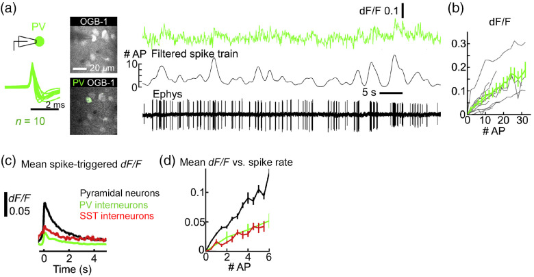
Fig. 2.
Somatic calcium transients correlate with firing rates of cortical GABAergic neurons. (a) Somatic calcium transients from a parvalbumin-expressing (PV) interneuron in layer 2/3 of the mouse visual cortex, imaged using a two-photon microscope. Cell type was identified based on tdTomato expression. Neurons were loaded with the synthetic calcium dye Oregon Green BAPTA-1 (OGB-1). Spiking activity was recorded under cell-attached condition. The left panel shows the mean normalized spike waveforms of 10 cells. The middle panel shows images including a recorded neuron. The right panel shows fluorescence and electrophysiological traces from an example cell. The filtered spike train was smoothed with a Gaussian filter (s.d. = 0.5 s). (b) Fluorescence versus number of action potentials was determined using the filtered spike train. Gray lines, individual cells. Black line, mean ± s.e.m. (c) Mean spike-triggered fluorescence for excitatory (), PV (), and SST neurons (). (d) Mean fluorescence versus number of action potentials for excitatory, PV, and SST neurons. The figure is adapted from Ref. 32. Reproduced with permission, courtesy of Elsevier.

