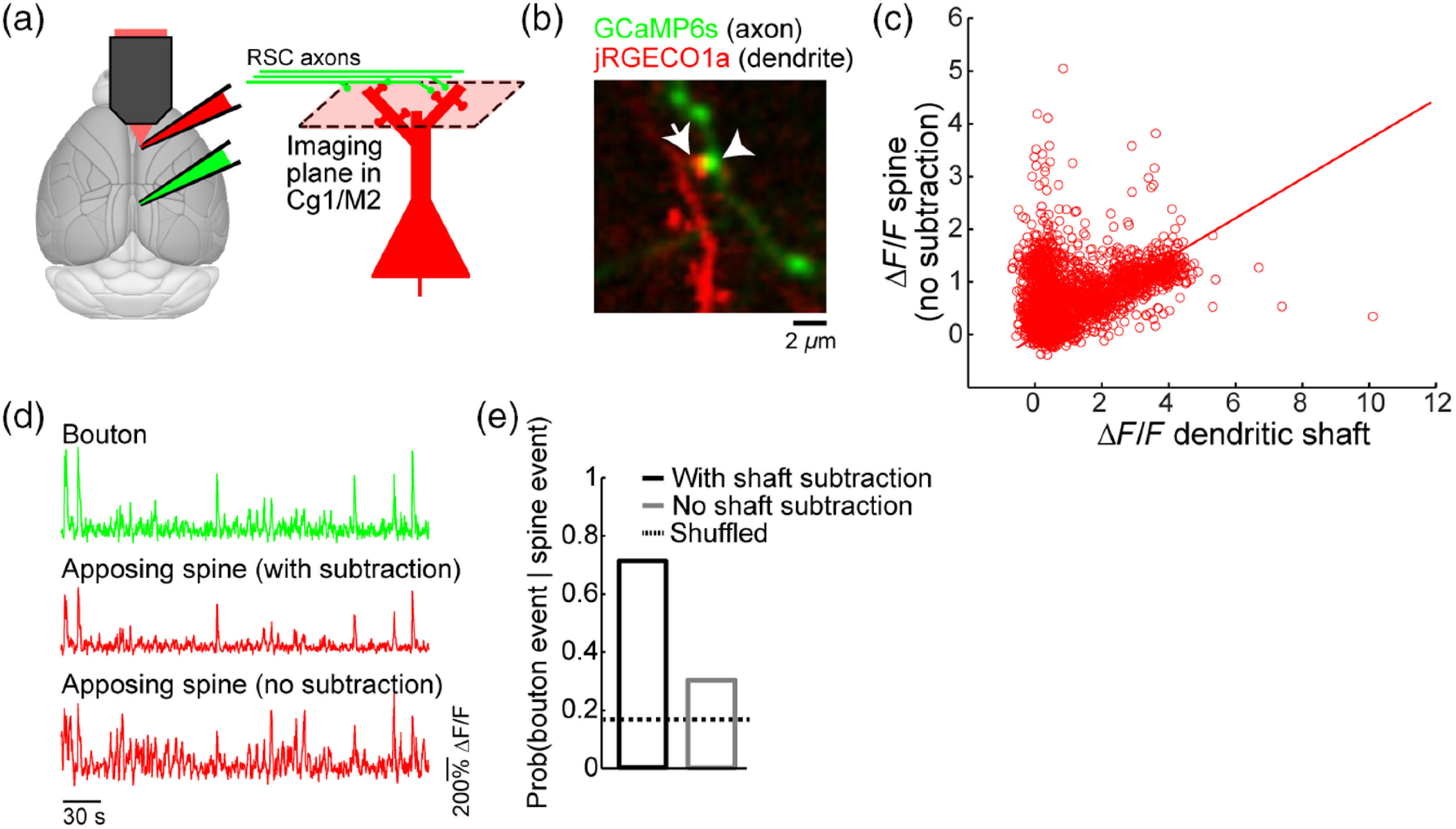
Fig. 8.

Dendritic calcium signals from synaptic inputs. (a) The experimental setup involving expression of GCaMP6s in RSC and jRGECO1a in Cg1/M2. (b) An in vivo two-photon image of GCaMP6s-expressing axonal boutons and jRGECO1a-expressing dendrites in Cg1/M2. (c) A scatter plot of the fluorescence transients () measured from spine indicated by arrow in (b) against the measured from the adjacent dendritic shaft. Each open circle represents an image frame. Line, a least-squares regression line forced through the origin. (d) Fluorescence traces for the bouton and apposing spine (either with subtraction or no subtraction of the shaft contribution) in (b). (e) The probability of detecting a presynaptic calcium event in the bouton within (i.e., ± the duration of one image frame) given a postsynaptic calcium event in the apposing spine, either with subtraction or no subtraction of the shaft contribution. Calcium events were determined from fluorescence traces using a peeling algorithm. Shuffled level was calculated by randomly shuffling the calcium event times of the bouton, and averaged across 100 replicates. These are unpublished data from the Kwan lab.
