Abstract
The mangosteen fruit is a popular Southeast Asian fruit consumed for centuries. There have been a variety of xanthones isolated from the fruit, bark, roots and leaves with each having unique chemical and physical properties. Previously, the most abundant xanthone α-mangostin has been shown to inhibit CDK4. Herein we describe the role of selected xanthones from the mangosteen inhibiting CDK4. The evidence we provide here is that key functional groups are required to inhibit the CDK4 protein to prevent the phosphorylation of downstream targets critical to inhibiting uncontrolled cell cycle progression. To define the properties of xanthones for inhibiting CDK4 we utilized a cell free biochemical assay to identify inhibitors of CDK4. The following xanthones were used for the analysis: α-mangostin, β-mangostin, γ-mangostin, gartanin, 8-desoxygartanin, garcinone C and garcinone D, 9-hydroxycalabaxanthone, and 3-isomangostin These results further substantiate the unique pharmacological properties of individual xanthones and how a mixture of xanthones may be responsible for a multi-targeted effect in cell based pharmacology systems.
Keywords: cancer, CDK4, garcinia mangostana, gartanin, mangosteen, mangostin, xanthone
Introduction
The purple mangosteen (Garcinia mangostana L.) is a fruit-producing tree that is native to Southeast Asia and has been used for hundreds of years for medicinal purposes1. All parts of the tree are used medicinally, as the roots, tree bark, and fruit are all consumed and can be commonly prepared as powders, ointments, and teas for treatment of dysentery, eczema, psoriasis, diarrhea, urinary disorders, and for healing wounds and ulcers1, 2. A class of biologically active compounds called xanthones can be found in all different parts of G. mangostana, including the outer pericarp rind, whole fruit, trunk, branches, and leaves1. Xanthones contain a C6-C1-C6 ring structure, with various isoprene and hydroxyl units branching off the foundational tricyclic structure. These units are thought to be important contributors to xanthones’ biological activities3. There have been more than 70 different xanthones isolated from G. mangostana3–5, with α-mangostin being the most abundant xanthone found and α-mangostin, β-mangostin, γ-mangostin, gartanin, and 8-desoxygartanin being the most studied xanthones1. These xanthones have been shown to have cytotoxic, anti-tumor and anti-cancer activities and here we will present data showing that xanthones inhibit cyclin dependent kinases (CDK’s)4–7.
Given the potential that xanthones have shown as promising new anti-cancer agents, we have further evaluated their role in prostate, colon, and breast cancers. In many cancers, including the three aforementioned, CDK’s have become increasingly important targets, as there is often abnormal activation and upregulation of CDK’s and their regulation of the cell cycle in cancer8, 9. CDK 4 is especially important as it controls the G1 checkpoint, where cells commit to entering the DNA synthesis phase of the cell cycle10. Recently, CDK 4/6 inhibitors have been tested in preclinical and clinical settings and have seen success as therapeutics in prostate and breast cancer10, 11. Previously, our lab has shown that α-mangostin not only inhibits the activity of CDK 4 in a cell free assay, but also decreases xenograft tumor growth in mice7. Here we furthered our studies of CDK 4, using eight different xanthones in vitro, and show them to be not only cytotoxic, but also to inhibit CDK 4 activity.
Materials and Methods
Cell culture
HCC 1937 and MDA-MB-231were cultured in RPMI1640 medium containing 10% fetal bovine serum and 1% penicillin/streptomycin. All cells were maintained under standard cell culture conditions as described previously12, 13.
Cell viability
Cell viability of xanthones (1–100 μM) isolated from the mangosteen fruit included: α-mangostin, β-mangostin, γ-mangostin, gartanin, 8-desoxygartanin, garcinone C and garcinone D, and 3-isomangostin. Cell viability following treatment with selected xanthones was determined by 3‐(4, 5‐dimethylthiazol‐2‐yl)‐2, 5‐diphenyltetrazolium bromide (MTT) assay as previously described7, 12. For this assay, 104 and 2×10⁴ cells of HCC1937 and MDA-MB-231 cell lines were plated in 96 well plate and incubated for 24h. Following incubation, media was replaced with media containing the test compounds in serial dilution ranging from 100 μM to 1 μM in duplicate. After incubating the cells with test compounds for 48 h, test media was replaced with media containing MTT reagent (Sigma-Aldrich) at a final concentration of 0.5 mg/mL. The cells were incubated for a period of 2 h and the media was discarded and the formazan crystals were solubilized in DMSO on a plate shaker protected from light at all times. The reading was taken at a wavelength of 570 nm and the viability was calculated with respect to control (0.01% DMSO). IC50 was calculated with Graphpad Prism 5.0. using Non-linear regression analysis.
CDK4 Inhibition assay
A cell free assay was utilized as previously described.7 Xanthones were diluted into the assay mixture with concentrations ranging from 10 to 40 μM of xanthones to determine inhibition of CyclinD1/CDK4 (Kinexus, Vancouver, British Columbia, Canada). The IC50 for each xanthone was determined against CyclinD1/CDK4. The addition of [33P]-ATP to the reaction mixture initiated the reaction. Following room temperature incubation the assay was terminated by spotting 10 μl of the reaction mixture onto multiscreen phosphocellulose P81 plate and washed three times for ~15 min each in a 1% phosphoric acid solution. The radioactivity on the P81 plate was counted in the presence of scintillation fluid in a Trilux scintillation counter. Protein kinase-specific activity of [33P]-ATP incorporated per minute per sample was determined. A total counts per minute for each reaction sample was determined for blanks (without substrate), control (without α-mangostin) and samples (with α-mangostin). The corrected activity for control samples (i.e. without α-mangostin) represented 100% kinase activity and was used to determine the percent of kinase activity.
Results
Selected xanthones inhibit HCC1937 and HCT116 cancer cells
A decrease in cell viability was observed following treatment of individual xanthones to HCC-1937 cells and HCT116 cells (Table 1). Following a 48 hour treatment with xanthones the IC50 (μM) for cell viability was determined for α-mangostin (13.0), β-mangostin (18.6), γ-mangostin (14.4), gartanin (31.2), 8-desoxygartanin (24.4), garcinone C (38.6), garcinone D (32.2), and 3-isomangostin (59.9). The colon cell line HCT 116 was treated with increasing concentrations of xanthones (0 – 100 μM) with the following EC50 calculated for each α-mangostin (23.4), β-mangostin (19.1), γ-mangostin (12.1), gartanin (6.84), 8-desoxygartanin (18.2), garcinone C (27.4), garcinone D (15.8), and 3-isomangostin (47.4).
Table 1.
IC50 (μM) of selected xanthones from Garcinia mangostana (mangosteen) against HCC1937 and HCT116
| Xanthone | HCC 1937 | HCT 116 |
|---|---|---|
| α-mangostin | 13.0 | 23.4 |
| β-mangostin | 18.6 | 19.1 |
| γ-mangostin | 14.4 | 12.1 |
| garcinone c | 38.6 | 27.4 |
| garcinone d | 32.2 | 15.8 |
| 3-isomangostin | 59.9 | 47.4 |
| gartanin | 31.2 | 6.8 |
| 8-desoxygartanin | 24.4 | 18.2 |
Cells were exposed to increasing concentrations of xanthone (0–100 uM) for 48 hours in triplicate. A mean and standard deviation was determined for each to establish the EC50. This was followed by determining the cell viability using an MTT assay.
Selected xanthones inhibit CDK4/Cyclin D1 in a cell free biochemical assay
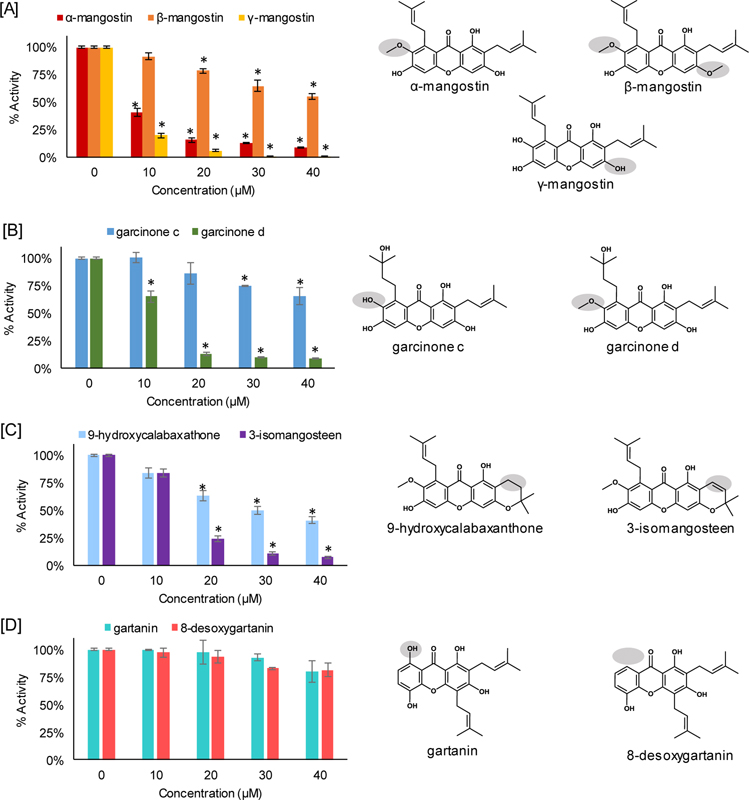
To determine if xanthones inhibited CDK4/Cyclin D1 in a cell free assay CDK4/Cyclin D1 was exposed to individual phytochemicals to determine if phosphorylation of the substrate was inhibited (Table 2 and Figure 1). Inhibitory activity as determined by an IC50 was determined for the following xanthones: α-mangostin (8.5 μM), γ-mangostin (6.2 μM), garcinone D (12.8 μM), and 3-isomangostin (15.6 μM). Several xanthones did not display any significant inhibition of CDK4/Cyclin D1 included: β-mangostin, garcinone C, gartanin, and 8-desoxygartanin.
Table 2.
Inhibitory activity of xanthones from Garcinia mangostana (mangosteen) against CDK4/Cyclin D1
| % Activity | |||||||
|---|---|---|---|---|---|---|---|
| Xanthone | Control | 10 μM | 20 μM | 30 μM | 40 μM | EC50 | |
| α-mangostin | 100 ± 1.38 | 41.29 ± 4.01 | 15.81 ± 1.89 | 13.05 ± 0.44 | 8.77 ± 0.42 | 8.5 | |
| β-mangostin | 100 ± 1.38 | 91.7 ± 3.40 | 78.60 ± 1.94 | 64.83 ± 5.67 | 54.97 ± 2.88 | – | |
| γ-mangostin | 100 ± 1.38 | 20.01 ± 2.38 | 6.10 ± 1.09 | 0.59 ± 0.72 | 0.51 ± 0.55 | 6.2 | |
| garcinone C | 100 ± 1.38 | 100.92 ± 4.91 | 86.07 ± 10.50 | 75.11 ± 0.85 | 65.65 ± 7.94 | – | |
| garcinone D | 100 ± 1.38 | 65.38 ± 5.33 | 12.89 ± 1.52 | 9.72 ± 0.39 | 9.02 ± 0.58 | 12.8 | |
| 9-hydroxycalabaxanthone | 100 ± 1.38 | 83.91 ± 5.18 | 63.14 ± 4.70 | 49.65 ± 3.88 | 83.53 ± 3.08 | 30.0 | |
| 3-isomangostin | 100 ± 1.38 | 83.53 ± 3.72 | 24.15 ± 2.48 | 10.77 ± 1.25 | 7.4 ± 1.38 | 15.6 | |
| gartanin | 100 ± 1.38 | 99.63 ± 0.14 | 97.85 ± 11.61 | 92.9 ± 3.75 | 80.21 ± 10.25 | – | |
| 8-desoxygartanin | 100 ± 1.38 | 97.25 ± 4.66 | 93.46 ± 6.45 | 82.87 ± 1.13 | 81.42 ± 6.38 | – | |
Values represent the mean of three values followed by the standard deviation.
Figure 1.
Cell free biochemical kinase assay established the IC50 of xanthones against CyclinD1/CDK4. Data points are represented by the average of three values with standard deviation. Statistical analysis was performed using GraphPad software by one way analysis of variance and statistical significance was performed by the Turkey test with *P < 0.01. All valued are compared to the control sample.
Discussion
Deregulation of the CDK4/cyclin D1-retinoblastoma (Rb) pathway is an important contributor to increased mitogenic potential of cancer cells14. Further, there is evidence suggesting that deregulation of this pathway contributes to endocrine therapy resistance in hormone dependent cancers including breast cancer15. Cyclin dependent kinase activity is regulated by several variables that include T-loop phosphorylation, abundance of cyclins, and association with CDK inhibitors (Cip/Kip) and/or INK family proteins16. The active complex of CDK4 is CDK4/Cyclin D1 that targets the retinoblastoma protein for phosphorylation. This post-translational modification releases E2F transcription factors that activate G1/S phase gene expression.
α-Mangostin represents the most abundant xanthone in the mangosteen and by default makes it the most studied xanthone. The mangosteen has more than 80 xanthones isolated from the whole plant suggesting a significant degree of chemical diversity may be responsible for its multi-targeted effect in cancer models.17, 18 Taken together this approach emphasizes the need to evaluate a variety of xanthones to more adequately describe the pharmacological properties of a complex extract of xanthones. Previously, we have described the inhibitory properties of α-mangostin against CDK4 using a combination of cell free biochemical, cell based models, in vivo animal model along with molecular modeling7.
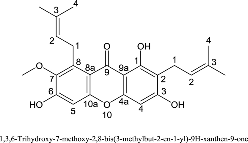
At present our working hypothesis is that α-mangostin and more specifically the isoprenyl group at 2 position is able to bind deep within the ATP binding pocket (Figure 2)7. It is possible that the other end of α-mangostin (i.e. carbons 5–8) is able to bind into the pocket, however, we consider this unlikely in that the pocket does not fill as effectively. Interestingly, we have previously shown that gartanin is a potent AR disruptor, however, it does not inhibit CDK47. Our data shown in Figure 1 interestingly, shows that when an isoprenyl group is at the 4 position (e.g. gartanin and 8-desoxygartanin) and is absent at the 8 position there is no inhibition of CDK4. Based on these results it could be that when an isoprenyl group is present at the 4 position (e.g. gartanin and 8-desoxygartanin), this prevents the xanthone from binding the ATP binding pocket of CDK4. Hydroxylation of key groups also appears to be important as shown in Figure 1A when comparing the CDK4 inhibition profiles of α-mangostin, β-mangostin, and γ-mangostin. Hydroxylation at the 3 and 7 positions provided the most potent of the three (i.e. γ-mangostin compared to α-mangostin and β-mangostin). Unfortunately, the experiments performed in a cell free biochemical assay do not consider phase I metabolism which may occur. In an in vivo model or real world setting it could be possible that methoxy groups such as those present in α-mangostin (i.e. 7 carbon), β-mangostin (i.e. 3 and 7 carbons) may undergo o-demethylation to generate γ-mangostin. When comparing γ-mangostin to garcinone D it is evident that the when the isoprenyl group is hydroxylated it can counter the methoxy group at the 7 position ultimately restoring CDK4 inhibition. We have described previously that α-mangostin undergoes Phase II metabolism to form mono-glucuronide and di-glucuronides after oral administration to mice.19, 20 More work is needed to understand the potential of o-demethylation of xanthones from the mangosteen following exposure to phase I enzymes (P450). This is especially significant as most individuals who consumer mangosteen will be exposed to a variety of xanthones.
Figure 2.
α-Mangostin
Conclusion
Taken together we provide an explanation as to how α-mangostin inhibits CDK4 using an additional 8 xanthones. Key functional groups appear to be the isoprenyl groups at the 2 and 8 positions with hydroxyl groups extending from 3 and 7 positions. Another important consideration is the lack of an isoprenyl group at the 4 position for inhibition of CDK4. Interestingly, we have shown that gartanin does not inhibit CDK4 while inhibiting AR functionality while α-mangostin inhibits CDK4. Future studies are clearly needed to characterize the key functional groups needed for specific pharmacological actions of xanthones isolated from the mangosteen fruit.
Acknowledgements
Johnson JJ is supported by the National Institutes of Health (R37 CA227101)
Footnotes
Conflicts of interest
The authors do not declare any conflicts of interest.
References
- 1.Pedraza-Chaverri J, Cardenas-Rodriguez N, Orozco-Ibarra M, Perez-Rojas JM. Medicinal properties of mangosteen (Garcinia mangostana). doi: 10.1016/j.fct.2008.07.024. Food Chem Toxicol. 2008;46(10):3227–39. [DOI] [PubMed] [Google Scholar]
- 2.Obolskiy D, Pischel I, Siriwatanametanon N, Heinrich M. Garcinia mangostana L.: a phytochemical and pharmacological review. doi: 10.1002/ptr.2730. Phytother Res. 2009;23(8):1047–65. [DOI] [PubMed] [Google Scholar]
- 3.Suksamrarn S, Komutiban O, Ratananukul P, Chimnoi N, Lartpornmatulee N, Suksamrarn A. Cytotoxic prenylated xanthones from the young fruit of Garcinia mangostana. Chem Pharm Bull (Tokyo). 2006;54(3):301–5. [DOI] [PubMed] [Google Scholar]
- 4.Shan T, Ma Q, Guo K, Liu J, Li W, Wang F, Wu E. Xanthones from mangosteen extracts as natural chemopreventive agents: potential anticancer drugs. Current molecular medicine. 2011;11(8):666–77. [DOI] [PMC free article] [PubMed] [Google Scholar]
- 5.Mohamed GA, Al-Abd AM, El-Halawany AM, Abdallah HM, Ibrahim SRM. New xanthones and cytotoxic constituents from Garcinia mangostana fruit hulls against human hepatocellular, breast, and colorectal cancer cell lines. doi: 10.1016/j.jep.2017.01.030. J Ethnopharmacol. 2017;198:302–12. [DOI] [PubMed] [Google Scholar]
- 6.Akao Y, Nakagawa Y, Iinuma M, Nozawa Y. Anti-cancer effects of xanthones from pericarps of mangosteen. Int J Mol Sci. 2008;9(3):355–70. [DOI] [PMC free article] [PubMed] [Google Scholar]
- 7.Johnson JJ, Petiwala SM, Syed DN, Rasmussen JT, Adhami VM, Siddiqui IA, Kohl AM, Mukhtar H. alpha-Mangostin, a xanthone from mangosteen fruit, promotes cell cycle arrest in prostate cancer and decreases xenograft tumor growth. doi: 10.1093/carcin/bgr291. Carcinogenesis. 2012;33(2):413–9. [DOI] [PMC free article] [PubMed] [Google Scholar]
- 8.Otto T, Sicinski P. Cell cycle proteins as promising targets in cancer therapy. doi: 10.1038/nrc.2016.138. Nature reviews Cancer. 2017;17(2):93–115. [DOI] [PMC free article] [PubMed] [Google Scholar]
- 9.Whittaker SR, Mallinger A, Workman P, Clarke PA. Inhibitors of cyclin-dependent kinases as cancer therapeutics. doi: 10.1016/j.pharmthera.2017.02.008. Pharmacology & therapeutics. 2017;173:83–105. [DOI] [PMC free article] [PubMed] [Google Scholar]
- 10.VanArsdale T, Boshoff C, Arndt KT, Abraham RT. Molecular Pathways: Targeting the Cyclin D-CDK4/6 Axis for Cancer Treatment. doi: 10.1158/1078-0432.Ccr-14-0816. Clin Cancer Res. 2015;21(13):2905–10. [DOI] [PubMed] [Google Scholar]
- 11.Vidula N, Rugo HS. Cyclin-Dependent Kinase 4/6 Inhibitors for the Treatment of Breast Cancer: A Review of Preclinical and Clinical Data. doi: 10.1016/j.clbc.2015.07.005. Clinical breast cancer. 2016;16(1):8–17. [DOI] [PubMed] [Google Scholar]
- 12.Li G, Petiwala SM, Yan M, Won JH, Petukhov PA, Johnson JJ. Gartanin, an isoprenylated xanthone from the mangosteen fruit (Garcinia mangostana), is an androgen receptor degradation enhancer. doi: 10.1002/mnfr.201600037. Mol Nutr Food Res. 2016;60(6):1458–69. [DOI] [PubMed] [Google Scholar]
- 13.Li G, Petiwala SM, Nonn L, Johnson JJ. Inhibition of CHOP accentuates the apoptotic effect of alpha-mangostin from the mangosteen fruit (Garcinia mangostana) in 22Rv1 prostate cancer cells. doi: 10.1016/j.bbrc.2014.09.054. Biochem Biophys Res Commun. 2014;453(1):75–80. [DOI] [PubMed] [Google Scholar]
- 14.Goel S, DeCristo MJ, McAllister SS, Zhao JJ. CDK4/6 Inhibition in Cancer: Beyond Cell Cycle Arrest. doi: 10.1016/j.tcb.2018.07.002. Trends in cell biology. 2018;28(11):911–25. [DOI] [PMC free article] [PubMed] [Google Scholar]
- 15.Spring L, Bardia A, Modi S. Targeting the cyclin D-cyclin-dependent kinase (CDK) 4/6-retinoblastoma pathway with selective CDK 4/6 inhibitors in hormone receptor-positive breast cancer: rationale, current status, and future directions. Discovery medicine. 2016;21(113):65–74. [PMC free article] [PubMed] [Google Scholar]
- 16.Sherr CJ. Cancer cell cycles. Science. 1996;274(5293):1672–7. [DOI] [PubMed] [Google Scholar]
- 17.Li G, Thomas S, Johnson JJ. Polyphenols from the mangosteen (Garcinia mangostana) fruit for breast and prostate cancer. doi: 10.3389/fphar.2013.00080. Front Pharmacol. 2013;4:80. [DOI] [PMC free article] [PubMed] [Google Scholar]
- 18.Ovalle-Magallanes B, Eugenio-Perez D, Pedraza-Chaverri J. Medicinal properties of mangosteen (Garcinia mangostana L.): A comprehensive update. doi: 10.1016/j.fct.2017.08.021. Food Chem Toxicol. 2017;109(Pt 1):102–22. [DOI] [PubMed] [Google Scholar]
- 19.Petiwala SM, Li G, Ramaiya A, Kumar A, Gill RK, Saksena S, Johnson JJ. Pharmacokinetic characterization of mangosteen (Garcinia mangostana) fruit extract standardized to alpha-mangostin in C57BL/6 mice. doi: 10.1016/j.nutres.2014.03.002. Nutr Res. 2014;34(4):336–45. [DOI] [PMC free article] [PubMed] [Google Scholar]
- 20.Ramaiya A, Li G, Petiwala SM, Johnson JJ. Single Dose Oral Pharmacokinetic Profile of alpha-Mangostin in Mice. Curr Drug Targets. 2012;13(14):1698–704. [DOI] [PubMed] [Google Scholar]


