

Abstract
The present paper describes four new species of virgulate xiphidiocercariae infecting the freshwater gastropod, Bithynia (Digoniostoma) pulchella (Benson, 1836) collected from Malabar, Kerala. Cercaria sp. XXII Malabar n. sp. has a spinose body and tail; four pairs of penetration glands; short, narrow oesophagus; a pair of short, unequal caeca; bicornuate excretory vesicle and 18 pairs of flame cells. Cercaria sp. XXIII Malabar n. sp. is characterized by an oval, spinose body; aspinose tail; dagger-shaped stylet; medially fused virgula; globular pharynx; short oesophagus; three pairs of penetration glands; bicornuate, ‘V’-shaped excretory bladder, and 18 pairs of flame cells. Cercaria sp. XXIV Malabar n. sp. has a spinose body and tail, medially fused virgula, muscular pharynx, short prepharynx and oesophagus; four pairs of penetration glands; bicornuate, ‘V’-shaped excretory bladder, and 12 pairs of flame cells. Cercaria sp. XXV Malabar n. sp. has a spinose body, aspinose tail, sac-like virgula; globular pharynx, narrow oesophagus; short caeca; four pairs of penetration glands; transversely elongated, bicornuate excretory bladder, and 12 pairs of flame cells. All four cercariae developed in sporocysts within the digestive gland of B. (D.) pulchella. Morphology and morphometry of the cercariae are compared with related species to establish their systematic position.
Keywords: Virgulate, Xiphidiocercaria, Sporocyst, Bithynia (Digoniostoma) pulchella, Malabar, Kerala
Introduction
The freshwater gastropod, Bithynia (Digoniostoma) pulchella (Benson, 1836) is a small, operculate snail distributed throughout the Indian subcontinent and inhabits the stagnant freshwater bodies and slow moving streams (Subba Rao 1989; Ramakrishna and Dey 2007). This snail species is a common inhabitant of paddy fields, streams, canals and ponds in the Malabar region of Kerala. Twenty-one cercariae have been reported from Bithynia (Digoniostoma) spp. from India, of which four (Cercaria indicae LX Sewell, 1922; C. indicae LXIX Murty, 1976 (= Cercaria of Pleurogenoides malampuzhensis Brinesh and Janardanan, 2014); Cercaria sp. XII Kerala Mohandas, 1977 and Cercaria of Pleurogenoides ovatus Janardanan and Prasadan, 1991) belong to virgulate xiphidiocercaria group. Information on the larval trematode infections in B. (D.) pulchella in Malabar is limited to five species (C. indicae VIII Sewell, 1922; C. indicae LXIX Murty, 1976 (= Cercaria of Pleurogenoides malampuzhensis Brinesh and Janardanan, 2014); Cercaria of Pleurogenoides ovatus Janardanan and Prasadan, 1991; Cercaria sp. XII Malabar Brinesh and Janardanan, 2009 and Cercaria sp. XIII Malabar Brinesh and Janardanan, 2009) During a survey on larval trematode infections in freshwater gastropods of Malabar, we came across four new species of virgulate xiphidiocercariae infecting B. (D.) pulchella. Following the system adopted by Sewell (1922), the cercariae are designated by Roman numerals, followed by Malabar to indicate the region of collection.
Materials and methods
Samples of B. (D.) pulchella were collected from various water bodies in Malabar during May 1989 to October 1990. The snails were collected either by hand or using hand nets, transported live to the laboratory, and separated into small glass containers. The containers were exposed to light and observed for 24 h. Naturally emerging cercariae were isolated and studied in detail under a phase-contrast research microscope (Leica, Germany), with or without vital staining (Neutral red or Nile blue sulphate). Lacto-acetic carmine was used to study the genital primordia. After completing the cercarial study, the infected snails were crushed, and digestive glands examined for sporocysts. The recovered sporocysts were stained live with neutral red and studied in detail. Morphometrics of the cercariae and sporocysts (expressed in micrometres [µm] as range, along with mean values in parentheses) were taken from 10% formalin-fixed specimens (n = 30). Details from live observations were incorporated into figures drawn with the help of a camera lucida.
Results and discussion
Cercaria sp. XXII Malabar n. sp.
Host: Bithynia (Digoniostoma) pulchella (Benson, 1836)
Site of infection: Hepatopancreas
Type locality: Kundayithode in Kozhikode district, Kerala
Collected by: N. K. Sanil
Holotype and paratype: Deposited in the parasite collections of Parasitology Laboratory, Department of Zoology, University of Calicut (Accession numbers: CV.1.1and CV(P). 1.1 respectively).
Etymology: Named after ‘Malabar’, the region from where it was recorded
Period of collection: May 1989–October 1990
Prevalence: 1.38% (fifteen of 1085 snails screened were infected)
Cercarial behavior Cercariae emerged throughout the daytime, with peak emergence during morning hours; change of water enhanced emission. Prolonged swimming activity was followed by brief periods of rest.
Description (Fig. 1a, b, c, d, Table 1)
Fig. 1.
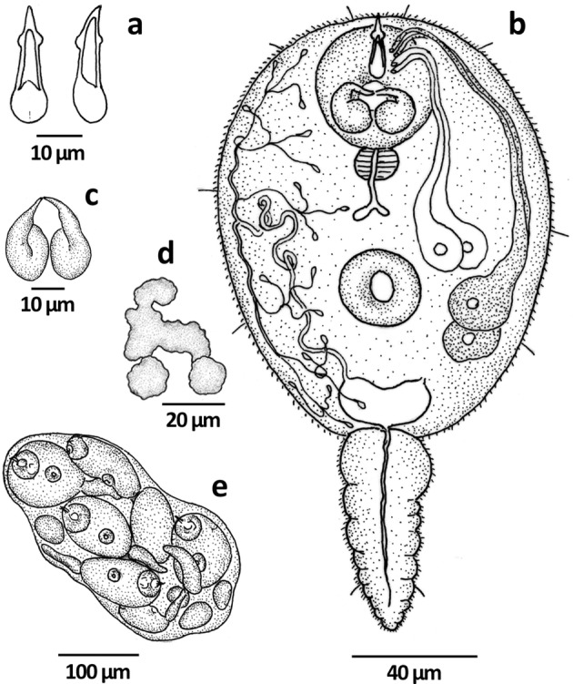
Cercaria sp. XXII Malabar n. sp. a stylet, b cercaria, c virgula organ, d genital primordium, e sporocyst
Table 1.
Showing comparative characters and measurements of Cercaria sp. XXII Malabar n. sp. and five related cercariae
| Cercaria (hosts) | Cercaria pseudotarda (Pike, 1967) Nasir, 1984Bithynia tentaculata (Linnaeus) | Cercaria halli Etges, Page and Bonner, 1969Anaplocamus dilatatus (Conrad) | Cercaria steenophrenezua Nasir and Diaz, 1973Stenophysa venezuelensis (von Martens) | Cercaria indicae LXVIII Murty, 1976Amnicola travancorica (Benson) | Cercaria melanopsi XXII Ismail and Bdair, 1987Melanopsis praemorsa (Linnaeus) | Cercaria sp. XXII Malabar n. sp. Bithynia (Digoniostoma) pulchella (Benson, 1836) |
|---|---|---|---|---|---|---|
| Body | Spinose, 120–260 | Spinose, 120–160 × 75–80 | Spinose, 105–135 × 54–64 | Spinose, 128–176 (146) × 64–90 (73) | Spinose, 118–135 × 68–80 | Spinose, 115.5–161.7 (139.3) × 92.4–118.8 (103.6) |
| Tail | Aspinose, 40–170 | Spinose, 85 × 22 | Spinose, 45–63 × 20–24 | Spinose, 76–96 (88) × 16–20 (18) | Spinose, 38–68 × 20–25 | Spinose, 39.6–135.3 (84.3) × 23.1–29.7 (25.9) |
| Oral sucker | 47.7 | 30–35 × 30–32 | 27–33 | 36–44 (39) | 38–63 | 36.3–46.2 (43.3) × 33.0–49.5 (40.5) |
| Ventral sucker | 29.5 | 19–22 | 15–20 | 24–28 (25) | 20 × 20 | 19.8–26.4 (24.1) × 23.1–29.7 (25.9) |
| Stylet | 20 | 17–20 × 5 | 14–18 × 4–5 | – | 25 × 3 | 24. 2–26.6 (26.4) × 5.0–6.6 (5.9) |
| Virgula organ | – | 35–41 | – | 11 × 20 | Bilobed | 24.2–26.5 (25.2) × 22.2–24.8 (23.6) |
| Prepharynx | Small | Absent | Short | Short | Absent | Absent |
| Pharynx | 11.4 | Absent | 5–9 | 12 × 10 | 10 × 10 | Globular, 9.9–15.5 (11.9) × 9.9–19.8 (13.8) |
| Oesophagus | Long | Absent | Long, narrow | Long | Long, narrow | Short, narrow |
| Caeca | Short | Absent | Dilated | Absent | Short | Short, unequal |
| Excretory bladder | ‘V’-shaped | ‘U’-shaped | Transversely elongate | ‘U’-shaped | Bicornuate | Bicornuate |
| Flame cell formula | 2 [(2 + 1+1 + 1+1) + 1 + 1+1 + 1+2)] = 24 | – | 2 [(2 + 2+2) + (2 + 2+2)] = 24 | 2 [(2 + 2+2) + (2 + 2+2)] = 24 | 2 [(3 + 2) + (1 + 3)] = 18 | 2 [(3 + 3+3) + (3 + 3+3)] = 36 |
Virgulate xiphidiocercaria with an oval, highly contractile body measuring 115.5–161.7 (139.3) × 92.4–118.8 (103.6). Cercarial body armed with small, backwardly directed spines, distributed evenly except at the posterior region where it is sparse. Four pairs of sensory hairs present on body. Highly contractile tail measured 39.6–135.3 (84.3) × 16.5–29.7 (24.1) in size, shorter than body, armed with small, lightly distributed spines, and a pair of sensory hairs on its posterior three-fourth. Cystogenous cells enclosing refractory materials present on body surface. Oral sucker longer than wide, 36.3–46.2 (43.3) × 33.0–49.5 (40.5) in size. Ventral sucker smaller than oral sucker, post-equatorial in position, wider than long, and measured 19.8–26.4 (24.1) × 23.1–29.7 (25.9). Stylet mounted on the roof of oral sucker, sharply tapering, with a pointed, inwardly curved tip, bulbous base, reinforced walls and shoulders; measured 24.2–26.6 (26.4) long and 5.0–6.6 (5.9) and 6.6 wide at base and shoulders respectively. Virgula organ positioned near the base of stylet, with two distinct, closely apposed lobes which open out ventrally through a pair of independent ducts, measured 24.2–26.5 (25.2) × 22.2–24.8 (23.6). Contents of virgula stain with neutral red; appeared alveolar when empty. Mouth at the center of oral sucker; prepharynx absent; pharynx globular, wider than long, measured 9.9–16.5 (11.9) × 9.9–19.8 (13.8). Short, narrow oesophagus is followed by a pair of short, unequal caeca with slightly swollen tips. Penetration glands four pairs, anterior two pairs smaller in size, positioned antero-lateral to ventral sucker, with clear, hyaline contents; gland ducts ran forward and emptied independently near the base of stylet; posterior two pairs larger, with round margins, coarsely granular contents and conspicuous nucleus; gland ducts opened near the mid-region of stylet. Genital primordium represented by a tri-lobed mass of cells, overlapping the ventral sucker. Excretory vesicle bicornuate; primary collecting vessels originated from the tip of each cornu, ran forward, formed a few coils and antero-lateral to ventral sucker, bifurcated into anterior and posterior collecting vessels. Flame cell formula: 2[(3 + 3+3) + (3 + 3+3)] = 36. Posteriorly, bladder continued as caudal excretory canal into the tail; canal opening not discernible.
Sporocyst (Fig. 1e)
Sporocysts developed in the digestive gland of infected B. (D.) pulchella. Body small, sac-like; 261.3–438.9 (351.2) × 130.9–200.2 (161.6); contained two to three cercariae, a few developing cercariae and numerous germ balls.
Remarks
The present virgulate xiphidiocercaria with spinose body and tail, and four pairs of penetration glands resembles Cercaria pseudotarda (Pike 1967) Nasir, 1984, Cercaria halli Etges, Page and Bonner, 1969, Cercaria steenophrenezua Nasir and Diaz, 1973, Cercaria indica LXVIII Murty, 1976 and Cercaria melanopsi XXII Ismail and Bdair, 1987. Detailed comparison of characters and measurements of the present cercaria with that of the above five cercariae is made in Table 1. It differs from all compared cercariae, except C. halli in the shape of stylet and from all cercariae except C. steenophrenezua in the position/arrangement and granular nature of penetration glands. C. steenophrenezua differs from present cercaria in the nature and arrangement of penetration gland ducts. C. melanopsi XII differs in the shape and size of virgula and C. halli and C. steenophrenezua differ in the shape of virgula; while C. pseudotarda distinguishes itself from the present cercaria in having an aspinose tail. The present cercaria differs from all the above cercariae in the number and arrangement of flame cells and in its snail host. In the light of the differences with related cercariae, the present cercaria is considerd as a new species and the name Cercaria sp. XXII Malabar n. sp. proposed.
Cercaria sp. XXIII Malabar n. sp.
Host: Bithynia (Digoniostoma) pulchella (Benson, 1836)
Site of infection: Hepatopancreas
Type locality: Kundayithode in Kozhikode district, Kerala
Collected by: N. K. Sanil
Holotype and paratype: Deposited in the parasite collections of Parasitology Laboratory, Department of Zoology, University of Calicut (Accession numbers: CV.1.2 and CV(P). 1.2 respectively).
Etymology: Named after ‘Malabar’, the region from where it was recorded
Period of collection: May 1989–October 1990
Prevalence: 0.92% (ten of 1085 snails screened were infected)
Cercarial behavior Cercariae emerged throughout the day, preferred to stay at the bottom; are active swimmers; swimming interrupted by brief intervals of rest.
Description (Fig. 2a, b, c, d; Table 2)
Fig. 2.
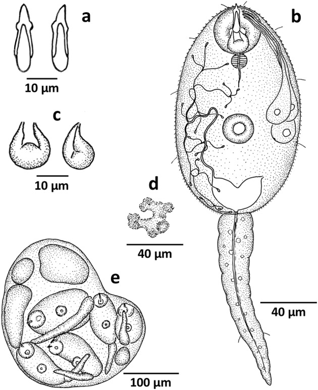
Cercaria sp. XXIII Malabar n. sp. a stylet, b cercaria, c virgula organ, d genital primordium, e sporocyst
Table 2.
Showing comparative characters and measurements of Cercaria sp. XXIII Malabar n. sp. and five related cercariae
| Cercaria (hosts) | Cercariae indicae LX Sewell, 1922Digoniostoma cerameopoma (Benson) | Cercariae indicae XLII Sewell, 1922Amnicola travancorica (Benson) | Cercaria nipponensis Faust, 1924Semisulcospira spp. Boettger | Cercaria dolomeda Hall and Groves, 1963Nitocris dilatatus (Conrad) | Cercaria leyteensis no. 14 Ito, 1977Melanoides. tuberculatus (Müller), Antimelania asperata (Lamarck), A. dactylus (Lea) | Cercaria sp. XXIII Malabar n. sp. Bithynia (Digoniostoma) pulchella (Benson) |
|---|---|---|---|---|---|---|
| Body | Spinose, 111.0 × 61.0 | Spinose, 139.0–196.0 × 28.0–75.0 | Spinose, 134.3–148.0 (140.3) × 49.2–69.2 (61.7) | Spinose, 102.0–162.0 × 44.0–77.0 | Spinose, 86.0–102.0 (94.0) × 52.0–67.0 (61.0) | Spinose, 133.5–163.5 (148.4) × 72.0–103.5 (87.3) |
| Tail | Aspinose, 39.0–111.0 × 18.0 | Aspinose, 89.0–143.0 × 17.0 | Aspinose, 55.9–83.8 (70.1) × 13.3–23.9 (19.7) | Spinose, 88.0–134.0 × 13.0–22.0 | Aspinose, 71.0–107.0 (85.0) × 14.0–19.0 (16.0) | Aspinose, 102.0–133.5 (118.6) × 15.0–21.0 (17.9) |
| Oral sucker | 36.0 | 39.0 | 39.9–50.5 (46.4) × 37.2–53.2 (46.1) | 28.0–35.0 × 26.0–35.0 | 31.0–46.0 (36.0) × 30.0–38.0 (35.0) | 30.0–34.5 (32.5) × 33.0–39.0 (35.6) |
| Ventral sucker | 18.0 | 25.0 | 17.3–22.6 (20.1) × 20.0–24.0 (21.7) | 14.0–19.0 | 17.0–21.0 (19.0) | 19.0–22.5 (21.5) × 21.0–27.0 (23.7) |
| Stylet | 18.0 | 14.0 | 13.6–16.0 (15.3) × 4.0–4.7 (4.3) | 20.0–24.0 × 3.0–5.0 | 15.0–20.0 (17.0) × 2.5–3.5 (3.0) | 22.2–22.7 (22.5) × 4.5 |
| Virgula organ | – | 21.0 × 14.0 | – | 9.0–18.0 × 8.0–18.0 | – | 12.0–18.0 (14.8) × 10.5–13.5 (12.5) |
| Prepharynx | Short | Short | Absent | Absent | Absent | Absent |
| Pharynx | Small | Rounded | 6.7–12.0 (9.5) × 10.6–21.0 (12.7) | 8–14 × 7–12 | 6.0–8.0 (7.0) × 7.0–10.0 (9.0) | Globular, 10.5–13.5 (11.5) × 10.5–13.5 (11.8) |
| Oesophagus | Absent | Long | Short | Long | Short | Short, narrow |
| Caeca | Absent | Absent | Very short | Very short | Absent | Absent |
| Excretory bladder | ‘U’-shaped | ‘V’-shaped | ‘Y’-shaped | Crescent or ‘V’-shaped | Cup-shaped | Bicornuate |
| Flame cell formula | – | – | 2 [(2 + 2+2) + (2 + 2+2)] = 24 | 2 [(3 + 3+3) + (3 + 3+3)] = 36 | 2 [(2 + 2) + (2 + 2)] = 16 | 2 [(3 + 3+3) + (3 + 3+3)] = 36 |
Virgulate xiphidiocercaria with an elongate-oval, spinose body and four pairs of sensory hairs, measured 133.5–163.5 (148.4) × 72.0–103.5 (87.3). Spines small, backwardly directed, uniformly distributed except on ventral sucker where they are inwardly directed. Posterior half of body covered with opaque, cystogenous glands. Aspinose, highly contractile tail with caudal bodies and a pair of sensory hairs on its posterior third; measured 102.0–133.5 (118.6) × 15.0–21.0 (17.9). Oral sucker sub-terminal, wider than long, 30.0–34.5 (32.5) × 33.0–39.0 (35.6). Ventral sucker post-equatorial, wider than long, measured 19.0–22.5 (21.5) × 21.0–27.0 (23.7). Dagger-shaped stylet inserted into the roof of oral sucker, with a slightly bent tip, round base and prominent shoulders; stylet walls reinforced except at base; measured 22.2–22.7 (22.5) long and 4.5 and 4.7 wide at shoulders and base respectively. Virgula organ in the posterior half of oral sucker, pear-shaped, medially fused, ducts opened out ventrally just behind the mouth, 12.0–18.0 (14.8) × 10.5–13.5 (12.5) in size. Mouth at the center of oral sucker; prepharynx absent; pharynx globular, 10.5–13.5 (11.5) × 10.5–13.5 (11.8); oesophagus short, tapering; caeca not discernible. Penetration glands three pairs, rounded in outline, with clear nucleus and fine, granular contents. Gland ducts ran forward and opened out individually near the shoulder of stylet. A ‘G’-shaped mass of cells overlapping ventral sucker represented the genital primordium. Excretory bladder bicornuate, ‘V’-shaped; primary collecting vessels originated from the antero-lateral tip of bladder, ran forward and lateral to ventral sucker, bifurcated into anterior and posterior collecting vessels. Flame cell formula: 2[(3 + 3+3) + (3 + 3+3)] = 36. Caudal excretory canal originated from the posterior region of excretory bladder, runs through tail; opening not discernible.
Sporocyst (Fig. 2e)
Sporocyst developed in the digestive gland of infected B. (D.) pulchella; body sac-like; 216.0–339.0 (271.0) × 140.0–189.0 (163.4); contained one to three cercariae, a few immature ones and germ balls.
Remarks
The present virgulate xiphidiocercaria has a spinose body, aspinose tail, dagger-shaped stylet, a digestive system without prepharynx and caeca, three pairs of penetration glands and 36 flame cells and is comparable to Cercariae indicae LX Sewell 1922, Cercariae indicae XLII Sewell 1922, Cercaria nipponensis Faust 1924, Cercaria dolomeda Hall and Groves 1963 and Cercaria leyteenensis no. 14 Ito 1977. Comparison of characters and measurements (Table 2) show that, in spite of the similarities, it differs from these cercariae in many aspects. It differs from C. nipponensis, C. leyteensis No. 14 and C. indicae LX in measurements of body and tail; from C. dolomeda and C. indicae LX in measurements of ventral sucker and from C. leyteensis No14 in measurements of oral sucker. C. dolomeda further differs from the present form in having a spinose tail with a tuft of spines at its tip. The present cercaria distinguishes itself again from C. nipponensi, C. leyteensis no. 14 and C. dolomeda in the position/arrangement and granulation of penetration glands; from C. indicae LX in granular nature of penetration glands and from C. indicae XLII in the arrangement of gland ducts. Except C. nipponensis it differs from all other cercariae in the shape of stylet and except C. dolomeda, differs from all others in the shape and size of virgula. Further, the present cercaria differs from all other compared forms in having a different snail host. Hence, the present cercaria is treated as a new species and the name Cercaria sp. XXIII Malabar n. sp. is proposed.
Cercaria sp. XXIV Malabar n. sp.
Host: Bithynia (Digoniostoma) pulchella (Benson, 1836)
Site of infection: Hepatopancreas
Type locality: Kundayithode in Kozhikode district, Kerala
Collected by: N. K. Sanil
Holotype and paratype: Deposited in the parasite collections of Parasitology Laboratory, Department of Zoology, University of Calicut (Accession numbers: CV.1.3 and CV(P). 1.3 respectively).
Etymology: Named after ‘Malabar’, the region from where it was recorded
Period of collection: May 1989–October 1990
Prevalence: 1.75% (nineteen of 1085 snails screened were infected)
Cercarial behavior Cercariae emerged during the morning hours and continued till noon. They swim actively, with jerky movements; swimming interrupted by brief intervals of rest.
Description (Fig. 3 a, b, c, d; Table 3)
Fig. 3.
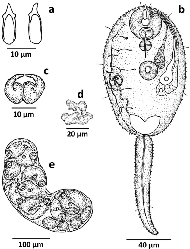
Cercaria sp. XXIV Malabar n. sp. a stylet, b cercaria, c virgula organ, d genital primordium, e sporocyst
Table 3.
Showing comparative characters and measurements of Cercaria sp. XXIV Malabar n. sp. and five related cercariae
| Cercaria (hosts) | Cercaria lahtinensis Probert, 1965Bithynia tentaculata (Linnaeus) | Cercaria halli Etges, Page and Bonner, 1969Anaplocamus dilatatus (Conrad) | Cercaria indicae LXVIII Murty, 1976Amnicola travancorica (Benson) | Cercaria of Pleurogenoides ovatus Rao, 1977 Bithynia (D.) pulchella (Benson) | Cercaria sp. XXV Malabar n. sp. Bithynia (D.) pulchella (Benson) | Cercaria sp. XXIV Malabar n. sp. Bithynia (D.) pulchella (Benson) |
|---|---|---|---|---|---|---|
| Body | Spinose, 170.0–220.0 (210.0) × 120.0–130.0 (120) | Spinose, 120–160 × 75–80 | Spinose, 128–176 (146) × 64–90 (73) | Spinose, 96.0–122.0 (109.0) × 66.0–84.0 (77.0) | Spinose, 102.0–142.0 (128.6) 76.0–82.5 (80.1) | Spinose, 90.0–138.0 (111.3) × 51.0–78.0 (61.5) |
| Tail | Spinose, 150.0–190.0 (180.0) × 20.0 | Spinose, 85 × 22 | Spinose, 76–96 (88) × 16–20 (18) | Aspinose, 45.0–74.0 (63) × 18.0–24.0 (22.0) | Aspinose, 88.5–130.5 (113.9) × 15.0–16.5 (15.5) | Spinose, 75.0–105.0 (94.4) × 12.0–18.0 (14.6) |
| Oral sucker | 53.0–65.0 (62.0) × 52.0–61.0 (60.0) | 30–35 × 30–32 | 36–44 (39) | 32.0–38.0 (36.0) × 31.0–35.0 (33.0) | 25.5–31.5 (29.6) × 30.0–39.0 (36.0) | 27.0–34.5 (30) × 30.0–34.5 (32.5) |
| Ventral sucker | 27.0–31.0 (29.0) × 27.0–32.0 (30.0) | 19–22 | 24–28 (25) | 20.0–21.0 (21) × 18.0–20.0 (19.0) | 19.5–22.5 (21.2) × 16.5–19.5 (18.0) | 19.5–24.0 (20.3) × 16.0–21.0 (18.0) |
| Stylet | 22–24 (23) × 6.5 | 17–20 × 5 | – | 21–24 (23) × 4 | 16.5–18 (17.4) × 3 | 13.5–15 (13.9) |
| Virgula organ | – | 35–41 | 11 × 20 | – | 9.0–21.5 (14.2) × 18.0–27.0 (21.0) | 10.5–16.5 (12.8) × 15.0–18.0 (15.7) |
| Prepharynx | Short | Absent | Short | Absent | Absent | Short |
| Pharynx | Present | Absent | 12 × 10 | 9 × 6 | Globular 10.5–13.5 (11.9) × 10.5–12 (11.1) | Muscular, 7.5–10.5 (8.6) × 9–10.5 (9.4) |
| Oesophagus | Short | Absent | Long | Absent | Narrow | Short, narrow |
| Caeca | Broad | Absent | Absent | Absent | Short, narrow | Absent |
| Excretory bladder | ‘U’-shaped | ‘U’-shaped | ‘U’-shaped | ‘V’-shaped | Transversely elongate, bicornuate | ‘V’-shaped, bicornuate |
| Flame cell formula | 2 [(1 + 1+1 + 1+1 +1) + (1 + 1+1 + 1)] = 20 | – | 2 [(2 + 2+2) + (2 + 2+2)] = 24 | 2 [(2 + 2+2) + (2 + 2+2)] = 24 | 2 [(2 + 2+2) + (2 + 2+2)] = 24 | 2 [(2 + 2+2) + (2 + 2+2)] = 24 |
Virgulate xiphidiocercaria with spinose, elongate-oval body and broader anterior half measured 90.0–138.0 (111.3) × 51.0–78.0 (61.5) in size. Spines minute, backwardly directed, those in the anterior region smaller; spines on ventral sucker larger, centrally directed. Four pairs of sensory hairs present on body. Cystogenous glands present, restricted to antero-lateral sides of ventral sucker. Tail spinose, highly contractile with a pair of sensory hairs at its posterior one-third, measured 75.0–105.0 (94.4) × 12.0–8.0 (14.6). Oral sucker sub-terminal, wider than long, larger than ventral sucker, measured 27.0–34.5 (30.0) × 30.0–34.5 (32.5). Ventral sucker equatorial, longer than wide, 19.5–24.0 (20.3) × 16.5–21.0 (18.0). Stylet, attached to the roof of oral sucker, stout with pointed, slightly curved tip; round base and prominent shoulder; stylet walls reinforced except at base; 13.5–15.0 (13.9) × 4.5 in size. Virgula organ in the posterior half of oral sucker; appeared as a pair of closely apposed, medially fused structures; duct from each half opened out individually near the mouth on the ventral side and measured 10.5–16.5 (12.8) × 15.0–18.0 (15.7). Mouth sub-terminal; prepharynx very short; pharynx muscular, wider than long; measured 7.5–10.5 (8.6) × 9.0–10.5 (9.4); oesophagus short, tapering; caeca not discernible. Penetration glands four pairs, with smooth outline and clear nucleus. Anterior pair of glands positioned close to ventral sucker in an antero-lateral position; posterior three pairs and their ducts arranged in a bundle, postero-lateral to ventral sucker. Contents of anterior pair appeared coarsely-granular, that of the middle one clear, hyaline while posterior two had fine granules. Penetration gland ducts opened out independently near the shoulder of stylet. Genital primordium overlaps ventral sucker, appeared as a ‘C’-shaped mass of cells with two to three lobes of cells posteriorly. Excretory bladder bicornuate, ‘V’-shaped; primary collecting vessels arose from the antero-lateral tip of bladder, took a convoluted course and postero-lateral to ventral sucker, bifurcated into anterior and posterior collecting vessels. Flame cell formula: 2 [(2 + 2+2) + (2 + 2+2)] = 24. Posteriorly, bladder continued as caudal excretory canal, runs through tail; opening not discernible.
Sporocyst (Fig. 3e)
Sporocysts recovered from the digestive gland of infected D. pulchella. Body elongate, thin-walled, 128–480 (282.3) × 62–192 (112.9); contained three to five mature cercariae, a few developing cercariae and germ balls.
Remarks
The present virgulate xiphidiocercaria having spinose body and tail and four pairs of penetration glands closely resembles Cercaria lahtinensis Probert, 1965, Cercaria halli Etges, Page and Bonner, 1969, Cercaria indicae LXVIII Murty, 1976, Cercaria of Pleurogenoides ovatus Janardanan and Prasadan, 1991 and Cercaria sp. XXV Malabar n. sp. However, it differs from the above five forms in many aspects; a comparison of selected characters and measurements is made in Table 3. It differs from C. lahtinensis in measurements of body, tail and suckers and in the arrangement of penetration gland ducts. It disagrees with C. halli in the shape, size, arrangement and granulation of penetration glands. C. indicae LXVIII differentiates itself from the present cercaria in measurements of suckers, having bristles on tail tip, in the shape of stylet and in the arrangement of penetration gland ducts. The present cercaria deviates from cercaria of P. ovatus in spination and measurements of tail and in the granular nature of penetration glands. Cercaria sp. XXV Malabar n. sp. differs from the present cercaria in having penetration gland ducts with swollen endings and aspinose tail. Further the present cercaria differs from C. halli, C. lahtinensis and cercaria of P. ovatus in the shape of virgula and stylet. Besides, the snail host in which the present cercaria develops is also different from all the other compared forms except Cercaria sp. XXV Malabar n. sp. Based on the above differences, the present cercaria is treated as new and the name Cercaria sp. XXIV Malabar n. sp. is proposed.
Cercaria sp. XXV Malabar n. sp.
Host: Bithynia (Digoniostoma) pulchella (Benson, 1836)
Site of infection: Hepatopancreas
Type locality: Nilambur in Malappuram district, Kerala
Collected by: N. K. Sanil
Holotype and paratype: Deposited in the parasite collections of Parasitology Laboratory, Department of Zoology, University of Calicut (Accession numbers: CV.1.4 and CV(P). 1.4 respectively).
Etymology: Named after ‘Malabar’, the region from where it was recorded
Period of collection: May 1989–October 1990
Prevalence: 0.46% (five of 1085 snails screened were infected)
Cercarial behavior Cercariae emerged during night hours, evenly distributed in the water column. Swim actively, propelled by the lashing movements of their tails, swimming interrupted by short intervals of rest.
Description (Fig. 4a, b, c, d; Table 4)
Fig. 4.
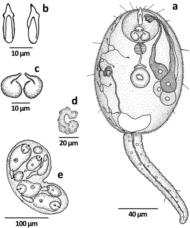
Cercaria sp. XXV Malabar n. sp. a cercaria, b stylet, c virgula organ, d genital primordium, e sporocyst
Table 4.
Showing comparative characters and measurements of Cercaria sp. XXV Kerala n. sp. and five related cercariae
| Cercaria (hosts) | Cercaria of Allasogonoporus vespertilionis Burns, 1961Fluminicola virens (Lea) | Cercaria leyteensis no. 4 Ito, Yasuraoka, Santos and Blas, 1977 Oncomelania quadrasi (Möllendorff) | Cercaria of Pleurogenoides orientalis Madhavi, Dhanumkumari and Ratnakumari, 1987Alocinma travancorica Benson | Cercaria of Pleurogenoides ovatus Janardanan and Prasadan, 1991Bithynia (D.) pulchella (Benson) | Cercaria sp. XXIV Malabar n. sp. Bithynia (D.) pulchella (Benson) | Cercaria sp. XXV Malabar n. sp. Bithynia (D.) pulchella (Benson) |
|---|---|---|---|---|---|---|
| Body | Spinose, 142.0–177.0 × 55.0–67.0 | Spinose, 94.0–101.0 (97.0) × 49.0–62.0 (55.0) | Spinose, 144.0–152.0 × 80.0–88.0 | Spinose, 96.0–122.0 (109) × 66.0–84.0 (77.0) | Spinose, 90.0–138.0 (111.3) × 51.0–78.0 (61.5) | Spinose, 102.0–142.5 (128.6) × 76.0–82.5 (80.1) |
| Tail | Aspinose, 75.0–105.0 × 15.0–20.0 | Aspinose, 59.0–81.0 (69.0) × 16.0–23.0 (18.0) | Aspinose, 98.0–116.0 × 20.0–28.0 | Aspinose, 45.0–74.0 (63.0) × 18.0–24.0 (22.0) | Spinose, 75.0–105.0 (94.4) × 12.0–18.0 (14.6) | Aspinose, 88.5–130.5 (113.9) × 15.0–16.5 (15.5) |
| Oral sucker | 45.0–47.0 × 32.0–40.0 | 33.0–39.0 (36.0) × 29.0–36.0 (33.0) | 36.0–44.0 × 32.0–40.0 | 32.0–38.0 (36.0) × 31.0–35.0 (33.0) | 27.0–34.5 (30) × 30.0–34.5 (32.5) | 25.5–31.5 (29.6) × 30.0–39.0 (36.0) |
| Ventral sucker | 17.0–22.0 | 16.0–20.0 (18.0) × 16.0–23.0 (19.0) | 20.0–24.0 | 20.0–21.0 (21.0) × 18.0–20.0 (19.0) | 19.5–24.0 (20.3) × 16.0–21.0 (18.0) | 19.5–22.5 (21.2) × 16.5–19.5 (18.0) |
| Stylet | 15.0–16.0 | 13.0–16.0 | 24.0 × 6.0 | 21–24 (23) × 4 | 13.5–15 (13.9) | 16.5–18.0 (17.4) × 3.0 |
| Virgula organ | 30.0–35.0 × 32.0–40.0 | – | 16.0–20.0 | – | 10.5–16.5 (12.8) × 15.0–18.0 (15.7) | 9.0–21.5 (14.2) × 18.0–27.0 (21.0) |
| Prepharynx | Absent | Long, narrow | Absent | Absent | Short | Absent |
| Pharynx | 7.0–12.0 × 10.0–12.0 | Small | 12.0 | 9.0 × 6.0 | Muscular, 7.5–10.5 (8.6) × 9–10.5 (9.4) | Globular, 10.5–13.5 (11.9) × 10.5–12.0 (11.1) |
| Oesophagus | Long, narrow | Short | Long, narrow | Absent | Short, narrow | Narrow |
| Caeca | Short | Faint | Short | – | Absent | Short, narrow |
| Excretory bladder | Vesicular | Cup-shaped | ‘U’-shaped | ‘V’-shaped | ‘V’-shaped, bicornuate | Transversely elongate, bicornuate |
| Flame cell formula | 2 [(2 + 2+2) + (2 + 2+2)] = 24 | – | 2 [(2 + 2+2) + (2 + 2+2)] = 24 | 2 [(2 + 2+2) + (2 + 2+2)] = 24 | 2 [(2 + 2+2) + (2 + 2+2)] = 24 | 2 [(2 + 2+2) + (2 + 2+2)] = 24 |
Virgulate xiphidiocercaria with a spinose, elongate-oval body measuring 102.0–142.5 (128.6) × 76.0–82.5 (80.1). Spines small, backwardly directed, uniformly distributed, with spine-size reducing from anterior to posterior. Six pairs of bristles present on body surface. Refractory cystogenous cells present except the anterior one-third of body. Tail aspinose, highly contractile; 88.5–130.5 (113.9) × 15.0–16.5 (15.5); with circular and longitudinal muscles; a pair of bristles present in posterior third; caudal bodies present. Oral sucker sub-terminal, wider than long, 25.5–31.5 (29.6) × 30.0–39.0 (36.0). Ventral sucker equatorial, smaller than oral sucker, longer than wide, 19.5–22.5 (21.2) × 16.5–19.5 (18.0). Stylet small, stout, dagger-shaped, inserted in the anterior half of oral sucker; 16.5–18.0 (17.4) × 3.0 (3.0); with an inwardly bent tip, round base, reinforced walls and shoulder. Virgula large, consisted of two distinct, sac-like structures which opened out independently on the ventral side of oral sucker; measured 9.0–21.5 (14.2) × 18.0–27.0 (21.0). Mouth at the center of oral sucker; prepharynx absent; pharynx globular, 10.5–13.5 (11.9) × 10.5–12.0 (11.1); oesophagus narrow, bifurcates to two short caeca. Penetration glands four pairs, with rounded outline and clear nucleus. Anterior two glands have coarse granules while the posterior ones have fine granules. Three glands and their ducts arranged in a bundle, postero-lateral to ventral sucker while the fourth was anterio-lateral with an independent duct. Gland ducts opened out near stylet; ducts of the anterior two glands have swollen tips. Genital primordium represented by a ‘C’ - shaped mass of cells, overlapping ventral sucker. Excretory bladder transversely elongated, bicornuate; primary collecting vessels arose from the antero-lateral corners of bladder, ran forward, coils and lateral to ventral sucker, divide into anterior and posterior collecting tubules. Flame cell formula: 2 [(2 + 2+2) + (2 + 2+2)] = 24. Caudal excretory canal originated from the posterior margin of excretory bladder, runs through tail; external opening not discernible.
Sporocyst (Fig. 4e)
Sporocysts were recovered from the digestive gland of infected D. pulchella; body elongate, thin-walled; 84.7–292.6 (173.5) × 77.0–254.1 (141.7); enclosed one to two cercariae, a few developing cercariae and germ balls.
Remarks
The present virgulate xiphidiocercaria with spinose body, aspinose tail, dagger-shaped stylet, a digestive system with narrow oesophagus and short caeca, four pairs of penetration glands and 24 flame cells is comparable to cercaria of Allasogonoporus vespertilionis Burns, 1961, Cercaria leyteensis no. 4 Ito, Yasuraoka, Santos and Blas, 1977, cercaria of Pleurogenoides orientalis Madhavi, Dhanumkumari and Ratnakumari, 1987, cercaria of Pleurogenoides ovatus Janardanan and Prasadan, 1991 and Cercaria sp. XXIV Malabar n. sp. Table 4 compares the characters and measurements of the present cercaria with that of the above five forms. It differs from cercaria of P. orientalis and C. leyteenensis no. 4 in measurements of body and tail; from cercaria of P. ovatus in the size of tail, from Cercaria sp. XXIV Malabar n. sp. in having an aspinose tail and from cercaria of A. vespertilionis in the size of body and oral sucker. The present cercaria differs from cercaria of P. orientalis, cercaria of P. ovatus and C. leyteensis no.4 in the shape and size of virgula and in the shape of stylet; from cercaria of A. vespertilionis in the size and shape of virgula and from all the above cercariae in the granulation of penetration glands and in having 2 swollen duct endings. Further, C. leyteenensis no. 4 differs in the position/arrangement of penetration glands while cercaria of P. orientalis differs in the arrangement of gland ducts. Based on the differences in characters and measurements between the present cercaria and its closely related forms, it is treated as a new species the name Cercaria sp. XXV Malabar n. sp. proposed.
Acknowledgements
The authors thank the Head, Department of Zoology, University of Calicut for providing necessary facilities. One of the authors (NKS) is grateful to the Council of Scientific and Industrial Research (CSIR) for financial support in the form of a Senior Research Fellowship and the study forms a part of his Ph.D. thesis.
Author’s contribution
The study was designed and guided by KPJ. NKS carried out the survey, collected and studied the cercariae in detail. The manuscript was written by both the authors.
Compliance with ethical standards
Conflict of interest
The authors declare that that there is no conflict of interest between them.
Footnotes
Publisher's Note
Springer Nature remains neutral with regard to jurisdictional claims in published maps and institutional affiliations.
References
- Brinesh R, Janardanan KP. Two new species of xiphidiocercariae from freshwater snail Digoniostoma pulchella (Bithyniidae) in Palakkad, Kerala, India. In: Tandon V, Yadav AK, Roy B, editors. Current trends in parasitology. Bangalore: Panima Publishing Corporation; 2009. pp. 169–174. [Google Scholar]
- Brinesh R, Janardanan KP. The life-history of Pleurogenoides malampuzhensis sp. nov. (Digenea: Pleurogenidae) from amphibious and aquatic hosts in Kerala. Indian J Helminthol. 2014;88:230–236. doi: 10.1017/S0022149X13000084. [DOI] [PubMed] [Google Scholar]
- Burns WC. Six virgulate xiphidiocercariae from Oregon, including redescription of Allassogonoporus vespertilionis and Acanthatrium oregonense. J Parasitol. 1961;47(6):919–925. doi: 10.2307/3275020. [DOI] [PubMed] [Google Scholar]
- Etges FJ, Page CR, Bonner TP. Two new species of xiphidiocercariae from Mountain Lake, Virginia. Trans Am Microsc Soc. 1969;88:84–88. doi: 10.2307/3224662. [DOI] [Google Scholar]
- Faust EC. Notes on larval flukes from China, II. Studies on some larval flukes from the central and south coast provinces of China. Am J Hyg. 1924;4:241–301. [Google Scholar]
- Hall E, Groves AE. Virgulate xiphidiocercariae from Nitocris dilatus Conrad. J Parasitol. 1963;49(2):249–263. doi: 10.2307/3275992. [DOI] [PubMed] [Google Scholar]
- Ismail NS, Bdair SM. Four new xiphidiocercariae from Melanopsis praemorsa (L. 1758) (Thiaridae) snails in Jordon. Jpn J Parasitol. 1987;36(3):135–141. [Google Scholar]
- Ito J. Studies on the freshwater cercariae in Leyte Island, Philippines 3. Cercariae from Thiaridae. Jpn J Exp Med. 1977;47(4):223–248. [PubMed] [Google Scholar]
- Ito J, Yasuraoka K, Santos AT, Blas BL. Studies on the fresh water cercariae in Leyte Island, Philippines. 1 Collection of snails. Japan J Exp Med. 1977;47(3):141–150. [PubMed] [Google Scholar]
- Janardanan KP, Prasadan PK. Studies on the life-cycle of Pleurogenoides ovatus Rao, 1977 (Trematoda: Pleurogenetinae) J Helminthol. 1991;65:43–50. doi: 10.1017/S0022149X00010427. [DOI] [PubMed] [Google Scholar]
- Madhavi R, Dhanumkumari C, Ratnakumari TB. The life-history of Pleurogenoides orientalis (Srivastava, 1934) (Trematoda: Lecithodendriidae) Parasitol Res. 1987;73(1):41–45. doi: 10.1007/BF00536334. [DOI] [PubMed] [Google Scholar]
- Mohandas A. On two new species of cercariae and the histopathology of digestive gland of their host, Digoniostoma pulchella (Benson) Acta Parasitol Pol. 1977;25(2):17–23. [Google Scholar]
- Murty AS. Studies on Indian cercariae III. Virgulate plagiorchioid cercariae. Proc Indian Acad Sci. 1976;83(1):12–17. [Google Scholar]
- Nasir P. British freshwater cercariae. Cumana: Universidad de Oriente; 1984. [Google Scholar]
- Nasir P, Diaz MT. Freshwater larval trematodes XXXII. Twenty new species of Venezuelan cercariae. Riv Parassit. 1973;34(1):1–44. [PubMed] [Google Scholar]
- Probert AJ. Studies on larval trematodes infecting freshwater molluscs of Llangorse Lake, South Wales. Part I. The xiphidio- and microcercous cercariae. J Helminthol. 1965;39:35–52. doi: 10.1017/S0022149X0002006X. [DOI] [PubMed] [Google Scholar]
- Ramakrishna, Dey A. Handbook on Indian freshwater molluscs. Kolkata: Zool Surv India; 2007. pp. 1–399. [Google Scholar]
- Sewell RBS. Cercariae indicae. Indian J Med Res. 1922;10:1–370. [Google Scholar]
- Subba Rao NV. Handbook of freshwater molluscs of India. Calcutta: Zool Surv India; 1989. pp. 1–289. [Google Scholar]