Abstract
Interstitial lung disease (ILD) is one of the extra-articular involvement forms of rheumatoid arthritis (RA), and it is associated with increased mortality. The presence of genetic susceptibility, smoking, rheumatoid factor positivity, and the presence of anticitrulline peptide antibodies are factors contributing to the development of ILD in patients with RA. Early diagnosis and treatment of ILD contribute to the reduction of morbidity and mortality. We herein evaluated the current literature for the diagnosis and treatment of RA-associated ILD.
Keywords: Interstitial lung disease, arthritis, life threatening
Introduction
Rheumatoid arthritis (RA) is a chronic systemic inflammatory disease manifested by articular and extra-articular features. Pulmonary involvement is a common extra-articular manifestation occurring roughly in approximately 60%–80% of patients with RA. The pulmonary involvement in RA may vary, including interstitial lung disease (ILD), pleural disease, rheumatoid nodules, bronchiectasis, and vasculitis. ILD is one particular type of pulmonary involvement associated with significant morbidity and mortality (1–3). The management of RA-associated ILD (RA-ILD) depends on patients’ clinical, functional, and radiologic findings. Several therapeutic agents have been suggested in the literature, but the optimal treatment has not been determined (4–6).
Risk factors
The mechanism behind ILD in RA is poorly understood, but there are certain risk factors believed to play an important role. Smoking is believed to play the major role, but male sex, high titers of the rheumatoid factor, and elevated anticyclic citrullinated protein antibodies are the prevailing factors.
Clinical features and diagnostic tools
In 2002, the American Thoracic Society/European Respiratory Society classification of acute and chronic parenchymal lung diseases (7) has also been adopted for the identification of ILD (Table 1). The clinical manifestations of RA-related ILD are dyspnea and dry cough. However, some patients may be asymptomatic, especially in early stages. Pleuritic chest pain, fever, hemoptysis, and tachypnea are other symptoms of the disease. The most frequent finding on physical examination were crepitant rallies in bilateral baselines in pulmonary auscultation. In the majority of patients, the pulmonary function test (PFT) has a low force vital capacity (FVC), low total lung capacity, low/normal carbon monoxide diffusion capacity (DLCO), and restrictive respiratory pattern with exercise or resting hypoxemia (8). Since the earliest functional disorder in patients with ILD is a DLCO decrease, DLCO is one of the important tests for the early diagnosis of ILD. FVC may not be affected until the late stage. The PFTs may also reveal a normal or high force vital capacity at 1 second/FVC (restrictive type lung disease finding) ratio. DLCO and FVC are the most commonly and most important used diagnostic markers in ILD. Generally, changes of 10% in FVC and 15% in DLCO are considered to be significant (8). X-ray has a low sensitivity in the ILD detection, and X-ray findings may be normal in early stages. Reticular and small nodular opacities can be seen in lower lung zones on plain chest X-ray. High-resolution computed tomography (HRCT) has been accepted as the gold standard noninvasive imaging method in the diagnosis of ILD in patients with RA (9). HRCT results were consistent with the results of open lung biopsy (10).
Table 1.
American Thoracic Society/European Respiratory Society Classification of Idiopathic Interstitial Pneumonia
| Histologic Pattern | Clinical/Radiologic/Pathologic Diagnosis |
|---|---|
| Usual interstitial pneumonia | Idiopathic pulmonary fibrosis/cryptogenic fibrosing alveolitis |
| Nonspecific interstitial pneumonia | Nonspecific interstitial pneumonia |
| Organizing pneumonia | Cryptogenic organizing pneumonia |
| Diffuse alveolar damage | Acute interstitial pneumonia |
| Respiratory bronchiolitis | Respiratory bronchiolitis interstitial lung disease |
| Desquamative interstitial pneumonia | Desquamative interstitial pneumonia |
| Lymphoid interstitial pneumonia | Lymphoid interstitial pneumonia |
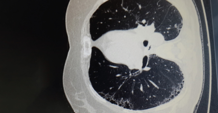
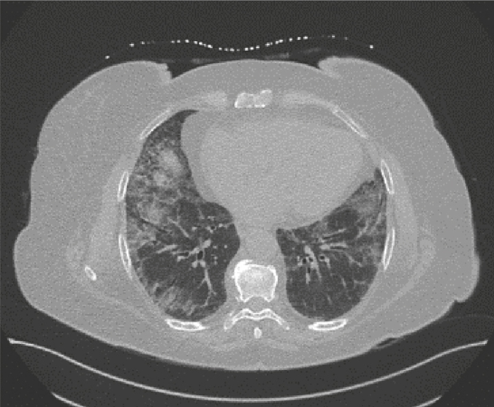
The most common HRCT findings in patients with RA-ILD are usual interstitial pneumonia (UIP) (Figure 1), nonspecific interstitial pneumonia (NSIP) (Figure 2), lymphocytic interstitial pneumonia, organizing pneumonia, diffuse alveolar damage, respiratory bronchiolitis, and desquamative interstitial pneumonia (11). Since there may be some alterations in bronchoalveolar lavage (BAL) fluid in the absence of ILD, BAL is not routinely used as a diagnostic tool to demonstrate the presence of ILD in RA. Neutrophil and macrophage predominance is a characteristic feature of RA-ILD. Although BAL may be useful in the exclusion of infection, it is not necessary for the diagnosis of RA-ILD. Surgical lung biopsy has been considered the gold standard in the histopathological diagnosis. However, due to the potential risks associated with the method, many patients are diagnosed without surgical biopsy and pathologic confirmation. Video-assisted thoracoscopic surgery is often preferred for open lung biopsy. Transbronchial biopsy in diagnosis is limited, and it is not required in patients with typical clinical and radiological findings.
Figure 1.
Usual Interstitial Pneumonia (UIP); Peripheral Septal Thickening, Bronchiectasis, and Honeycombing
Figure 2.
Nonspecific Interstitial Pneumonia (NSIP); Bilateral Ground-Glass Opacities with Associated Fine Reticulations and Traction Bronchiectasis
Recent developments in diagnosis
Studies on biomarkers that can be used to confirm the diagnosis and evaluate the treatment response are still ongoing. In recent years, a serologic marker known as KL-6 (Krebsvon del Lungen-6, proliferating Type 2 pneumocytes and high-molecular-weight glycoprotein expressed in epithelial cells) has been investigated. Serum KL-6 levels were elevated in interstitial pneumonia, hypersensitivity pneumonitis, tuberculosis, sarcoidosis, and pulmonary alveolar proteinosis. It was also observed in patients with RA-ILD (11, 12). A significant increment in the serum KL-6 levels reflects the extensity of HRCT lesions. The grade of alveolitis and severity of pulmonary fibrosis, however, may not be very sensitive in early-stage pulmonary disease. It can also be helpful in the detection of active and progressive pulmonary disease in RA-ILD (12). Oyama et al. (13) reported that KL-6 was elevated in 88.9% of patients with active ILD, and in only 0.6% of patients without active ILD. Recently, studies indicating that transthoracic ultrasonography is a useful method in the diagnosis of early-stage ILD have been published. In a study involving patients with RA, systemic sclerosis, and systemic lupus erythematosus, a significant proportion of patients who showed ILD on HRCT also showed pathological changes in transthoracic ultrasonography (14).
The 18-flurodeoxyglucose positron emission tomography/computed tomography (18 FDG PET-CT) is an alternative method that can be used in the diagnosis of ILD. A recent study evaluating patients with connective tissue disease reported that deep-inspiration breath-hold 18F-FDG-PET/CT could be a useful method in the diagnosis of ILD (15).
Management
Treatment regimens must be tailored to the individual patient; both the supportive treatment and medical treatment should be emphasized. The promotion of smoking cessation comes at the beginning of supportive therapies, which is considered to play a pivotal role in the pathogenesis of RA, and also known to increases both joint symptoms and lung damage. Pneumococcal and influenza vaccines should be routinely administered. Oxygen should be administered to patients with low oxygen saturation (16, 17).
Treatment of RA-ILD is becoming more complicated and challenging, since almost all drugs used in the treatment of RA can cause ILD or have already mediated ILD. Hence, treatment depends on the severity of the disease and the clinical condition of the patient. At this point, radiological findings and functional capacity of the lungs are as important as the clinical condition of the patient. There are some cases where disease symptoms are absent (e.g., breathlessness and/or cough) that are incidentally detected with radiologic imaging of ILD findings, and these patients do not need additional treatment if they are functionally stable (18). It is recommended to follow these patients with the respiratory function test (PFT) every 3 months, and HRCT and PFT/DLCO every 6–12 months for the first 2 years, if the symptoms progress. Treatment of asymptomatic RA-associated patients with ILD without progressive disease should be continued as usual, and the use of any DMARD that controls joint symptoms is not contraindicated in those patients. During the follow-up period, patients in who progression is indicated according to clinical, radiological, or PFT findings should be given additional ILD treatment. Despite the lack of controlled studies in the RA-ILD treatment, corticosteroids are considered as the first-line therapy. The initial dose is usually 0.5–1 mg/kg prednisolone (usually 40–60 mg/daily). Once the response to corticosteroids is achieved, the dose is gradually reduced to the lowest possible dose. Some ILD subtypes, such as the NSIP and OP, have a better response to corticosteroids than other subtypes. When corticosteroids are reduced, additional immunosuppressive agents may be required. For this purpose, corticosteroids may be combined with azathioprine, cyclophosphamide, or mycophenolate mofetil (MMF) (19, 20). As there is a risk of respiratory failure, patients with a rapidly progressive ILD pattern should be approached aggressively.
Corticosteroids at high doses (prednisolone at 10 mg/kg/day) and intravenous pulse cyclophosphamide (6 cycles at 15 mg/kg) every 3–4 weeks are recommended in these patients. At the end of the cyclophosphamide treatment, MMF or azathioprine therapy are recommended as an indwelling therapy. Cyclophosphamide has been reported to be useful in active, rapidly progressing disease (19), although there are no randomized controlled trials of its use in RA-ILD, and limited efficacy has been reported. In a cohort of mixed connective tissue diseases involving a small number of patients with RA-ILD, it has been reported that mycophenolate mofetil contributes to symptomatic recovery and stabilization or improvement in PFTs, and it is also useful in reducing steroid doses (20, 21). Although few studies have shown positive effects on lung findings, since there is no curative effect on joint symptoms, the use of mycophenolate mofetil is limited.
Rituximab (RTX) has been one of the most commonly used drugs in the treatment of RA-ILD in recent years. It is a monoclonal antibody against the B-cell marker CD20 used to treat patients with RA who do not respond to anti-TNF therapy or who cannot use these drugs. After the demonstration of follicular B-cell hyperplasia and interstitial plasma cell infiltration in patients with RA-ILD, a potential role of B-cells in pathogenesis is suggested. This led to an increased interest in RTX for the treatment of RA-ILD (22, 23). In contrast to other biological therapies, there is a concern about the potential pulmonary RTX toxicity. The reported cases are largely due to lymphoproliferative diseases. However, there are several reports on the development or worsening of ILD in RA patients using rituximab (24–26). It has been shown that antitumor necrosis factor (anti-TNF) agents have great efficacy in the suppression of RA joint symptoms and improvement of the disease progression, and they have been used in many RA patients who have not responded to conventional DMARD therapy in recent years.
However, with the widespread use of these drugs, they have caused concerns about potential pulmonary toxicity. There have been reports of new-onset ILD or worsening of preexisting disease after starting an anti-TNF treatment (27–31). In a study by Ramos-Casals et al. (32), it was reported that ILD develops in 10% of 226 patients (83% of RA) who received anti-TNF therapy.
On the other hand, some studies show that there is no correlation between the use of anti-TNF and the development or exacerbation of ILD in RA. A report from the British Society for Rheumatology Biologics Register on patients with RA-ILD taking either anti-TNF agents or traditional DMARDs showed no difference in mortality rates. However, in the same report, the ILD mortality was 21% in patients treated with anti-TNF therapy compared to 7% in conventional DMARDs (33). In another study, Herrinton et al. (34) compared anti-TNF therapy with non-TNF biologic therapy in their study of more than 8.000 patients; there was no difference in the development of ILD.
However, anti-TNF drugs have been shown to have potentially positive effects in stabilizing or ameliorating pulmonary disease (35). Experimental studies indicate that TNF-alpha may have both profibrotic and antifibrotic effects; the imbalance between these two roles may trigger fibrosis or stable ILD in vulnerable individuals, but further studies are needed to confirm this hypothesis (36). Patients with preexisting RA-ILD should be followed closely during anti-TNF treatment. Since methotrexate has a potential risk of pulmonary toxicity, the combination of anti-TNF and methotrexate may have a greater risk of developing ILD or worsening the preexisting disease. Therefore, in patients with ILD, the combination of methotrexate and anti-TNF should not be preferred or should be closely monitored in case of necessity. Although the non-TNF biological agents such as tocilizumab and abatacept are reported to be beneficial in RA patients with ILD, there are some reports that suggest new ILD developments or worsening of the preexisting ILD under these agents (37–40). Lung transplantation is recommended as an option in case of progressive and severe pulmonary disease, despite medical treatment.
Prognosis
Patients with RA-associated ILD have an increased mortality compared to patients without ILD. The male sex, an advanced age, high seropositivity, and greater impairment of the pulmonary function at diagnosis have been associated with worse prognosis (30). Histopathologic and/or radiologic phenotype plays an important role in the prognosis of RA-associated ILD. The cases with the UIP pattern have a worse prognosis compared to those without the UIP pattern (3, 11).
Conclusion
Interstitial lung disease is a common extra-articular manifestation of RA, associated with increased morbidity and mortality. At baseline, an assessment of the severity and the extent of ILD using objective pulmonary parameters is required to make decisions with regard to therapy. The treatment of RA-associated ILD should be tailored for each patient. It should also be kept in mind that drugs used for RA may exacerbate ILD.
Footnotes
Peer-review: Externally peer-reviewed.
Conflict of Interest: The author has no conflict of interest to declare.
Financial Disclosure: The author declared that this study has received no financial support.
References
- 1.Olson AL, Swigris JJ, Sprunger DB, Fischer A, Fernandez-Perez ER, Solomon J, et al. Rheumatoid arthritis-interstitial lung disease-associated mortality. Am J Respir Crit Care Med. 2011;183:372–8. doi: 10.1164/rccm.201004-0622OC. [DOI] [PMC free article] [PubMed] [Google Scholar]
- 2.Ascherman DP. Interstitial lung disease in rheumatoid arthritis. Curr Rheumatol Rep. 2010;12:363–9. doi: 10.1007/s11926-010-0116-z. [DOI] [PubMed] [Google Scholar]
- 3.Kim EJ, Elicker BM, Maldonado F, Webb WR, Ryu JH, Uden JHV, et al. Usual interstitial pneumonia in rheumatoid arthritis-associated interstitial lung disease. Eur Respir J. 2010;35:1322–8. doi: 10.1183/09031936.00092309. [DOI] [PubMed] [Google Scholar]
- 4.Mori S, Koga Y, Sugimoto M. Different risk factors between interstitial lung disease and airway disease in rheumatoid arthritis. Respir Med. 2012;106:1591–9. doi: 10.1016/j.rmed.2012.07.006. [DOI] [PubMed] [Google Scholar]
- 5.Grutters JC, du Bois RM. Genetics of fibrosing lung diseases. Eur Respir J. 2005;25:915–27. doi: 10.1183/09031936.05.00133404. [DOI] [PubMed] [Google Scholar]
- 6.Saag G, Kolluri S, Koehnke RK, Georgou TA, Rachow JW, Hunninghake GW, et al. Rheumatoid arthritis lung disease: Determinants of radiographic and physiologic abnormalities. Arthritis Rheum. 1996;39:1711–9. doi: 10.1002/art.1780391014. [DOI] [PubMed] [Google Scholar]
- 7.American Thoracic Society; European Respiratory Society. American Thoracic Society/European Respiratory Society International Multidisciplinary Consensus Classification of the Idiopathic Interstitial Pneumonias. This joint statement of the American Thoracic Society (ATS), and the European Respiratory Society (ERS) was adopted by the ATS board of directors, June 2001 and by the ERS Executive Committee, June 2001. Am J RespirCrit Care Med. 2002;165:277–304. doi: 10.1164/ajrccm.165.2.ats01. [DOI] [PubMed] [Google Scholar]
- 8.Behr J, Furst DE. Pulmonary Function Tests. Rheumatology (Oxford) 2008;47(Suppl 5):v65–7. doi: 10.1093/rheumatology/ken313. [DOI] [PubMed] [Google Scholar]
- 9.Biederer J, Schnabel A, Muhle C, Gross WL, Heller M, Reuter M. Correlation between HRCT findings, pulmonary function tests and bronchoalveolar lavage cytology in interstitial lung disease associated with rheumatoid arthritis. Eur Radiol. 2004;14:272–80. doi: 10.1007/s00330-003-2026-1. [DOI] [PubMed] [Google Scholar]
- 10.Salaffi F, Manganelli P, Carotti M, Baldelli S. The differing patterns of subclinical pulmonary involvement in connective tissue diseases as shown by application of factor analysis. Clin Rheumatol. 2000;19:35–41. doi: 10.1007/s100670050008. [DOI] [PubMed] [Google Scholar]
- 11.Tsuchiya Y, Takayanagi N, Sugiura H, Miyahara Y, Tokunaga D, Kawabata Y, et al. Lung diseases directly associated with rheumatoid arthritis and their relationship to outcome. Eur Respir J. 2011;37:1411–7. doi: 10.1183/09031936.00019210. [DOI] [PubMed] [Google Scholar]
- 12.Kinoshita F, Hamano H, Harada H, Kinoshita T, Igishi T, Hagino H, et al. Role of KL-6 in evaluating the disease severity of rheumatoid lung disease: comparison with HRCT. Respir Med. 2004;98:1131–7. doi: 10.1016/j.rmed.2004.04.003. [DOI] [PubMed] [Google Scholar]
- 13.Oyama T, Kohno N, Yokoyama A, Hirasawa Y, Hiwada K, Oyama H, et al. Detection of interstitial pneumonitis in patients with rheumatoid arthritis by measuring circulating levels of KL-6, a humanMUC1 mucin. Lung. 1997;175:379–85. doi: 10.1007/PL00007584. [DOI] [PubMed] [Google Scholar]
- 14.Moazedi-Fuerst FC, Kielhauser S, Brickmann K, Tripolt N, Meilinger M, Lufti A, et al. Sonographic assessment of interstitial lung disease in patients with rheumatoid arthritis, systemic sclerosis and systemic lupus erythematosus. Clin Exp Rheumatol. 2015;33:87–91. [PubMed] [Google Scholar]
- 15.Uehara T, Takeno M, Hama M, Yoshimi R, Suda A, Ihata A, et al. Deepinspiration breath-hold 18F-FDG-PET/CT is useful for assessment of connective tissue disease associated interstitial pneumonia. Mod Rheumatol. 2016;26:121–7. doi: 10.3109/14397595.2015.1054099. [DOI] [PubMed] [Google Scholar]
- 16.Demoruelle MK, Mittoo S, Solomon JJ. Connective tissue disease-related interstitial lung diseases. Best Pract Res Clin Rheumatol. 2016;30:39–52. doi: 10.1016/j.berh.2016.04.006. [DOI] [PubMed] [Google Scholar]
- 17.Aparicio IJ, Lee JS. Connective tissue disease-associated interstitial lung diseases: unresolved Issues. Semin Respir Crit Care Med. 2016;37:468–76. doi: 10.1055/s-0036-1580689. [DOI] [PubMed] [Google Scholar]
- 18.Mathai SC, Danoff SK. Management of interstitial lung disease associated with connective tissue disease. BMJ. 2016;352:h6819. doi: 10.1136/bmj.h6819. [DOI] [PMC free article] [PubMed] [Google Scholar]
- 19.Chan E, Chapman K, Kelly C. Interstitial lung disease in rheumatoid arthritis: a review. Arthritis Research UK; 2013. [Google Scholar]
- 20.O’Dwyer DN, Armstrong ME, Cooke G, Dodd JD, Veale DJ, Donnelly SC. Rheumatoid Arthritis (RA) associated Interstitial Lung Disease (ILD) Eur J Intern Med. 2013;24:597–603. doi: 10.1016/j.ejim.2013.07.004. [DOI] [PubMed] [Google Scholar]
- 21.Saketkoo L, Espinoza L. Rheumatoid arthritis interstitial lung disease: mycophenolate mofetil as an antifibrotic and disease-modifying anti rheumatic drug. Arch Intern Med. 2008;168:1718–9. doi: 10.1001/archinte.168.15.1718. [DOI] [PubMed] [Google Scholar]
- 22.Atkins S, Turesson C, Myers J, Tazelaar H, Ryu J, Matteson E, et al. Morphologic and quantitative assessment of CD20+ B cell infiltrates in rheumatoid arthritis-associated nonspecific interstitial pneumonia and usual interstitial pneumonia. Arthritis Rheum. 2006;54:635–41. doi: 10.1002/art.21758. [DOI] [PubMed] [Google Scholar]
- 23.Yusof MY, Kabia A, Darby M, Lettieri G, Beirne P, Vital EM, et al. Effect of rituximab on the progression of rheumatoid arthritis-related interstitial lung disease: 10 years’ experience at a single centre. Rheumatology (Oxford) 2017;56:1348–57. doi: 10.1093/rheumatology/kex072. [DOI] [PMC free article] [PubMed] [Google Scholar]
- 24.Liote H, Liote F, Seroussi B, Mayaud C, Cadranel J. Rituximab-induced lung disease: a systematic literature review. Eur Resp J. 2010;35:681–7. doi: 10.1183/09031936.00080209. [DOI] [PubMed] [Google Scholar]
- 25.Soubrier M, Jeannin G, Kemeny J, Tournadre A, Caillot N, Caillaud D, et al. Organizing pneumonia after rituximab therapy: two cases. Joint Bone Spine. 2008;75:362–5. doi: 10.1016/j.jbspin.2007.10.009. [DOI] [PubMed] [Google Scholar]
- 26.Panopoulos S, Sfikakis P. Biological treatments and connective tissue disease associated interstitial lung disease. Curr Opin Pulmon Med. 2011;17:362–7. doi: 10.1097/MCP.0b013e3283483ea5. [DOI] [PubMed] [Google Scholar]
- 27.Mori S, Imamura F, Kiyofuji C, Sugimoto M. Development of interstitial pneumonia in a rheumatoid arthritis patient treated with infliximab, an antitumor necrosis factor alpha-neutralizing antibody. Mod Rheumatol. 2006;16:251–5. doi: 10.3109/s10165-006-0491-5. [DOI] [PubMed] [Google Scholar]
- 28.Takeuchi T, Tatsuki Y, Nogami Y, Ishiguro N, Tanaka Y, Yamanaka H, et al. Post marketing surveillance of the safety profile of infliximab in 5000 Japanese patients with rheumatoid arthritis. Ann Rheum Dis. 2008;67:189–94. doi: 10.1136/ard.2007.072967. [DOI] [PubMed] [Google Scholar]
- 29.Yamazaki H, Isogai S, Sakurai T, Nagasaka K. A case of adalimumab-associated interstitial pneumonia with rheumatoid arthritis. Mod Rheumatol. 2010;20:518–21. doi: 10.3109/s10165-010-0308-4. [DOI] [PubMed] [Google Scholar]
- 30.Hadjinicolaou A, Nisar M, Bhagat S, Parfrey H, Chilvers E, Ostor A. Noninfectious pulmonary complications of newer biological agents for rheumatic diseases-a systematic literature review. Rheumatology (Oxford) 2011;50:2297–305. doi: 10.1093/rheumatology/ker289. [DOI] [PubMed] [Google Scholar]
- 31.Pearce F, Johnson S, Courtney P. Interstitial lung disease following certolizumab pegol. Rheumatology (Oxford) 2012;51:578–80. doi: 10.1093/rheumatology/ker309. [DOI] [PubMed] [Google Scholar]
- 32.Ramos-Casals M, Brito-Zeron P, Munoz S, Soria N, Galiana D, Bertolaccini L, et al. Autoimmune diseases induced by TNF-targeted therapies: analysis of 233 cases. Medicine (Baltimore) 2007;86:242–51. doi: 10.1097/MD.0b013e3181441a68. [DOI] [PubMed] [Google Scholar]
- 33.Dixon W, Hyrich K, Watson K, Lunt M, Symmons D. Influence of anti-TNF therapy on mortality in patients with rheumatoid arthritis associated interstitial lung disease: results from the British Society for Rheumatology Biologics Register. Ann Rheum Dis. 2010;69:1086–91. doi: 10.1136/ard.2009.118935. [DOI] [PMC free article] [PubMed] [Google Scholar]
- 34.Herrinton L, Harrold L, Liu L, Raebel M, Taharka A, Winthrop K, et al. Association between anti-TNF-α therapy and interstitial lung disease. Pharmacoepidemiol Drug Saf. 2013;22:394–402. doi: 10.1002/pds.3409. [DOI] [PMC free article] [PubMed] [Google Scholar]
- 35.Horai Y, Miyamura T, Shimada K, Takahama S, Minami R, Yamamoto M, et al. Eternacept for the treatment of patients with rheumatoid arthritis and concurrent interstitial lung disease. J Clin Pharm Ther. 2012;37:117–21. doi: 10.1111/j.1365-2710.2010.01234.x. [DOI] [PubMed] [Google Scholar]
- 36.Olivas-Flores E, Bonilla-Lara D, Gamez-Nava J, Rocha-Muñoz A, GonzalezLopez L. Interstitial lung disease in rheumatoid arthritis: current concepts in pathogenesis, diagnosis and therapeutics. World J Rheumatol. 2015;5:1–22. doi: 10.5499/wjr.v5.i1.1. [DOI] [Google Scholar]
- 37.Curtis JR, Sarsour K, Napalkov P, Costa LA, Schulman KL. Incidence and complications of interstitial lung disease in users of tocilizumab, rituximab, abatacept and anti-tumor necrosis factor α agent, a retrospective cohort study. Arthritis Res Ther. 2015;17:319. doi: 10.1186/s13075-015-0835-7. [DOI] [PMC free article] [PubMed] [Google Scholar]
- 38.Mera-Varela A, Perez-Pampin E. Abatacept therapy in rheumatoid arthritis with interstitial lung disease. J Clin Rheumatol. 2014;20:445–6. doi: 10.1097/RHU.0000000000000084. [DOI] [PubMed] [Google Scholar]
- 39.Roubille C, Haraoui B. Interstitial lung diseases induced or exacerbated by DMARDS and biologic agents in rheumatoid arthritis: a systematic literature review. Semin Arthritis Rheum. 2014;43:613–26. doi: 10.1016/j.semarthrit.2013.09.005. [DOI] [PubMed] [Google Scholar]
- 40.Gauhar UA, Gaffo AL, Alarcón GS. Pulmonary manifestations of rheumatoid arthritis. Semin Respir Crit Care Med. 2007;28:430–40. doi: 10.1055/s-2007-985664. [DOI] [PubMed] [Google Scholar]


