CASE PRESENTATION
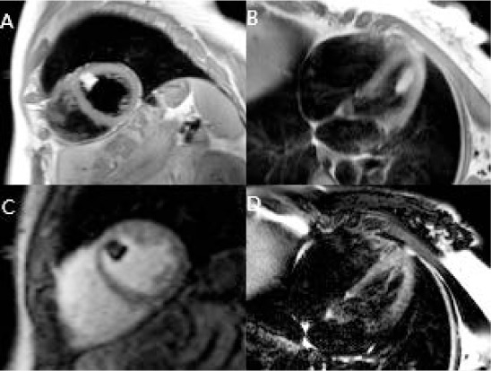
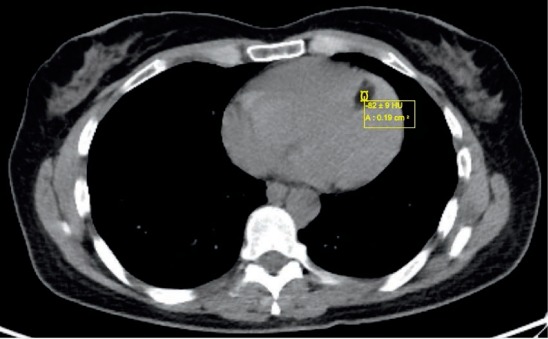
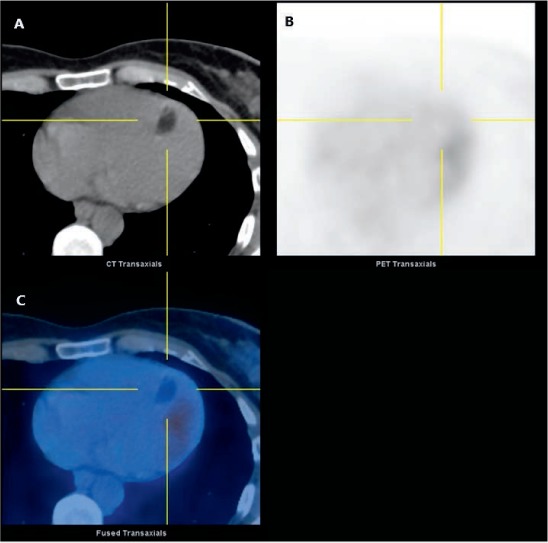
A 59-year-old woman with a 6-month history of exertional dyspnea and worsening fatigue underwent cardiac magnetic resonance imaging (CMR) to further characterize a left ventricular mass incidentally noted on transthoracic echocardiography. The mass attached to the mid-anterior wall, measured 2.6 cm in its major axis, and displayed the following tissue characteristics: hyperintensity to myocardium with T1 weighted imaging (Figure 1 A, B), lack of perfusion with first-pass imaging of gadolinium-based contrast agent (Figure 1 C), and signal loss with T2 weighted fat saturation sequences (Figure 1 D). Combined computed tomography (CT) and positron emission tomography (PET) imaging revealed a hypodense mass that did not enhance with contrast administration, Hounsfield units of −82 consistent with adipose density, and low uptake of fluorodeoxyglucose (Figures 2, 3). In summation, the findings were virtually diagnostic of an intracardiac lipoma.
Figure 1.

Cardiac magnetic resonance imaging showing hyperintense signal of the left ventricular mass on dark blood T1 weighted imaging in (A) short axis and (B) 4-chamber long axis views, (C) lack of perfusion of the mass with first-pass administration of gadolinium-based contrast, and (D) complete signal void with T2 fat suppression imaging.
Figure 2.

Axial computed tomography slice showing low Hounsfield units of −82 when the left ventricular mass is sampled, consistent with adipose tissue.
Figure 3.

Combined positron emission (PET) and computed tomography (CT) images showing (A) a hypodense left ventricular mass with virtually no uptake of fluorodeoxyglucose (18F-FDG), suggesting (B) a benign etiology due to lack of metabolic activity. (C) A hybrid PET/CT axial slice.
Cardiac lipomas are primary tumors of the heart that consist of an encapsulated collection of adipocytes. Although classified as benign lesions, they may lead to symptoms depending on their size, location, and compressive effects on cardiac chambers or interference with the conduction system. Whether cardiac1,2 or extracardiac,3 lipomas display characteristic CMR findings, with the elegant demonstration of hypointensity on fat suppression imaging being its sina qua non feature. Supportive CT findings include an encapsulated mass with fat density Hounsfield units usually between −90 and −110 and homogeneous hypoenhancement with the administration of iodine-based contrast.1,2 Although the role of PET is less well established for cardiac lipomas, extracardiac lipomas demonstrate reduced metabolic activity as evidenced by low avidity for fluorodeoxyglucose with correspondingly low standardized uptake values.4,5
Our case demonstrates classic noninvasive cardiac imaging findings of this tumor type. A conservative “watch and wait” strategy was recommended, and repeat CMR 3 months later did not demonstrate interval growth of the lipoma.
REFERENCES
- 1.Sivrioglu AK, Ozturk E, Geceer G, Incedayi M, Kara K. Incidental right atrial lipoma: appearance on multidetector computer tomography imaging. Hellenic J Cardiol. 2014;55:422–3. [PubMed] [Google Scholar]
- 2.Barbuto L, Ponsiglione A, Del Vecchio W et al. Humongous right atrial lipoma: a correlative CT and MR case report. Quant Imaging Med Surg. 2015 Oct;5(5):774–7. doi: 10.3978/j.issn.2223-4292.2015.01.02. [DOI] [PMC free article] [PubMed] [Google Scholar]
- 3.Filli L, Huber A, Husain NA. Symptomatic lipoma of the internal auditory canal: CT and MRI findings. A case report. Neuroradiol J. 2014 Sep;27(4):479–81. doi: 10.15274/NRJ-2014-10077. [DOI] [PMC free article] [PubMed] [Google Scholar]
- 4.Costelloe CM, Chuang HH, Chasen BA et al. Bone windows for distinguishing malignant from benign primary bone tumors on FDG PET/CT. J Cancer. 2013 Aug 9;4(7):524–30. doi: 10.7150/jca.6259. [DOI] [PMC free article] [PubMed] [Google Scholar]
- 5.Shin DS, Shon OJ, Han DS, Choi JH, Chun KA, Cho IA. The clinical efficacy of 18F-FDG-PET/CT in benign and malignant musculoskeletal tumors. Ann Nucl Med. 2008 Aug;22(7):603–9. doi: 10.1007/s12149-008-0151-2. [DOI] [PubMed] [Google Scholar]


