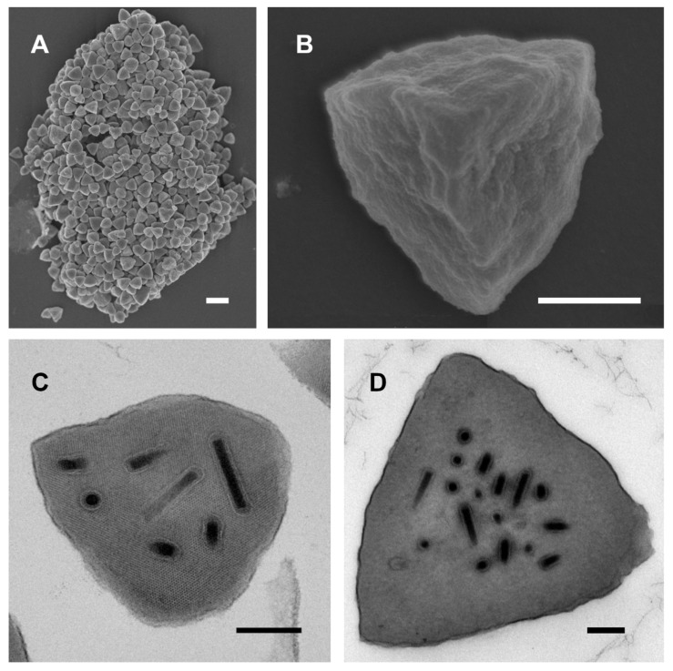
Figure 1.
Scanning and transmission electron micrographs of ChinNPV#1 occlusion bodies (OBs): (A) A group of ChinNPV#1 OBs; (B) high-magnification view of a single OB; (C) section through a single OB, showing the lattice lines of the paracrystalline polyhedrin matrix surrounding the occluded virions; (D) section through a larger OB. Scale bars: (A) 2 µm, (B) 500 nm, (C) and (D) 200 nm.

