This cohort study investigates the association of biologic therapy with coronary inflammation in patients with psoriasis as assessed by the perivascular fat attenuation index, an imaging biomarker that assesses coronary inflammation by mapping spatial changes of perivascular fat composition via coronary computed tomography angiography.
Key Points
Question
Is biologic therapy for psoriasis associated with a change in coronary inflammation as assessed by the perivascular fat attenuation index?
Findings
In this cohort study of 134 consecutive patients with moderate to severe psoriasis, biologic therapy was associated with a significant decrease in coronary inflammation as assessed by perivascular fat attenuation index, a marker of coronary inflammation associated with cardiovascular outcomes. Patients not receiving biologic therapy had no change in perivascular fat attenuation index at 1 year.
Meaning
The findings suggest that biologic therapy for moderate to severe psoriasis is associated with a reduction in coronary inflammation assessed as perivascular fat attenuation index and thus that perivascular fat attenuation index may be used to track response to interventions in the coronary artery.
Abstract
Importance
Psoriasis is a chronic inflammatory skin disease associated with increased coronary plaque burden and cardiovascular events. Biologic therapy for psoriasis has been found to be favorably associated with luminal coronary plaque, but it is unclear whether these associations are attributable to direct anti-inflammatory effects on the coronary arteries.
Objective
To investigate the association of biologic therapy with coronary inflammation in patients with psoriasis using the perivascular fat attenuation index (FAI), a novel imaging biomarker that assesses coronary inflammation by mapping spatial changes of perivascular fat composition via coronary computed tomography angiography (CCTA).
Design, Setting, and Participants
This prospective cohort study performed from January 1, 2013, through March 31, 2019, analyzed changes in FAI in patients with moderate to severe psoriasis who underwent CCTA at baseline and at 1 year and were not receiving biologic psoriasis therapy at baseline.
Exposures
Biologic therapy for psoriasis.
Main Outcomes and Measures
Perivascular FAI mapping was performed based on an established method by a reader blinded to patient demographics, visit, and treatment status.
Results
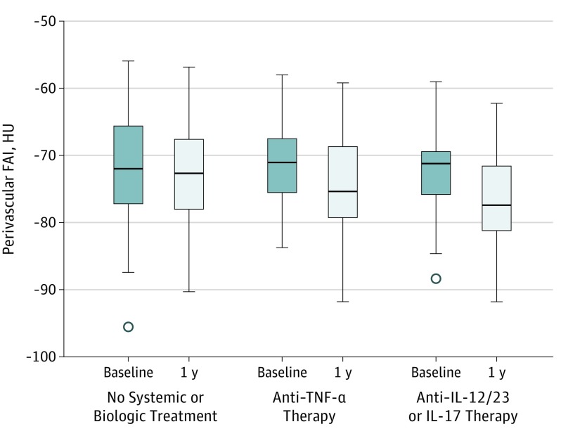
Of the 134 patients (mean [SD] age, 51.1 [12.1] years; 84 [62.5%] male), most had low cardiovascular risk by traditional risk scores (median 10-year Framingham Risk Score, 3% [interquartile range, 1%-7%]) and moderate to severe skin disease. Of these patients, 82 received biologic psoriasis therapy (anti–tumor necrosis factor α, anti–interleukin [IL] 12/23, or anti–IL-17) for 1 year, and 52 did not receive any biologic therapy and were given topical or light therapy (control group). At baseline, 46 patients (27 in the treated group and 19 in the untreated group) had a focal coronary atherosclerotic plaque. Biologic therapy was associated with a significant decrease in FAI at 1 year (median FAI −71.22 HU [interquartile range (IQR), −75.85 to −68.11 HU] at baseline vs −76.09 HU [IQR, −80.08 to −70.37 HU] at 1 year; P < .001) concurrent with skin disease improvement (median PASI, 7.7 [IQR, 3.2-12.5] at baseline vs 3.2 [IQR, 1.8-5.7] at 1 year; P < .001), whereas no change in FAI was noted in those not receiving biologic therapy (median FAI, −71.98 [IQR, −77.36 to −65.64] at baseline vs −72.66 [IQR, −78.21 to −67.44] at 1 year; P = .39). The associations with FAI were independent of the presence of coronary plaque and were consistent among patients receiving different biologic agents, including anti–tumor necrosis factor α (median FAI, −71.25 [IQR, −75.86 to −66.89] at baseline vs −75.49 [IQR, −79.12 to −68.58] at 1 year; P < .001) and anti–IL-12/23 or anti–IL-17 therapy (median FAI, −71.18 [IQR, −75.85 to −68.80] at baseline vs −76.92 [IQR, −81.16 to −71.67] at 1 year; P < .001).
Conclusions and Relevance
In this study, biologic therapy for moderate to severe psoriasis was associated with reduced coronary inflammation assessed by perivascular FAI. This finding suggests that perivascular FAI measured by CCTA may be used to track response to interventions for coronary artery disease.
Introduction
Chronic inflammatory diseases, including psoriasis, are associated with increased cardiovascular (CV) risk, which may be reduced when treating the underlying psoriasis.1,2,3,4 This finding has opened interest to whether treating areas of low-grade inflammation in the body may be associated with downstream CV risk. A study2 of patients with moderate to severe psoriasis recently found that treatment of psoriasis with biologic therapy, including anti–tumor necrosis factor α (TNF-α), anti–interleukin (IL) 17, or anti–IL-12/23 therapy, may modulate coronary plaque compared with no biologic therapy.
The perivascular fat attenuation index (FAI) is a computed tomography (CT)–based, novel, noninvasive imaging technique that allows for direct visualization and quantification of coronary inflammation using differential mapping of attenuation gradients in pericoronary fat.5,6 Coronary inflammation may provide clues about the risk of developing future atherosclerosis and the stability of existing atherosclerotic plaques; abnormal perivascular FAI was recently found to be associated with a 6- to 9-fold increased risk of major adverse cardiac events.6 Inhibition of perivascular adipose tissue (PVAT) adipogenesis by local proinflammatory cytokines released from within the diseased coronary arteries results in lipolysis, inhibits adipogenesis, and triggers edema within PVAT,5 thus leading to a higher water-lipid ratio closer to the inflamed vascular wall compared with non-PVAT that is distant from the vascular wall.7 Coronary computed tomography angiography (CCTA) allows 3-dimensional assessment of these gradients of PVAT composition, captured by mapping the weighted attenuation gradients within the perivascular space.5
Given that biologic therapy has been found to be associated with reduced noncalcified coronary plaque burden in patients with psoriasis,2 we hypothesized that treatment with biologic psoriasis therapy would also be associated with a reduction in coronary inflammation assessed as perivascular FAI after 1 year of treatment. We also hypothesized that the association would be seen even in the absence of coronary atherosclerotic plaque if these treatments are to be associated with a reduced risk of developing coronary atherosclerosis prospectively.
Methods
Study Design and Population
In this prospective cohort study, 310 participants were recruited from an ongoing cohort study to assess the association between psoriasis and cardiometabolic disease under the Psoriasis, Atherosclerosis and Cardiometabolic Disease Initiative from January 1, 2013, through March 31, 2019, with 257 completing 1 year of follow-up. Study protocols were approved by the institutional review board at the National Institutes of Health, and all participants provided written informed consent. All data were deidentified. The study was performed in accordance with the Declaration of Helsinki.8 The Strengthening the Reporting of Observational Studies in Epidemiology Guidelines (STROBE) were followed for reporting the findings.
We analyzed patients with moderate to severe psoriasis who were not undergoing biologic treatment for at least 3 months before starting biologic therapy at baseline for a 1-year interval. Participants who elected to not receive biologic therapy during this 1-year interval were used as a control group and were treated with topical and/or light therapies only. Treatment was initiated by a referring dermatologist (J.A.R., B.L.) based on the physician’s recommendation. Our protocol stipulates that patients with severe psoriasis who have not started biologic therapy are invited to join our 4-year cohort study. All patients included in this study qualified for biologic therapy at baseline until and unless their disease was resistant to biologic therapy, they had contraindications, or they declined this treatment. Of 310 participants, 134 consecutive patients with psoriasis were included in our analyses based on the inclusion criteria and in whom FAI has been analyzed to date (eFigure in the Supplement).
CCTA Acquisition
All patients underwent CCTA at baseline and at 1 year on the same day as blood sample obtainment using the same CT scanner (320-detector row Aquilion ONE ViSION; Toshiba) following Society of Cardiovascular Computed Tomography acquisition guidelines.9 Guidelines implemented by the National Institutes of Health Radiation Exposure Committee were followed. Scanning was performed with prospective electrocardiographic gating, with a 100- or 120-kV tube potential, tube current of 100 to 850 mA adjusted to the patient’s body size, and a gantry rotation time of 275 milliseconds. Images were acquired at a section thickness of 0.5 mm with a section increment of 0.25 mm.4
Coronary Artery Perivascular FAI Analysis
Perivascular FAI was measured around the proximal right coronary artery and defined as the weighted mean attenuation of all adipose tissue–containing voxels (−190 to −30 Hounsfield units [HU]) located within a radial distance from the outer vessel wall equal to the diameter of the respective vessel. Analyses were performed using the CaRi-HEART algorithm (Caristo Diagnostics) adjusted for scan-specific settings, as previously described and validated.5,6 To avoid the effects of the aortic wall, we excluded the most proximal 10 mm of the right coronary artery and analyzed the proximal 10 to 50 mm of the vessel, as described previously.5,6 The selection of the right coronary artery segment was based on a prior histologic and gene expression study5 that found an association of the perivascular attenuation gradients around this standardized segment with subclinical atherosclerosis and coronary inflammation. In stable patients, perivascular FAI around the right coronary artery is associated with the perivascular FAI around the left anterior descending artery but offers a more reliable metric of coronary inflammation owing to the absence of major branches and abundance of perivascular fat in the right atrioventricular groove.5,6 All scans were anonymized, and the reader was blinded to demographics, treatment, and time of the scan. Intraobserver and interobserver agreement for the perivascular FAI were very good (intraclass correlation coefficients, 0.987 [P < .001] for intraobserver agreement and 0.980 [P < .001] for interobserver agreement).
Statistical Analysis
Skewness and kurtosis measures were considered to assess normality. Data were reported as mean (SD) for parametric variables, median with interquartile range (IQR) for nonparametric variables, and percentages for categorical variables. Parametric variables were compared between 2 groups using the t test, whereas the paired t test was used for longitudinal analyses. Nonparametric variables were compared using the Wilcoxon signed rank test for paired and Mann-Whitney test for independent groups, whereas the Pearson χ2 test was performed for categorical variables between 2 groups. To examine the interaction between the treatment group and change in perivascular FAI during 1 year, we also performed a 2-factor mixed analysis of variance with a treatment × time point interaction (normality of the dependent variable was first confirmed using a Shapiro-Wilk test). Post hoc analyses for between–time point comparisons in each treatment group were also performed and adjusted for multiple comparisons using the Bonferroni method. To assess the robustness of our observations given the nonrandomized nature of our study, we repeated the analysis after propensity score matching of patients allocated to treatment with vs without biologics based on relevant clinical variables (age, sex, race/ethnicity, body mass index, hypertension, hypercholesterolemia, diabetes mellitus, smoking, 10-year Framingham Risk Score, total cholesterol level, high-density lipoprotein level, systolic and diastolic blood pressure, antihyperlipidemic therapy, Psoriasis Area and Severity Index [PASI] severity score [which combines the severity of lesions and the area affected into a single score, considering erythema, induration, and desquamation within each lesion], and the time since psoriasis diagnosis) and a tolerance level of 0.1. A 2-tailed P < .05 was considered to be statistically significant. Statistical analysis was performed using Stata, version 12 (StataCorp), SPSS Statistics for Windows, version 22.0 (IBM Corp), and GraphPad Prism 7.0 (GraphPad Software Inc).
Results
Baseline Characteristics of Study Groups
The 134 patients with psoriasis were middle-aged (mean [SD] age, 51.1 [12.1] years), predominantly male ([62.5%]), at low CV risk by traditional risk scores (median 10-year Framingham Risk Score, 3% [IQR, 1%-7%]), and had moderate to severe skin disease. Study participants had low CV risk by traditional risk scores, with moderate to severe skin disease involvement at baseline (Table 1). Of the 134 patients, 82 received biologic (anti–TNF-α, anti–IL-12/23, or anti–IL-17) psoriasis therapy (treatment group) at 1 year, whereas 52 did not receive any systemic or biologic therapy (control group). No significant differences were found in demographic characteristics, baseline medication use, or laboratory values between the 2 groups. Baseline coronary inflammation as assessed by perivascular FAI was similar between groups (Table 1). In addition, 46 patients (27 in the treated group and 19 in the untreated group) had a coronary atherosclerotic plaque at baseline.
Table 1. Baseline Characteristics of Patients With Psoriasisa.
| Characteristic | Patients Treated With Systemic or Biologic Therapy (n = 82) | Patients Not Treated With Systemic or Biologic Therapy (n = 52) | P Valueb |
|---|---|---|---|
| Age, mean (SD), y | 49.7 (12.3) | 51.4 (12.4) | .41 |
| Male | 48 (58.5) | 36 (69.2) | .85 |
| BMI, mean (SD) | 29.7 (6.4) | 28.5 (5.3) | .19 |
| Hypertension | 24 (29.3) | 14 (26.9) | .84 |
| Hyperlipidemia | 35 (42.7) | 26 (50.0) | .35 |
| Statin treatment | 23 (28.0) | 17 (32.7) | .52 |
| Type 2 diabetes | 9 (11.0) | 5 (9.6) | .92 |
| Current smoker | 10 (12.2) | 4 (7.7) | .15 |
| Cholesterol level, mean (SD), mg/dL | |||
| Total | 180.8 (35.9) | 184.0 (39.5) | .25 |
| HDL-C | 56.2 (16.4) | 55.9 (15.8) | .31 |
| LDL-C | 102.7 (28.9) | 106.0 (30.9) | .17 |
| Framingham risk score, median (IQR) | 3 (1 to 6) | 3 (1 to 7) | .41 |
| C-reactive protein level, median (IQR), mg/L | 2.2 (0.8 to 5.5) | 2.0 (0.6 to 3.5) | .68 |
| HOMA-IR, median (IQR) | 3.0 (1.8 to 5.4) | 2.2 (1.5 to 4.7) | .43 |
| PASI score, median (IQR) | 7.7 (3.2 to 12.5) | 6.0 (3.7 to 10.0) | .19 |
| Disease duration, mean (SD), y | 22.8 (14.1) | 21.0 (14.1) | .44 |
| Topical therapy | 49 (59.8) | 36 (69.2) | .52 |
| Light therapy | 14 (17.1) | 7 (13.5) | .23 |
| Perivascular FAI, median (IQR), HU | −71.22 (−75.85 to −68.11) | −71.98 (−77.36 to −65.64) | .70 |
Abbreviations: BMI, body mass index (calculated as weight in kilograms divided by square of height in meters); FAI, fat attenuation index; HDL-C, high-density lipoprotein cholesterol; HOMA-IR, homeostasis model assessment of insulin resistance; HU, Hounsfield units; IQR, interquartile range; LDL-C, low-density lipoprotein cholesterol; PASI, Psoriasis Area and Severity Index.
SI conversion factors: To convert C-reactive protein to nanomoles per liter, multiply by 9.524; cholesterol to millimoles per liter, multiply by 0.0259.
Data are reported as number (percentage) of patients unless otherwise indicated.
Two-tailed P < .05 deemed to be statistically significant.
Modulation of Coronary Inflammation After Treatment
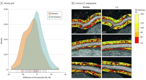
At 1-year follow-up (Table 2), no significant change was found in body mass index, lipids, or glucose. A significant PASI score improvement between baseline and 1 year was seen in the treatment group (58% improvement, P < .001), whereas a nonsignificant reduction in PASI score was seen in the control group (median score, 6.0 [IQR, 3.7-10.0] vs 4.6 [IQR, 2.1-6.9]; P = .10). Moreover, a reduction in high-sensitivity C-reactive protein level was seen only in the treatment group (median, 2.2 mg/L [IQR, 0.8-5.5 mg/L] vs 1.3 mg/L [IQR, 0.7-3.7 mg/L]; P = .03). In the treatment group, a significant decrease was found in coronary inflammation as assessed by perivascular FAI after 1 year of therapy (median, −71.22 HU [IQR, −75.85 to −68.11 HU] vs −76.09 HU [IQR, −80.08 to −70.37 HU]; P < .001), whereas no significant change in coronary inflammation was noted in the control group (median, –71.98 HU [IQR, –77.36 to –65.64 HU] vs –72.66 HU [IQR, –78.21 to –67.44 HU]; P = .39) (Table 2 and Figure 1). This association of treatment with a decrease in FAI was consistent among different biologic agents, including anti–TNF-α, anti–IL-12/23, and anti–IL-17 (Figure 1). The median change in perivascular FAI in the treated group was –3.73 HU (IQR, –8.72 to –0.25 HU) (Figure 2A). The range of observed responses is presented in Figure 2B, which shows perivascular FAI heatmaps for patients who had a great response, moderate response, and modest response to biologic therapy.
Table 2. One-Year Follow-up Characteristics and Treatment Effects in Patients With Psoriasis .
| Characteristic | Patients Treated With Systemic or Biologic Therapy (n = 82) | Patients Not Treated With Systemic or Biologic Therapy (n = 52) | ||||
|---|---|---|---|---|---|---|
| Baseline | 1 y | P Value | Baseline | 1 y | P Value | |
| Cholesterol level, mean (SD), mg/dL | ||||||
| Total | 180.8 (35.9) | 184. 9 (42.4) | .18 | 184.0 (39.5) | 184.5 (44.2) | .47 |
| HDL-C | 56.2 (16.4) | 57.6 (22.0) | .49 | 55.9 (15.8) | 57.9 (19.3) | .13 |
| LDL-C | 102.7 (28.9) | 103.2 (34.3) | .60 | 106.0 (30.9) | 103.8 (38.8) | .42 |
| Framingham risk score, median (IQR) | 3 (1 to 6) | 2 (1 to 5) | .82 | 3 (1 to 7) | 4 (1 to 6) | .56 |
| C-reactive protein level, median (IQR), mg/L | 2.2 (0.8 to 5.5) | 1.3 (0.7 to 3.7) | .03 | 2.0 (0.6 to 3.5) | 1.5 (0.6 to 3.2) | .12 |
| HOMA-IR, median (IQR) | 3.0 (1.8 to 5.4) | 3.7 (2.0 to 5.7) | .31 | 2.2 (1.5 to 4.7) | 2.6 (1.3 to 5.2) | .23 |
| PASI score, median (IQR) | 7.7 (3.2 to 12.5) | 3.2 (1.8 to 5.7) | <.001 | 6.0 (3.7 to 10.0) | 4.6 (2.1 to 6.9) | .10 |
| Topical therapy | 49 (59.8) | 36 (43.9) | .03 | 36 (69.2) | 37 (71.2) | .78 |
| Light therapy | 14 (17.1) | 9 (11.0) | .13 | 7 (13.5) | 8 (15.4) | .66 |
| Perivascular FAI, median (IQR), HU | −71.22 (−75.85 to −68.11) | −76.09 (−80.08 to −70.37) | <.001 | −71.98 (−77.36 to −65.64) | −72.66 (−78.21 to −67.44) | .39 |
Abbreviations: FAI, fat attenuation index; HDL-C, high-density lipoprotein cholesterol; HOMA-IR, homeostasis model assessment of insulin resistance; HU, Hounsfield units; IQR, interquartile range; LDL-C, low-density lipoprotein cholesterol; PASI, Psoriasis Area and Severity Index.
SI conversion factors: To convert C-reactive protein to nanomoles per liter, multiply by 9.524; cholesterol to millimoles per liter, multiply by 0.0259.
Data are reported as number (percentage) of patients unless otherwise indicated.
Figure 1. Perivascular Fat Attenuation Index (FAI) After 1 Year of Treatment.
Box-and-whisker Tukey plots depicting change in perivascular FAI in patients not receiving any systemic or biologic therapy during 1 year or receiving biologic therapy (P = .99) during 1 year with anti–tumor necrosis factor α (TNF-α) therapy (P < .001) or anti–interleukin (IL) 12/23 and anti–IL-17 therapies (P < .001) (P = .004 for interaction). HU indicates Hounsfield units.
Figure 2. Response to Biologic Therapy During 1 Year in Patients With Psoriasis.
A, Density plot depicting the changes in perivascular fat attenuation index (FAI) in patients with treated and untreated psoriasis during 1 year (FAI at follow-up minus FAI at baseline) (P = .001). B, Coronary computed tomography (CT) angiography images of the coronary arteries depicting the perivascular FAI before and after biologic therapy at 1 year of follow-up for patients with excellent response (top), moderate response (middle), and modest response (bottom) to biologic therapy. HU indicates Hounsfield units.
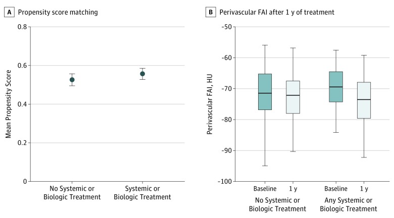
To further assess the robustness of our observations for confounders and variables that may have affected the allocation of patients to treatment with vs without biologics, we applied propensity score matching. After the identification of matched groups of patients treated with vs without systemic or biologic therapy (45 patients in each group) (Figure 3A), we replicated our previous observation that biologic therapy but not topical therapy was associated with a significant reduction in perivascular FAI.
Figure 3. Propensity Score Matching.
A, Propensity score matching for the use of biologics that included several clinical variables (given in the Statistical Analysis subsection of the Methods section) (P = .31). Dots indicate means, and the error bars indicate the SE of the observations. B, Box-and-whisker Tukey plots depicting change in perivascular fat attenuation index (FAI) in patients not receiving any systemic or biologic therapy (P = .95) and matched patients receiving biologic therapy (P < .001) during 1 year (45 in each group) (P = .003 for interaction). HU indicates Hounsfield units.
Finally, we performed a subgroup analysis to investigate the change in FAI in patients with and without coronary plaque-based treatment during 1 year. Perivascular FAI was significantly attenuated with biologic treatment during 1 year in patients with and without coronary artery disease (eTable in the Supplement).
Discussion
In this study, we found that biologic therapy in patients with moderate to severe treatment-naive psoriasis at baseline was associated with a reduction in coronary inflammation at 1 year. Those not treated with biologic therapy but with solely topical and/or light therapies did not have a change in coronary inflammation. In addition, when stratified by the presence of coronary plaque, the observed association was independent of the presence of coronary atherosclerosis. In this small group of patients, subgroup analyses among different commonly used biologic agents for psoriasis, including anti–TNF-α and anti–IL-12/23 or anti–IL-17 therapy, revealed a similar association with each biologic therapy.
Atherosclerosis is an immune-mediated phenomenon with inflammation that accounts for 20% to 30% of residual risk for CV events,10,11,12 which is not captured by traditional risk factors.10 In addition, those with chronic inflammatory diseases have a disproportional rate of CV events evidenced by increased subclinical aortic vascular inflammation, coronary artery calcium, and coronary plaque burden compared with age- and sex-matched counterparts.4,10,13,14 A recent study2 found that elevated coronary plaque burden in this population may be mitigated by use of biologic therapies; however, whether this was a direct effect of quelling coronary inflammation was not investigated.
Perivascular FAI is a novel, validated technique that allows direct detection of coronary inflammation on CCTA.5 Perivascular FAI was associated with all-cause mortality, cardiac mortality, and nonfatal acute myocardial infarction events in a large population study, independently of traditional risk factors, the extent of coronary atherosclerosis, and the presence of high-risk plaque features.6 Perivascular FAI can also be used to reliably separate unstable from stable lesions in patients with acute coronary syndromes and allows the detection of residual inflammatory risk not captured by current traditional CV risk factors.6,15
After the reporting of the prognostic value of perivascular FAI mapping for adverse cardiac events,6 one of the main questions remains whether perivascular FAI can be modified using anti-inflammatory interventions. Although this was a small study and needs to be confirmed with larger randomized clinical trials, to our knowledge, it is the first to assess the association of biologic therapy with coronary inflammation in patients with psoriasis as assessed by FAI. Our finding that initiation of biologic psoriasis therapy was associated with improvement in coronary inflammation suggests that these therapies may have a direct association with coronary vasculature and its microenvironment, with the potential to prevent the development of atherosclerotic plaques. We believe that the strength of perivascular FAI in risk stratifying patients with increased coronary inflammation will allow for better identification of patients at increased risk of future myocardial events that are not captured by traditional CV risk factors. The utility of this modality is that it is noninvasive, relatively cost-effective, and reliable in early studies to potentially identify patients at greater risk of future CV events and evaluate whether pharmacologic interventions, particularly anti-inflammatory therapies, may improve coronary inflammation. Thus, assessment of coronary inflammation through the perivascular FAI may serve to guide therapy intensity to reduce CV risk in patients.
Limitations
This was an observational study and thus subject to error introduced by treatment selection bias and unmeasured confounding. For example, a previous observational study10 found improvements in aortic vascular inflammation with TNF inhibitors, but no change was observed in adequately powered randomized clinical trials.16 Therefore, these findings should be confirmed in prospective randomized clinical trials. However, we tried to minimize the bias inherent to all observational studies by adding a propensity score matching analysis, which we believe strengthens our findings. Biologic therapy was administered in an open-label, nonrandomized fashion in a small sample size during 1 year. Also, we did not study hard CV end points to suggest that responsiveness of perivascular FAI to biologic treatment is associated with reduced CV risk in psoriasis. Our protocol stipulates that patients are followed up for 4 years; after enrollment, we plan to perform plaque-specific analyses to further understand the association between FAI and coronary plaque to examine whether frank coronary plaque will develop where there is no plaque but abnormal FAI and to elucidate possible causal mechanisms for these findings. Future studies will be needed to explore whether the residual CV risk detected by perivascular FAI can be attenuated using targeted anti-inflammatory interventions.
Conclusions
The findings suggest that biologic therapy in psoriasis is associated with a decrease in coronary inflammation as assessed by perivascular FAI. This observation warrants further follow-up to understand the potential association of these treatments with future CV events. Our study was the first, to our knowledge, to introduce FAI as a biomarker of coronary inflammation that enables tracking of response to anti-inflammatory treatments in the human coronaries.
eFigure. Recruitment Scheme
eTable. Change in Perivascular FAI (HU) in the Presence or Absence of Coronary Plaque
References
- 1.Wu JJ, Poon K-YT, Channual JC, Shen AY-J. Association between tumor necrosis factor inhibitor therapy and myocardial infarction risk in patients with psoriasis. Arch Dermatol. 2012;148(11):1244-1250. doi: 10.1001/archdermatol.2012.2502 [DOI] [PubMed] [Google Scholar]
- 2.Elnabawi YA, Dey AK, Goyal A, et al. Coronary artery plaque characteristics and treatment with biologic therapy in severe psoriasis: results from a prospective observational study. Cardiovasc Res. 2019;115(4):721-728. doi: 10.1093/cvr/cvz009 [DOI] [PMC free article] [PubMed] [Google Scholar]
- 3.Gelfand JM, Neimann AL, Shin DB, Wang X, Margolis DJ, Troxel AB. Risk of myocardial infarction in patients with psoriasis. JAMA. 2006;296(14):1735-1741. doi: 10.1001/jama.296.14.1735 [DOI] [PubMed] [Google Scholar]
- 4.Lerman JB, Joshi AA, Chaturvedi A, et al. Coronary plaque characterization in psoriasis reveals high-risk features that improve after treatment in a prospective observational study. Circulation. 2017;136(3):263-276. doi: 10.1161/CIRCULATIONAHA.116.026859 [DOI] [PMC free article] [PubMed] [Google Scholar]
- 5.Antonopoulos AS, Sanna F, Sabharwal N, et al. Detecting human coronary inflammation by imaging perivascular fat. Sci Transl Med. 2017;9(398):eaal2658. doi: 10.1126/scitranslmed.aal2658 [DOI] [PubMed] [Google Scholar]
- 6.Oikonomou EK, Marwan M, Desai MY, et al. Non-invasive detection of coronary inflammation using computed tomography and prediction of residual cardiovascular risk (the CRISP CT study): a post-hoc analysis of prospective outcome data. Lancet. 2018;392(10151):929-939. doi: 10.1016/S0140-6736(18)31114-0 [DOI] [PMC free article] [PubMed] [Google Scholar]
- 7.Oikonomou EK, Antoniades C. The role of adipose tissue in cardiovascular health and disease. Nat Rev Cardiol. 2019;16(2):83-99. doi: 10.1038/s41569-018-0097-6 [DOI] [PubMed] [Google Scholar]
- 8.World Medical Association World Medical Association Declaration of Helsinki: ethical principles for medical research involving human subjects. JAMA. 2013;310(20):2191-2194. doi: 10.1001/jama.2013.281053 [DOI] [PubMed] [Google Scholar]
- 9.Abbara S, Blanke P, Maroules CD, et al. SCCT guidelines for the performance and acquisition of coronary computed tomographic angiography: a report of the society of Cardiovascular Computed Tomography Guidelines Committee: endorsed by the North American Society for Cardiovascular Imaging (NASCI). J Cardiovasc Comput Tomogr. 2016;10(6):435-449. doi: 10.1016/j.jcct.2016.10.002 [DOI] [PubMed] [Google Scholar]
- 10.Dey AK, Joshi AA, Chaturvedi A, et al. Association between skin and aortic vascular inflammation in patients with psoriasis: a case-cohort study using positron emission tomography/computed tomography. JAMA Cardiol. 2017;2(9):1013-1018. doi: 10.1001/jamacardio.2017.1213 [DOI] [PMC free article] [PubMed] [Google Scholar]
- 11.Ridker PM, Everett BM, Thuren T, et al. ; CANTOS Trial Group . Antiinflammatory therapy with canakinumab for atherosclerotic disease. N Engl J Med. 2017;377(12):1119-1131. doi: 10.1056/NEJMoa1707914 [DOI] [PubMed] [Google Scholar]
- 12.Ridker PM. Residual inflammatory risk: addressing the obverse side of the atherosclerosis prevention coin. Eur Heart J. 2016;37(22):1720-1722. doi: 10.1093/eurheartj/ehw024 [DOI] [PubMed] [Google Scholar]
- 13.Naik HB, Natarajan B, Stansky E, et al. Severity of psoriasis associates with aortic vascular inflammation detected by FDG PET/CT and neutrophil activation in a prospective observational study. Arterioscler Thromb Vasc Biol. 2015;35(12):2667-2676. doi: 10.1161/ATVBAHA.115.306460 [DOI] [PMC free article] [PubMed] [Google Scholar]
- 14.Mansouri B, Kivelevitch D, Natarajan B, et al. Comparison of coronary artery calcium scores between patients with psoriasis and type 2 diabetes. JAMA Dermatol. 2016;152(11):1244-1253. doi: 10.1001/jamadermatol.2016.2907 [DOI] [PubMed] [Google Scholar]
- 15.Goeller M, Achenbach S, Cadet S, et al. Pericoronary adipose tissue computed tomography attenuation and high-risk plaque characteristics in acute coronary syndrome compared with stable coronary artery disease. JAMA Cardiol. 2018;3(9):858-863. doi: 10.1001/jamacardio.2018.1997 [DOI] [PMC free article] [PubMed] [Google Scholar]
- 16.Mehta NN, Shin DB, Joshi AA, et al. Effect of 2 psoriasis treatments on vascular inflammation and novel inflammatory cardiovascular biomarkers: a randomized placebo-controlled trial. Circ Cardiovasc Imaging. 2018;11(6):e007394. doi: 10.1161/CIRCIMAGING.117.007394 [DOI] [PMC free article] [PubMed] [Google Scholar]
Associated Data
This section collects any data citations, data availability statements, or supplementary materials included in this article.
Supplementary Materials
eFigure. Recruitment Scheme
eTable. Change in Perivascular FAI (HU) in the Presence or Absence of Coronary Plaque


