Abstract
Long-term changes in glutamate transmission in the nucleus accumbens core (NAcore) contribute to the reinstatement of drug seeking after extinction of cocaine self-administration. Whether similar adaptations in glutamate transmission occur during heroin and cue-induced reinstatement of heroin seeking is unknown. After 2 weeks of heroin self-administration and 2 weeks of subsequent extinction training, heroin seeking was induced by a noncontingent injection of heroin or by presentation of light/tone cues previously paired with heroin infusions. Microdialysis was conducted in the NAcore during reinstatement of heroin seeking in animals extinguished from heroin self-administration or in subjects receiving parallel (yoked) noncontingent saline or heroin. Reinstatement by either heroin or cue increased extracellular glutamate in the NAcore in the self-administration group, but no increase was elicited during heroin-induced reinstatement in the yoked control groups. The increase in glutamate during heroin-induced drug seeking was abolished by inhibiting synaptic transmission in the NAcore with tetrodotoxin or by inhibiting glutamatergic afferents to the NAcore from the prelimbic cortex. Supporting critical involvement of glutamate release, heroin seeking induced by cue or heroin was blocked by inhibiting AMPA/kainate glutamate receptors in the NAcore. Interestingly, although a heroin-priming injection increased dopamine equally in animals trained to self-administer heroin and in yoked-saline subjects, inhibition of dopamine receptors in the NAcore also blocked heroin- and cue-induced drug seeking. Together, these findings show that recruitment of the glutamatergic projection from the prelimbic cortex to NAcore is necessary to initiate the reinstatement of heroin seeking.
Keywords: self-administration, dialysis, dopamine, reinstatement, addiction, prelimbic
Introduction
Despite the debilitating personal and societal impact of heroin addiction, the mechanisms underlying relapse to heroin use are not clear (Kreek et al., 2005). Recent findings using the reinstatement animal model of relapse indicate that components of the brain circuitry involved in cocaine seeking, such as the prelimbic cortex and the nucleus accumbens core (NAcore), are also necessary for heroin seeking (Rogers et al., 2008). However, other components of heroin- and cocaine-seeking circuitry are distinct, such as involvement of the amygdala, accumbens shell, and infralimbic cortex in heroin- but not cocaine-induced drug seeking (McFarland and Kalivas, 2001; Rogers et al., 2008). It is thought that a key feature of the circuitry underlying cocaine seeking is enhanced glutamate release into the NAcore by afferents from prelimbic cortex. Thus, not only does inactivation of either the NAcore or prelimbic cortex prevent cue-, stress-, or cocaine-induced reinstatement of cocaine seeking (McFarland and Kalivas, 2001; McLaughlin and See, 2003; Di Ciano and Everitt, 2004; Fuchs et al., 2004; Di Ciano et al., 2008), but both cocaine- and stress-induced cocaine seeking are associated with a remarkable overflow of synaptic glutamate into the NAcore from prelimbic afferents (McFarland et al., 2003, 2004; Xi et al., 2006; Miguéns et al., 2008). Also, both cocaine- and cue-induced cocaine seeking are blocked by inhibiting AMPA-type glutamate receptors in the NAcore (Cornish and Kalivas, 2000; Di Ciano and Everitt, 2001; Park et al., 2002).
Although heroin and cocaine share the prelimbic cortex and NAcore as part of the circuitry necessary to elicit drug seeking, the aforementioned distinctions in the required circuitry make it unclear whether increased glutamate release into the NAcore by prelimbic afferents is necessary for heroin seeking. Given the importance of activation of this glutamate projection in cocaine seeking, it is important to determine whether this mechanism may extend to heroin seeking as an antecedent for parallel drug development for heroin addiction (LaRowe et al., 2007). The present experiments used rats trained to self-administer heroin, followed by extinction training. Microdialysis was conducted in the NAcore during heroin seeking to assess whether glutamate levels in the NAc core increase during such seeking and whether the projection from the prelimbic cortex to the NAcore is responsible for the increase. Also, the role of neurotransmission in the NAcore was examined by microinjecting glutamate or dopamine receptor antagonists into the NAcore. These experiments confirmed a critical role for glutamate release in prelimbic afferents to the NAcore in the initiation of heroin seeking, posing neuroadaptations in this projection as a common mediator of both heroin and cocaine relapse.
Materials and Methods
Subjects.
Male Sprague Dawley rats (∼300 g at the time of surgery; n = 81; Charles River, Wilmington, MA) were used in this study. They were individually housed, maintained in a temperature-controlled environment (22°C) on a 12 h light/dark cycle (lights on at 7:00 A.M.) with food and water ad libitum, and given 6–7 d to acclimatize to the vivarium before undergoing surgery. Behavioral procedures began 5–6 d after surgery. All methods used were in compliance with National Institutes of Health guidelines for care of laboratory animals and were approved by the Medical University of South Carolina Institutional Animal Care and Use Committee.
Surgery.
The rats were anesthetized with ketamine HCl (87.5 mg/kg, i.m.) and xylazine (5 mg/kg, i.m.). Ketorolac (3 mg/kg, i.p.) was administered before surgery to provide analgesia. For catheter implantation, a guide cannula (22 ga; C313G; Plastics One, Wallingford, CT) was bent at a 90° angle and attached to SILASTIC tubing (0.025 inner diameter, 0.047 outer diameter; Bio-Sil; Bio-Rad, Hercules, CA) via superglue. It was then attached to Prolite mesh via dental cement. The catheter was implanted subcutaneously between the shoulder blades and exited the skin via a dermal biopsy hole (3 mm). The other end of the tube was threaded under the skin above the shoulders and into the jugular vein.
The rats were then placed in a small animal stereotaxic instrument (Kopf Instruments, Tujunga, CA). Three surgical screws were implanted into the skull as anchors, and depending on the experiment, guide cannulas were implanted and secured with dental cement. The nose bar was maintained at −3.5 mm relative to the interaural line. For the NAcore dialysis experiments, cannulas (20 ga; Plastics One) were implanted bilaterally aimed at the NAcore [anteroposterior (AP), +1.7 mm; mediolateral (ML), ±2.5 mm at 6° angle; dorsoventral (DV), −5.5 mm; all coordinates from Paxinos and Watson (2005)]. For the experiments with NAcore dialysis and prelimbic cortex microinjections, the AP coordinates for the cannulas aimed at the core were adjusted [AP, +1.3 mm (Paxinos and Watson, 2005)] to allow room for the prelimbic cannulas. A single double-barreled cannula (26 ga; Plastics One) was implanted, aimed at the prelimbic bilaterally [AP, +3.2 mm; ML, ±0.6 mm for each cannula; DV, −2.8 mm (Paxinos and Watson, 2005)]. For microinjection experiments, the guide cannulas were constructed of 23 ga stainless steel tubing cut to a length of 15.00 ± 0.02 mm and were implanted bilaterally aimed at the NAcore [AP, +1.7 mm; ML, ±1.6 mm; DV, −5.6 mm (Paxinos and Watson, 2005)]. After the surgery, the rats were retained in an incubation chamber until they recovered from the anesthesia. Obdurators were placed in all cannulas and maintained through the reinstatement testing. The rats were then returned to their home cages and checked on the days after surgery to ensure that their wounds healed. Rats were flushed with heparinized cefazolin via the catheter for 7 d after surgery and then heparin alone throughout the remaining self-administration.
Self-administration, extinction, and reinstatement procedures.
All self-administration experiments occurred in standard operant chambers with two retractable levers, a house light, and a cue light and tone generator (Med Associates, Fairfield, VT). Before drug self-administration, all rats were food deprived for 24 h and then underwent a single 15 h food-training session in which the rats were trained to press the active lever (the right lever) for a single food pellet (45 mg; Noyes, Lancaster, NH) on a fixed-ratio 1 (FR1) schedule. After that training, the rats were given a limited quantity of food (∼20 g) immediately after every self-administration session. One day after the food training, the rats began heroin self-administration, and each session lasted 3 h or until the rats had taken a maximum of 200 infusions. The self-administration program was an FR1 schedule with a 20 s timeout to prevent overdose, although the animal could still press the active lever during the timeout. All active lever presses, including those during the timeout, were recorded and are reported as “active lever presses.” Each active lever press, except for those during the timeout, produced a 0.05 ml infusion of either 50 μg (days 1 and 2) or 22.5 μg (days 3–12) of heroin (dissolved in 0.9% sterile saline; heroin kindly provided by National Institute on Drug Abuse). Concurrent with the drug infusion, a cue tone (2900 Hz) and cue light immediately above the active lever turned on for 5 s. Any rats that did not have 10+ infusions per day for the last 3 d of self-administration were excluded from analysis.
After 12 d of self-administration, rats began extinction training. Active lever presses produced no drug infusion or light/tone cues. For microinjection experiments, rats underwent a minimum of 12 d of extinction and were required to meet a criterion of an average of <30 lever presses for 2 consecutive days before undergoing reinstatement testing. For microdialysis experiments, rats underwent 12–13 d of extinction before beginning the dialysis reinstatement. For heroin prime-induced reinstatement, rats received a single injection of heroin (0.25 mg/kg, s.c.) before beginning the 3 h session, and the session program was identical to the extinction program. For cue-induced reinstatement, no injection was given, but the session program was identical to the self-administration session program (i.e., cue tone and light turned on with an active lever press). Rats did not receive intravenous drug infusions during either reinstatement.
During self-administration, some rats were yoked to heroin self-administering rats and received noncontingent infusions of heroin (yoked-heroin) or saline (yoked-saline) in the same temporal pattern as their heroin self-administering partners. Yoked rats received identical food training and the same number of self-administration and extinction sessions as their partners.
Microinjection procedures.
For the NAcore microinjections, injector needles were constructed of 30 ga dental needles connected to PE20 tubing, which was attached to 10 μl Hamilton syringes controlled by an infusion pump. The dental needles were bent to 17 mm so that they extended 2 mm beyond the end of the cannula and into the NAcore. All microinjections (0.3 μl) into the NAcore occurred over 60 s, and the injector needles were left in place for an additional 60 s to allow for diffusion from the site of the injection. After the microinjection, rats then underwent their appropriate reinstatement testing. For the drug prime reinstatement, the following drugs were used in the microinjections: CNQX (1 nmol per side), fluphenazine (Flu; 10 nmol per side), SCH 23390 (0.6 and 6 nmol per side), and sulpiride (0.3 and 3 nmol per side). For the cue-induced reinstatement, CNQX and Flu were used. Within each experiment, each rat received every dose of the drug or vehicle in a counterbalanced manner, with a minimum of 3 d of extinction and the same extinction criteria between each reinstatement trial. All drugs were dissolved in PBS, except SCH 23390, which was dissolved in sterile saline. Doses of all four drugs were chosen based on previous work indicating a lack of effect of the high dose of the drug on locomotor activity and/or reinstatement lever pressing (Neisewander et al., 1995; Cornish and Kalivas, 2000; McFarland and Kalivas, 2001; Anderson et al., 2003, 2006).
Microdialysis procedures.
Hollow fiber microdialysis tubing (molecular weight cutoff, 13000) was inserted and glued into a 24 ga internal cannula (Plastics One). The end of the tubing was sealed with epoxy glue and cut so that 2 mm of active membrane extended beyond the end of the cannula. The probes were then constructed with inlet and outlet tubing made of fused silica inserted and glued into the other end of the cannula. On the night before reinstatement testing, the probes were inserted into the NAcore, with the internal cannula extending 1 mm beyond the end of the guide cannula. The rats were left in the operant chambers overnight, with dialysis buffer (5 mm glucose, 2.5 mm KCl, 140 mm NaCl, 1.4 mm CaCl2, 1.2 mm MgCl2, and 0.15% PBS, pH 7.4) perfusing through the probe (0.1 μl/min). The levers were retracted throughout the night and were not extended until the reinstatement session began. The following morning, the flow rate was increased to 2 μl/min. After 2 h, baseline sample collection began at 20 min intervals. All samples were collected in 10 μl of 0.05N HCl. After 2 h of baseline collection, rats underwent reinstatement testing. For heroin-induced reinstatement, all rats were given a single systemic injection of heroin (0.25 mg/kg, s.c.). For the cue-primed reinstatement, no injection was made. Immediately before the heroin injection, some rats also received a microinjection (0.3 μl) of either baclofen–muscimol (B/M; 1 and 0.1 mm, respectively) or aCSF into the prelimbic cortex. This dose combination has been shown previously to be effective at inactivating the prelimbic cortex without affecting locomotor activity (McFarland and Kalivas, 2001). For the microinjections, double-barreled injectors (33 ga; Plastics One) were inserted into the guide cannula, extending 1 mm beyond the end of the guide cannula and into the prelimbic cortex. Injections were made over 60 s, and injector needles were left in place for an additional 60 s to allow for diffusion. Rats then received their heroin-prime injection and were returned to the operant chamber. The reinstatement session and sample collection lasted for 3 h. For some rats that had undergone heroin self-administration, during dialysis, tetrodotoxin (TTX; 1 μm) was continuously reverse dialyzed into the NAcore beginning 1 h before the heroin prime to determine whether the glutamate release was activity dependent. Samples were stored at −80°C. Three rats underwent dialysis during cue-primed reinstatement followed by dialysis (on the other side) during heroin-primed reinstatement, with 4 d of extinction between the two reinstatement sessions. Otherwise, no rats received both a cue-prime and heroin-prime reinstatement.
Quantification of dopamine and glutamate.
Before analysis, samples were thawed and divided so that they could be used to measure levels of glutamate and dopamine. Of the 50 μl in each sample, 24 μl were used for quantification of dopamine, and 20 μl were used for quantification of glutamate. (The n is lower for dopamine measurements because some samples had to be run twice for glutamate analysis because of problems encountered during the first run; e.g., see Fig. 1, compare A, B). Dopamine was quantified using an ESA (Chelmsford, MA) model 540 autosampler connected to an HPLC system with electrochemical detection (mobile phase: 150 NaH2PO4·H2O, 4.76 mm citric acid, 50 μm EDTA, 2.5 mm SDS, 10% methanol, and 17% acetonitrile, pH 5.6). Separation was achieved by pumping the samples through HR-80 reversed-phase column (3 μm × 80 mm × 3.2 mm; ESA), and then samples were reduced-oxidized using coulometric detection. Three electrodes were used: a guard cell (+400 mV), a reduction analytical electrode (−100 mV), and an oxidation analytical electrode (+220 mV). Peaks were recorded, and the area under the curve was measured by a computer running ESA501 Chromatography Data System. These values were normalized by comparison with an external standard curve for dopamine.
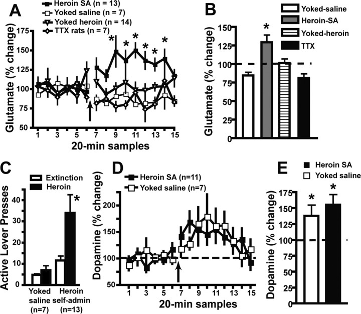
Figure 1.
Increased extracellular glutamate in the NAcore during heroin seeking (3 h reinstatement test). A, Extracellular glutamate before and after a noncontingent heroin injection (0.25 mg/kg, s.c.; arrow). The TTX (1 μm) group consisted of animals trained to self-administer heroin that had TTX administered through the dialysis probe beginning 1 h after the start of sample collection (sample 4). Data were evaluated using a two-way ANOVA with repeated measures over time, and there was a significant main effect of group (F(3,518) = 8.351; p < 0.001), no effect of time, and a significant interaction (F(42,518) = 2.128; p < 0.0001). Data are shown as mean ± SEM percentage change from the average of all six baseline samples. B, The data from A were averaged over the baseline and during the first hour after administration of heroin. Data were evaluated using a two-way ANOVA with repeated measures over time, and there was a significant main effect of group (F(3,38) = 7.318; p < 0.001), no effect of time, and a significant interaction (F(3,38) = 7.362; p < 0.001). The dashed line refers to the average 100% baseline for all groups. C, Active lever pressing during heroin seeking induced by the heroin injection in animals shown in A. Rats receiving TTX via reverse dialysis did not increase active lever pressing after a heroin prime (active lever presses for extinction, 30.6 ± 6.9; reinstatement, 49.6 ± 14.4). D, Extracellular dopamine was elevated in both heroin self-administration and yoked-saline groups by a heroin injection. These data were obtained from a portion of the samples measured for glutamate in A. A significant main effect of time (F(14,224) = 4.148; p < 0.0001) was measured, with no significant effect of group and no interaction. E, The data from D were averaged over the baseline and during the first hour after administration of heroin. Data were evaluated using a two-way ANOVA with repeated measures over time, and there was a significant main effect of time (F(1,16) = 14.12; p < 0.01), no effect of group, and no interaction. The dashed line refers to the average 100% baseline for all groups. *p < 0.05, post hoc comparison between heroin self-administration animals and all other groups at individual times after heroin administration for dialysis (A), comparison between group and baseline (B, E), or comparison between heroin-induced reinstatement and extinction levels of lever pressing (C). SA, Self-administration.
Glutamate was measured using an autosampler (Gilson Medical Electric, Middleton, WI) that performed a precolumn derivatization with ο-phthalaldehyde. The concentration of glutamate in samples was determined using HPLC with fluorometric detection (mobile phase: 0.1 m Na2HPO4, 0.09 mm EDTA-Na4, and 11% acetonitrile, pH 6.04). Amino acids were then separated with a Velosep RP-18 column (3 μm × 100 mm × 3.2 mm; Perkin-Elmer). Glutamate was detected using a fluorescence spectrophotometer (RF-10A Spectrofluorometric Detector; Shimadzu, Tokyo, Japan) using an excitation wavelength of 340 nm and an emission wavelength of 450 nm. A chart recorder recorded peaks, and peak heights were measured. The values were normalized by comparison with an external standard curve.
Histology and statistics.
Rats were overdosed with sodium pentobarbital (100 mg/kg, i.p.) and intracardially perfused with 0.9% saline. The brains were removed and stored in 10% formalin for at least 24 h. Coronal sections (75 μm thick) were made using a vibratome. Sections were mounted on gel-coated slides and then stained with cresyl violet. Sites of dialysis probes or injector needles were verified with a light microscope.
Dialysis data were analyzed using one-way or two-way repeated-measures ANOVA. Post hoc least significant difference tests were used to determine the source of significant differences between the yoked-saline, yoked-heroin, TTX, and heroin self-administration groups (p < 0.05). Lever presses during microinjection reinstatement tests were compared using Bonferroni's multiple-comparison post hoc tests, whereas those during dialysis-reinstatement tests were compared with extinction baseline using a nonparametric Mann–Whitney U test, because of the nonparametric distribution of the reinstatement data acquired during dialysis (p < 0.05).
Results
Baseline behavioral data
Table 1 outlines the mean ± SEM number of infusions of heroin administered and active lever presses averaged over the last 2 d of self-administration. Extinction and reinstatement active lever presses for individual experiments are illustrated in Figures 1–4, and inactive lever presses for individual experiments are listed in supplemental Table 1 (available at www.jneurosci.org as supplemental material).
Table 1.
Average infusions and active lever presses (which includes those presses during the 20 s timeout) across the last 2 d of self-administration (mean ± SEM)
| Experiment | Infusions | Active lever presses |
|---|---|---|
| Heroin dialysis | 26.3 ± 4.2 | 122.2 ± 40.9 |
| Heroin TTX dialysis | 16.7 ± 2.7 | 30.7 ± 4.1 |
| Yoked-heroin | 21.5 ± 4.0 | 7.7 ± 1.3 |
| Prelimbic infusions–dialysis | 47.8 ± 11.3 | 231.2 ± 82.8 |
| Cue–dialysis | 30.0 ± 5.6 | 83.7 ± 24.5 |
| CNQX | 48.9 ± 22.7 | 266.8 ± 194.0 |
| Fluphenazine | 21.0 ± 3.5 | 74.9 ± 29.8 |
| SCH | 28.6 ± 12.1 | 126.5 ± 83.5 |
| Sulpiride | 20.1 ± 3.2 | 55.4 ± 33.0 |
| Cue–fluphenazine + CNQX | 35.5 ± 6.8 | 102.8 ± 39.0 |
Figure 2.
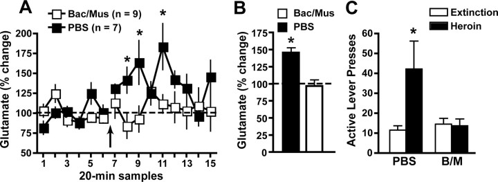
Inhibition of prelimbic cortex prevents the increase in glutamate release in the NAcore accompanying reinstatement (3 h test). A, Extracellular glutamate levels in the NAcore after prelimbic administration of B/M (1 and 0.1 mm, respectively) or PBS administered 5 min before a noncontingent heroin injection (0.25 mg/kg, s.c.; arrow). A two-way ANOVA revealed a trend toward a main effect of group (F(1,196) = 4.218; p < 0.06), a significant effect of time (F(14,196) = 2.448; p < 0.01), and a significant interaction (F(14,196) = 2.300; p < 0.01). Data are shown as mean ± SEM percentage change from the average of all six baseline samples. B, The data from A were averaged over the baseline and during the first hour after administration of heroin. Data were evaluated using a two-way ANOVA with repeated measures over time, and there was a significant main effect of group (F(1,14) = 16.11; p < 0.01), a significant effect of time (F(1,14) = 12.02; p < 0.01), and a significant interaction (F(1,14) = 15.64; p < 0.01). The dashed line refers to the baseline for all groups normalized to 100%. C, Active lever pressing during the reinstatement session shown in A. Data are shown as mean ± SEM active lever presses over 3 h. *p < 0.05, comparing PBS to B/M and, in B, between PBS and baseline. Bac/Mus, Baclofen/muscimol.
Figure 3.
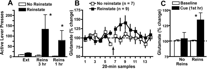
Extracellular glutamate in the NAcore is increased during cue-induced reinstatement (3 h test). A, Reinstatement of heroin seeking in animals during microdialysis shown in B. Animals were divided into two groups, those in which cues induced >15 lever presses over the 3 h reinstatement session (reinstate group), and those emitting <15 lever presses (no reinstate group). The third pair of bars indicates the number of active lever presses during the first hour of reinstatement. B, Extracellular glutamate in the NAcore before and during a cue-induced reinstatement session. C, For statistical comparison, the data from A were averaged over the baseline and during the first hour after beginning cue presentation. Thus, the bars in C indicate the means for the baseline and the first hour of the reinstatement session. A two-way ANOVA revealed that glutamate significantly increased above baseline in the reinstate group [time (F(1,14) = 5.174; p < 0.05), group (F(1,14) = 3.370; p < 0.09), interaction (F(1,14) = 3.631; p < 0.08)]. *p < 0.05 comparing first hour of glutamate release (C) or active lever presses (A) to baseline glutamate or extinction responding, respectively. Ext, Extinction; Reins, reinstatement.
Figure 4.
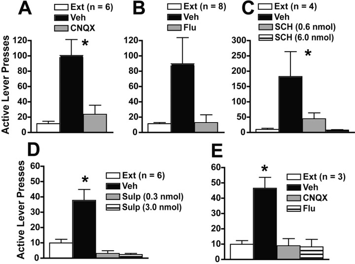
Blockade of AMPA/kainate or dopamine receptors in the NAcore prevents heroin seeking (3 h reinstatement test). A, Injection of the AMPA receptor antagonist CNQX (1 nmol/side) into the NAcore prevented heroin-induced reinstatement (F(2,10) = 9.795; p < 0.01). B, Injection of the general DA receptor antagonist Flu (10 nmol/side) reduced heroin-induced reinstatement (F(2,14) = 4.405; p < 0.05). C, Microinjection of the D1 receptor antagonist SCH 23390 (SCH; 6 nmol dose/side but not 0.6 nmol dose/side) prevented heroin-induced reinstatement (F(3,9) = 5.573; p < 0.05). D, Microinjection of the D2 receptor antagonist sulpiride (Sulp; 6 nmol and 0.6 nmol dose/side) prevented heroin-induced reinstatement (F(3,15) = 23.59; p < 0.0001). E, CNQX (1 nmol/side) or fluphenazine (10 nmol/side) into the NAcore prevents cue-induced reinstatement (F(3,6) = 10.49; p < 0.01). *p < 0.05 comparing vehicle treatment to extinction baseline and to drug treatments, except the low dose of SCH. Ext, Extinction; Veh, vehicle.
Heroin increases NAcore glutamate only in animals trained to self-administer heroin
Figure 1A shows that extracellular NAcore glutamate levels significantly increased in rats trained to self-administer heroin, and Figure 1B shows that this group of animals also demonstrated significant reinstatement of active lever pressing. In contrast, administration of a heroin-priming injection in the yoked-saline or -heroin groups did not elevate glutamate (Fig. 1A). Figure 1A also shows that perfusion of the NAcore with TTX via reverse dialysis prevented the increase in extracellular glutamate during the reinstatement of drug seeking in the heroin self-administration group, indicating that the increase relied on action potential-mediated neuronal depolarization. Reverse dialysis with TTX was begun at sample 4, and consistent with previous reports (Timmerman and Westerink, 1997; Baker et al., 2002), TTX did not affect the basal level of glutamate before injection of heroin in sample 7. The basal level of extracellular glutamate was equivalent in all four groups shown in Figure 1A (heroin self-administration, 43.9 ± 5.4 pmol/sample; yoked-saline, 45.3 ± 6.3; yoked-heroin, 40.3 ± 5.3; TTX in heroin self-administration, 57.1 ± 10.5). The mean of the first hour of glutamate levels after the heroin prime is shown in Figure 1B. Glutamate levels were significantly higher in the animals trained to self-administer heroin compared with the baseline and the other three groups. There was a trend toward a decrease in glutamate levels in the yoked-saline group compared with the baseline (p < 0.06), but no significant differences in glutamate levels compared with baseline for the other two groups.
Dopamine levels were measured in a portion of the glutamate samples obtained in the experiments shown in Figure 1A, and Figure 1D shows that in contrast to glutamate, acute heroin administration induced an equivalent increase in dopamine in both the heroin self-administration and yoked-saline groups. There was a trend toward a reduction in basal levels of dopamine in the heroin self-administration group (heroin self-administration, 6.9 ± 0.6 fmol/sample; yoked-saline, 12.1 ± 3.6; p < 0.1). Figure 1E shows the mean of the first hour of dopamine levels after the heroin prime.
The increase in glutamate during heroin seeking depends on afferents from prelimbic cortex
To determine the source of synaptic glutamate release in the NAcore during heroin seeking, rats previously trained to self-administer heroin were pretreated with either B/M or PBS vehicle in the prelimbic cortex before inducing heroin seeking with a noncontingent heroin injection. Figure 2A shows that extracellular glutamate was increased in the NAcore after vehicle injection into prelimbic cortex, and this increase was abolished by inhibiting neuronal activity in the prelimbic cortex with B/M. The mean of the first hour of glutamate levels after the heroin prime is shown in Figure 2B. Glutamate levels were significantly higher in the animals given PBS microinjections compared with rats with B/M microinjections and compared with baseline. Rats that received intraprelimbic vehicle showed significant reinstatement of heroin seeking, and akin to the rise in glutamate, heroin seeking was abolished by inhibiting prelimbic cortex (Fig. 2C). The basal level of extracellular glutamate was equivalent in both groups in Figure 2A (PBS, 39.5 ± 7.2 pmol/sample; B/M, 43.4 ± 6.2).
Cue-induced reinstatement of heroin seeking is associated with increased glutamate
Figure 3 shows that the reinstatement of heroin seeking by restoring heroin-associated conditioned cues (tone/light) to active lever pressing was associated with an increase in extracellular glutamate in the NAcore. Figure 3A shows that only 9 of the 15 animals examined reinstated >15 active lever presses over 3 h, and that in these 9 animals the lever pressing occurred predominantly in the first hour of the trial. This reinstatement criterion was used to divide the measurement of glutamate, because presentation of the cues during such reinstatement is contingent on lever pressing, and therefore any increase in glutamate caused by cue presentation should not occur in nonreinstating rats. Figure 3B illustrates the time course of glutamate change during heroin seeking and reveals that the majority of rise was during the first hour after beginning cue presentation. Figure 3C shows that the mean for this first hour of glutamate measurement was significantly greater than baseline in animals reinstating to heroin seeking, whereas there was no increase in animals not demonstrating reinstatement in response to cues. The basal level of extracellular glutamate was equivalent in both groups in Figure 3B (reinstating rats, 57.3 ± 9.8 pmol/sample; nonreinstating, 81.5 ± 22.7).
AMPA/kainate glutamate or dopamine receptor blockade in the NAcore prevents heroin seeking
Figures 4A–D show the active lever presses of rats that were microinjected into the NAcore with CNQX, fluphenazine, SCH 23390, or sulpiride before a noncontingent heroin injection to reinstate drug seeking. All drug treatments significantly reduced heroin seeking compared with intra-NAcore vehicle microinjection (F scores from one-way repeated-measures ANOVA shown in figure legend). Post hoc analysis revealed that in all instances, except in Figure 4B, heroin significantly increased active lever pressing after intra-NAcore pretreatment with vehicle. Figure 4E shows that, akin to heroin-induced reinstatement in Figures 4A–D, heroin seeking induced by the tone/light cue was blocked by either CNQX or fluphenazine.
Histology
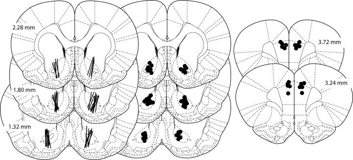
Figure 5 illustrates the location of dialysis probes and tips of the microinjection cannula for animals used in this study. Figure 5A shows a diagram of 20 randomly selected dialysis-probe tracks through the NAcore. Figure 5B shows the tip of the needle tracks in the NAcore from the microinjection experiments. Figure 5C shows the tip of the needle tracks in the prelimbic cortex.
Figure 5.
Diagrams showing 20 randomly selected dialysis tracks through the NAcore, the termination of needle tracks for microinjections into the NAcore, and the termination of needle tracks for microinjections into the prelimbic cortex, respectively (left to right). The images are adapted from Paxinos and Watson (2005), and AP coordinates (in mm) are given relative to bregma.
Discussion
The present findings indicate that the glutamatergic projection from the prelimbic cortex to the NAcore is critically involved in heroin-induced reinstatement to heroin seeking. Similarly, cue-induced heroin seeking is also associated with increased extracellular glutamate in the NAcore. In rats that were trained to self-administer heroin followed by extinction training, an acute heroin injection that reinstated drug seeking increased glutamate levels in the NAcore, but not in yoked-heroin or yoked-saline control rats. Inhibiting neuronal activity in the prelimbic cortex by microinjecting B/M prevented the increase in NAcore glutamate, implicating the prelimbic projection to the NAcore as the primary source of increased glutamate during heroin seeking. Further supporting synaptic origin, the heroin-induced increase in NAcore glutamate was absent when voltage-dependent sodium channels were blocked by TTX. The importance of increased glutamate transmission for the reinstatement of heroin seeking is further buttressed by the fact that blocking AMPA/kainate receptors in the NAcore prevented heroin- or cue-induced heroin seeking.
The role of glutamate transmission in drug seeking
It has long been postulated that glutamate transmission, especially at AMPA receptors, is necessary for the expression of behaviors associated with repeated administration of addictive drugs, such as behavioral sensitization (Pierce and Kalivas, 1997; Wolf, 1998). More recently, a number of studies have linked the glutamatergic projection from the prefrontal cortex to the NAcore with the expression of cocaine-seeking behavior, using dialysis and pharmacological approaches akin to those in the present study (Cornish and Kalivas, 2000; Di Ciano and Everitt, 2001; Park et al., 2002; McFarland et al., 2003, 2004; Xi et al., 2006; Miguéns et al., 2008). The fact that activation of the prelimbic to NAcore glutamate projection is also critical for heroin seeking supports a contention that activation of this projection may be an essential mechanism mediating drug seeking by many classes of addictive drugs. Although this possibility needs to be directly verified in animal models of drug seeking for each class of addictive drug, neuroimaging of addicts dependent on cocaine, amphetamines, heroin, alcohol, and nicotine reveals strong metabolic activation of the anterior cingulate cortex and nucleus accumbens in response to cues that the addict associates with drug taking (George et al., 2001; Goldstein and Volkow, 2002; Daglish et al., 2003; Franklin et al., 2007). Importantly, the anterior cingulate in humans is homologous to the prelimbic cortex in rats in terms of afferent and efferent projections (Ongur and Price, 2000), and not only has activation of the anterior cingulate been positively correlated with drug craving or desire, but this region of cortex is engaged during motivated behavior in general (Bush et al., 2002; Shidara and Richmond, 2002).
In contrast to withdrawal from cocaine (Pierce et al., 1996; Baker et al., 2003), the basal levels of extracellular glutamate were not significantly altered after either self-administered or yoked-heroin administration. The reduction in basal glutamate after chronic cocaine results from downregulation of the cystine–glutamate exchanger (Baker et al., 2003). In this regard, it is interesting that the activator of cystine–glutamate exchange, N-acetylcysteine, is effective at inhibiting the reinstatement of both cocaine and heroin seeking in the reinstatement animal model (Baker et al., 2003; Zhou and Kalivas, 2008). These data indicate that impairment of cystine–glutamate exchange may be occurring after heroin self-administration and not be detectable with microdialysis, or that rather than activation of cystine–glutamate exchange per se, it is the increase in glutathione that results from N-acetylcysteine-induced increase in cystine–glutamate exchange that is ameliorating both heroin and cocaine seeking (McBean, 2002). Alternatively, because the N-acetylcysteine is administered systemically (Baker et al., 2003; Zhou and Kalivas, 2008), it may inhibit reinstatement by acting in brain regions outside of the NAcore.
For the first time, the present findings demonstrate that reinstating drug seeking with cues previously associated with drug delivery increased extracellular glutamate in the NAcore. Consistent with increased excitatory drive into the NAcore during cue-induced drug seeking, the presentation of cocaine-associated cues after 1 month of abstinence increases NAcore neuronal activation (Hollander and Carelli, 2007). Moreover, intra-NAcore microinjections of the AMPA receptor antagonist CNQX or the dopamine receptor antagonist fluphenazine prevented cue-induced reinstatement. These results are consistent with previous findings showing that blockade of AMPA receptors prevents cue-induced reinstatement to cocaine seeking (Backstrom and Hyytia, 2007). In addition, activation of inhibitory mGluR2/3 autoreceptors in the NAcore or shell, which reduces glutamate release (Manzoni et al., 1997; Dietrich et al., 2002), prevents context-induced reinstatement to heroin seeking (Bossert et al., 2006).
Role of dopamine in the NAcore in heroin seeking
Consistent with previous reports, we found that that acute administration of heroin increases extracellular dopamine in the NAcore (Gerasimov et al., 1999; Sorge and Stewart, 2006). Moreover, it was found that activation of dopamine receptors was necessary for heroin seeking because blockade of either D1 or D2 receptors prevented the reinstatement. Similarly, D1 receptor blockade in the NAcore, but not NAshell, was recently shown to prevent cue-induced reinstatement of heroin seeking (Bossert et al., 2007). Although heroin alone increases NAcore dopamine levels, the increased NAcore glutamate levels found in reinstating rats may contribute to releasing dopamine, as activation of AMPA/kainate and metabotropic glutamate receptors modulates dopamine levels (Sakai et al., 1997). Regardless of the mechanism(s) regulating heroin-induced dopamine release in animals trained to self-administer heroin, the necessary role for dopamine receptors in heroin seeking is in contrast to previous studies with cocaine, in which blockade of dopamine receptors in the NAcore did not inhibit cocaine seeking (Cornish and Kalivas, 2000; McFarland and Kalivas, 2001; Anderson et al., 2003, 2006).
The reason for the distinct involvement of dopamine in the NAcore between heroin and cocaine seeking is unclear. However, there are a number of differences in the effect each class of drugs has on accumbens dopamine transmission. For example, the rise in dopamine produced by acute cocaine is much larger than heroin because the cocaine-induced increase arises from blocking dopamine transporters (Seiden et al., 1993), whereas heroin-induced dopamine relies largely on disinhibition of dopamine neurons in the ventral tegmental area (Kelley et al., 1980; Tanda et al., 1997). Also, in the limited access self-administration model, the rise in dopamine is usually augmented by repeated cocaine administration (Hooks et al., 1994; McFarland et al., 2003), whereas the current findings indicate that the increase in heroin-induced extracellular dopamine was equivalent in rats trained to self-administer heroin and yoked-saline subjects. Unfortunately, it is not clear how these distinctions result in dopamine in the NAcore being necessary for heroin- but not cocaine-induced drug seeking.
Conclusions
Heroin seeking in the reinstatement model depends on increased glutamate release in the NAcore from glutamatergic afferents arising in the prelimbic cortex. A parallel necessary involvement of this projection has also been shown for cocaine seeking. These data not only support the possibility that the glutamate projection from the prelimbic cortex to the NAcore is an important mediator of relapse for multiple chemical classes of addictive drugs, but also point to the possibility that drugs manipulating glutamate transmission may have utility in treating heroin addiction.
Footnotes
This work was supported by National Institutes of Health Grant USPHS 015369 (P.W.K.) and National Research Service Award USPHS DA 021460 (R.T.L.). We thank Jesse Smith, Jay Hutson, Kerranna Williamson, Kristin Trzcinski, Kyle Smith, Kate Niehoff, Hayden Alewine, Stephanie Kehoe, and Louisa Young for their excellent technical assistance.
References
- Anderson et al., 2003.Anderson SM, Bari AA, Pierce RC. Administration of the D1-like dopamine receptor antagonist SCH-23390 into the medial nucleus accumbens shell attenuates cocaine priming-induced reinstatement of drug-seeking behavior in rats. Psychopharmacology (Berl) 2003;168:132–138. doi: 10.1007/s00213-002-1298-5. [DOI] [PubMed] [Google Scholar]
- Anderson et al., 2006.Anderson SM, Schmidt HD, Pierce RC. Administration of the D2 dopamine receptor antagonist sulpiride into the shell, but not the core, of the nucleus accumbens attenuates cocaine priming-induced reinstatement of drug seeking. Neuropsychopharmacology. 2006;31:1452–1461. doi: 10.1038/sj.npp.1300922. [DOI] [PubMed] [Google Scholar]
- Backstrom and Hyytia, 2007.Backstrom P, Hyytia P. Involvement of AMPA/kainate, NMDA, and mGlu5 receptors in the nucleus accumbens core in cue-induced reinstatement of cocaine seeking in rats. Psychopharmacology (Berl) 2007;192:571–580. doi: 10.1007/s00213-007-0753-8. [DOI] [PubMed] [Google Scholar]
- Baker et al., 2002.Baker DA, Xi ZX, Shen H, Swanson CJ, Kalivas PW. The origin and neuronal function of in vivo nonsynaptic glutamate. J Neurosci. 2002;22:9134–9141. doi: 10.1523/JNEUROSCI.22-20-09134.2002. [DOI] [PMC free article] [PubMed] [Google Scholar]
- Baker et al., 2003.Baker DA, McFarland K, Lake RW, Shen H, Tang XC, Toda S, Kalivas PW. Neuroadaptations in cystine-glutamate exchange underlie cocaine relapse. Nat Neurosci. 2003;6:743–749. doi: 10.1038/nn1069. [DOI] [PubMed] [Google Scholar]
- Bossert et al., 2006.Bossert JM, Gray SM, Lu L, Shaham Y. Activation of group II metabotropic glutamate receptors in the nucleus accumbens shell attenuates context-induced relapse to heroin seeking. Neuropsychopharmacology. 2006;31:2197–2209. doi: 10.1038/sj.npp.1300977. [DOI] [PMC free article] [PubMed] [Google Scholar]
- Bossert et al., 2007.Bossert JM, Poles GC, Wihbey KA, Koya E, Shaham Y. Differential effects of blockade of dopamine D1-family receptors in nucleus accumbens core or shell on reinstatement of heroin seeking induced by contextual and discrete cues. J Neurosci. 2007;27:12655–12663. doi: 10.1523/JNEUROSCI.3926-07.2007. [DOI] [PMC free article] [PubMed] [Google Scholar]
- Bush et al., 2002.Bush G, Vogt BA, Holmes J, Dale AM, Greve D, Jenike MA, Rosen BR. Dorsal anterior cingulate cortex: a role in reward-based decision making. Proc Natl Acad Sci USA. 2002;99:523–528. doi: 10.1073/pnas.012470999. [DOI] [PMC free article] [PubMed] [Google Scholar]
- Cornish and Kalivas, 2000.Cornish JL, Kalivas PW. Glutamate transmission in the nucleus accumbens mediates relapse in cocaine addiction. J Neurosci. 2000;20(RC89):1–5. doi: 10.1523/JNEUROSCI.20-15-j0006.2000. [DOI] [PMC free article] [PubMed] [Google Scholar]
- Daglish et al., 2003.Daglish MR, Weinstein A, Malizia AL, Wilson S, Melichar JK, Lingford-Hughes A, Myles JS, Grasby P, Nutt DJ. Functional connectivity analysis of the neural circuits of opiate craving: “more” rather than “different”? NeuroImage. 2003;20:1964–1970. doi: 10.1016/j.neuroimage.2003.07.025. [DOI] [PubMed] [Google Scholar]
- Di Ciano and Everitt, 2001.Di Ciano P, Everitt BJ. Dissociable effects of antagonism of NMDA and AMPA/KA receptors in the nucleus accumbens core and shell on cocaine-seeking behavior. Neuropsychopharmacology. 2001;25:341–360. doi: 10.1016/S0893-133X(01)00235-4. [DOI] [PubMed] [Google Scholar]
- Di Ciano and Everitt, 2004.Di Ciano P, Everitt BJ. Direct interactions between the basolateral amygdala and nucleus accumbens core underlie cocaine-seeking behavior by rats. J Neurosci. 2004;24:7167–7173. doi: 10.1523/JNEUROSCI.1581-04.2004. [DOI] [PMC free article] [PubMed] [Google Scholar]
- Di Ciano et al., 2008.Di Ciano P, Robbins TW, Everitt BJ. Differential effects of nucleus accumbens core, shell, or dorsal striatal inactivations on the persistence, reacquisition, or reinstatement of responding for a drug-paired conditioned reinforcer. Neuropsychopharmacology. 2008 doi: 10.1038/sj.npp.1301522. in press. [DOI] [PubMed] [Google Scholar]
- Dietrich et al., 2002.Dietrich D, Kral T, Clusmann H, Friedl M, Schramm J. Presynaptic group II metabotropic glutamate receptors reduce stimulated and spontaneous transmitter release in human dentate gyrus. Neuropharmacology. 2002;42:297–305. doi: 10.1016/s0028-3908(01)00193-9. [DOI] [PubMed] [Google Scholar]
- Franklin et al., 2007.Franklin TR, Wang Z, Wang J, Sciortino N, Harper D, Li Y, Ehrman R, Kampman K, O'Brien C P, Detre JA, Childress AR. Limbic activation to cigarette smoking cues independent of nicotine withdrawal: a perfusion fMRI study. Neuropsychopharmacology. 2007;32:2301–2309. doi: 10.1038/sj.npp.1301371. [DOI] [PubMed] [Google Scholar]
- Fuchs et al., 2004.Fuchs RA, Evans KA, Parker MC, See RE. Differential involvement of the core and shell subregions of the nucleus accumbens in conditioned cue-induced reinstatement of cocaine seeking in rats. Psychopharmacology (Berl) 2004;176:459–465. doi: 10.1007/s00213-004-1895-6. [DOI] [PubMed] [Google Scholar]
- George et al., 2001.George MS, Anton RF, Bloomer C, Teneback C, Drobes DJ, Lorberbaum JP, Nahas Z, Vincent DJ. Activation of prefrontal cortex and anterior thalamus in alcoholic subjects on exposure to alcohol-specific cues. Arch Gen Psychiatry. 2001;58:345–352. doi: 10.1001/archpsyc.58.4.345. [DOI] [PubMed] [Google Scholar]
- Gerasimov et al., 1999.Gerasimov MR, Ashby CR, Jr, Gardner EL, Mills MJ, Brodie JD, Dewey SL. Gamma-vinyl GABA inhibits methamphetamine, heroin, or ethanol-induced increases in nucleus accumbens dopamine. Synapse. 1999;34:11–19. doi: 10.1002/(SICI)1098-2396(199910)34:1<11::AID-SYN2>3.0.CO;2-5. [DOI] [PubMed] [Google Scholar]
- Goldstein and Volkow, 2002.Goldstein RA, Volkow ND. Drug addiction and its underlying neurobiological basis: neuroimaging evidence for the involvement of the frontal cortex. Am J Psychiatry. 2002;159:1642–1652. doi: 10.1176/appi.ajp.159.10.1642. [DOI] [PMC free article] [PubMed] [Google Scholar]
- Hollander and Carelli, 2007.Hollander JA, Carelli RM. Cocaine-associated stimuli increase cocaine seeking and activate accumbens core neurons after abstinence. J Neurosci. 2007;27:3535–3539. doi: 10.1523/JNEUROSCI.3667-06.2007. [DOI] [PMC free article] [PubMed] [Google Scholar]
- Hooks et al., 1994.Hooks MS, Duffy P, Striplin C, Kalivas PW. Behavioral and neurochemical sensitization following cocaine self-administration. Psychopharmacology (Berl) 1994;115:265–272. doi: 10.1007/BF02244782. [DOI] [PubMed] [Google Scholar]
- Kelley et al., 1980.Kelley AE, Stinus L, Iversen SD. Interactions between D-ala-met-enkephalin, A10 dopaminergic neurones, and spontaneous behaviour in the rat. Behav Brain Res. 1980;1:3–24. doi: 10.1016/0166-4328(80)90043-1. [DOI] [PubMed] [Google Scholar]
- Kreek et al., 2005.Kreek MJ, Bart G, Lilly C, LaForge KS, Nielsen DA. Pharmacogenetics and human molecular genetics of opiate and cocaine addictions and their treatments. Pharmacol Rev. 2005;57:1–26. doi: 10.1124/pr.57.1.1. [DOI] [PubMed] [Google Scholar]
- LaRowe et al., 2007.LaRowe SD, Myrick H, Hedden S, Mardikian P, Saladin M, McRae A, Brady K, Kalivas PW, Malcolm R. Is cocaine desire reduced by N-acetylcysteine? Am J Psychiatry. 2007;164:1115–1117. doi: 10.1176/ajp.2007.164.7.1115. [DOI] [PubMed] [Google Scholar]
- Manzoni et al., 1997.Manzoni O, Michel JM, Bockaert J. Metabotropic glutamate receptors in the rat nucleus accumbens. Eur J Neurosci. 1997;9:1514–1523. doi: 10.1111/j.1460-9568.1997.tb01506.x. [DOI] [PubMed] [Google Scholar]
- McBean, 2002.McBean GJ. Cerebral cystine uptake: a tale of two transporters. Trends Pharmacol Sci. 2002;23:299–302. doi: 10.1016/s0165-6147(02)02060-6. [DOI] [PubMed] [Google Scholar]
- McFarland and Kalivas, 2001.McFarland K, Kalivas PW. The circuitry mediating cocaine-induced reinstatement of drug-seeking behavior. J Neurosci. 2001;21:8655–8663. doi: 10.1523/JNEUROSCI.21-21-08655.2001. [DOI] [PMC free article] [PubMed] [Google Scholar]
- McFarland et al., 2003.McFarland K, Lapish CC, Kalivas PW. Prefrontal glutamate release into the core of the nucleus accumbens mediates cocaine-induced reinstatement of drug-seeking behavior. J Neurosci. 2003;23:3531–3537. doi: 10.1523/JNEUROSCI.23-08-03531.2003. [DOI] [PMC free article] [PubMed] [Google Scholar]
- McFarland et al., 2004.McFarland K, Davidge SB, Lapish CC, Kalivas PW. Limbic and motor circuitry underlying footshock-induced reinstatement of cocaine-seeking behavior. J Neurosci. 2004;24:1551–1560. doi: 10.1523/JNEUROSCI.4177-03.2004. [DOI] [PMC free article] [PubMed] [Google Scholar]
- McLaughlin and See, 2003.McLaughlin J, See RE. Selective inactivation of the dorsomedial prefrontal cortex and the basolateral amygdala attenuates conditioned-cued reinstatement of extinguished cocaine-seeking behavior in rats. Psychopharmacology (Berl) 2003;168:57–65. doi: 10.1007/s00213-002-1196-x. [DOI] [PubMed] [Google Scholar]
- Miguéns et al., 2008.Miguéns M, Del Olmo N, Higuera-Matas A, Torres I, García-Lecumberri C, Ambrosio E. Glutamate and aspartate levels in the nucleus accumbens during cocaine self-administration and extinction: a time course microdialysis study. Psychopharmacology (Berl) 2008;196:303–313. doi: 10.1007/s00213-007-0958-x. [DOI] [PubMed] [Google Scholar]
- Neisewander et al., 1995.Neisewander JL, O'Dell LE, Redmond JC. Localization of dopamine receptor subtypes occupied by intra-accumbens antagonists that reverse cocaine-induced locomotion. Brain Res. 1995;671:201–212. doi: 10.1016/0006-8993(94)01317-b. [DOI] [PubMed] [Google Scholar]
- Ongur and Price, 2000.Ongur D, Price JL. The organization of networks within the orbital and medial prefrontal cortex of rats, monkeys and humans. Cereb Cortex. 2000;10:206–219. doi: 10.1093/cercor/10.3.206. [DOI] [PubMed] [Google Scholar]
- Park et al., 2002.Park WK, Bari AA, Jey AR, Anderson SM, Spealman RD, Rowlett JK, Pierce RC. Cocaine administered into the medial prefrontal cortex reinstates cocaine-seeking behavior by increasing AMPA receptor-mediated glutamate transmission in the nucleus accumbens. J Neurosci. 2002;22:2916–2925. doi: 10.1523/JNEUROSCI.22-07-02916.2002. [DOI] [PMC free article] [PubMed] [Google Scholar]
- Paxinos and Watson, 2005.Paxinos G, Watson C. Amsterdam: Elsevier Academic; 2005. The rat brain in stereotaxic coordinates, Ed 5. [Google Scholar]
- Pierce and Kalivas, 1997.Pierce RC, Kalivas PW. A circuitry model of the expression of behavioral sensitization to amphetamine-like psychostimulants. Brain Res Brain Res Rev. 1997;25:192–216. doi: 10.1016/s0165-0173(97)00021-0. [DOI] [PubMed] [Google Scholar]
- Pierce et al., 1996.Pierce RC, Bell K, Duffy P, Kalivas PW. Repeated cocaine augments excitatory amino acid transmission in the nucleus accumbens only in rats having developed behavioral sensitization. J Neurosci. 1996;16:1550–1560. doi: 10.1523/JNEUROSCI.16-04-01550.1996. [DOI] [PMC free article] [PubMed] [Google Scholar]
- Rogers et al., 2008.Rogers JL, Ghee S, See RE. The neural circuitry underlying reinstatement of heroin-seeking behavior in an animal model of relapse. Neuroscience. 2008;151:579–588. doi: 10.1016/j.neuroscience.2007.10.012. [DOI] [PMC free article] [PubMed] [Google Scholar]
- Sakai et al., 1997.Sakai K, Akiyama K, Kashihara K, Tsuchida K, Ujike H, Kuroda S, Shohmori T. AMPA receptors modulate dopamine release in the striatum, as measured by brain microdialysis. Neurochem Int. 1997;30:329–336. doi: 10.1016/s0197-0186(96)00047-2. [DOI] [PubMed] [Google Scholar]
- Seiden et al., 1993.Seiden LS, Sabol KE, Ricuarte GA. Amphetamine: effects on catecholamine systems and behavior. Annu Rev Pharmacol Toxicol. 1993;33:639–677. doi: 10.1146/annurev.pa.33.040193.003231. [DOI] [PubMed] [Google Scholar]
- Shidara and Richmond, 2002.Shidara M, Richmond BJ. Anterior cingulate: single neuronal signals related to degree of reward expectancy. Science. 2002;296:1709–1711. doi: 10.1126/science.1069504. [DOI] [PubMed] [Google Scholar]
- Sorge and Stewart, 2006.Sorge RE, Stewart J. The effects of long-term chronic buprenorphine treatment on the locomotor and nucleus accumbens dopamine response to acute heroin and cocaine in rats. Pharmacol Biochem Behav. 2006;84:300–305. doi: 10.1016/j.pbb.2006.05.013. [DOI] [PubMed] [Google Scholar]
- Tanda et al., 1997.Tanda G, Pontieri FE, Di Chiara G. Cannabinoid and heroin activation of mesolimbic dopamine transmission by a common mu1 opioid receptor mechanism. Science. 1997;276:2048–2050. doi: 10.1126/science.276.5321.2048. [DOI] [PubMed] [Google Scholar]
- Timmerman and Westerink, 1997.Timmerman W, Westerink BH. Brain microdialysis of GABA and glutamate: what does it signify? Synapse. 1997;27:242–261. doi: 10.1002/(SICI)1098-2396(199711)27:3<242::AID-SYN9>3.0.CO;2-D. [DOI] [PubMed] [Google Scholar]
- Wolf, 1998.Wolf ME. The role of excitatory amino acids in behavioral sensitization to psychomotor stimulants. Prog Neurobiol. 1998;54:679–720. doi: 10.1016/s0301-0082(97)00090-7. [DOI] [PubMed] [Google Scholar]
- Xi et al., 2006.Xi ZX, Gilbert JG, Peng XQ, Pak AC, Li X, Gardner EL. Cannabinoid CB1 receptor antagonist AM251 inhibits cocaine-primed relapse in rats: role of glutamate in the nucleus accumbens. J Neurosci. 2006;26:8531–8536. doi: 10.1523/JNEUROSCI.0726-06.2006. [DOI] [PMC free article] [PubMed] [Google Scholar]
- Zhou and Kalivas, 2008.Zhou W, Kalivas PW. N-acetylcysteine reduces extinction responding and induces enduring reductions in cue- and heroin-induced drug-seeking. Biol Psychiatry. 2008;63:338–340. doi: 10.1016/j.biopsych.2007.06.008. [DOI] [PMC free article] [PubMed] [Google Scholar]


