Abstract
Buckling surgery is one of the common procedures performed by the retinal surgeons for visual rehabilitation at the earliest in cases of retinal detachment. The optimal surgical skill in this section can only be achieved with repeated practices and clinical experiences. Here, we describe an easy and inexpensive way to perform, practice, and refine surgical skills by demonstrating this complicated surgery in a simple manner on goat's eyes. The advantages of this technique are real-tissue handling experiences and repeatability of the procedure with almost similar practical implications. Thus, whenever feasible, every attempt should be made to refine the residents or budding ophthalmologists surgical skills by undertaking this technique in their routine curriculum.
Keywords: Buckling surgery, goat's eye, resident's surgical training, simple technique
Skill enhancement of residents during the residency training program is of paramount importance. Among the surgeries performed by a retinal surgeon, buckling surgery for retinal detachment is one of the basic and important surgeries to be learnt.[1] In ophthalmology, experimental models are available for surgical skill enhancement for cataract and corneal surgeries, whereas such a model is yet not available for buckling surgery. In this article, the authors elaborate a technique of buckling surgery on goat's eyes.
Technique
In the national surgical skill development laboratory at our center, cataract surgeries on goat's eyes are routinely performed by the residents to enhance their surgical skills. Similarly, corneal and scleral suturing techniques and other corneal surgeries are also daily practised. Goat's eyes are obtained from a local slaughterhouse. The obtained goat's eyes are carefully inspected for intactness and regularity of the ocular surface. Grossly deformed globes with lack of tissue rigidity are usually discarded. For this particular procedure, intact globes with identifiable extraocular muscles were selected for surgical training. The extraocular muscles were identified and sutured onto a defined anatomical position from the limbus. In case of missing extraocular muscles, muscles from other eyes were removed and sutured on to a single globe. Following this, under the supervision of a senior resident (retina speciality), the junior residents performed the surgery in a stepwise manner.
The muscles were secured at two points, first along a defined insertion that is around 5 mm from the limbus and second one along the posterior pole (15 mm from the limbus) that is around the optic nerve in a curvilinear fashion so as to get a space for manipulation under the muscle. After this, the globe was mounted on a mannequin head for surgical manipulation.
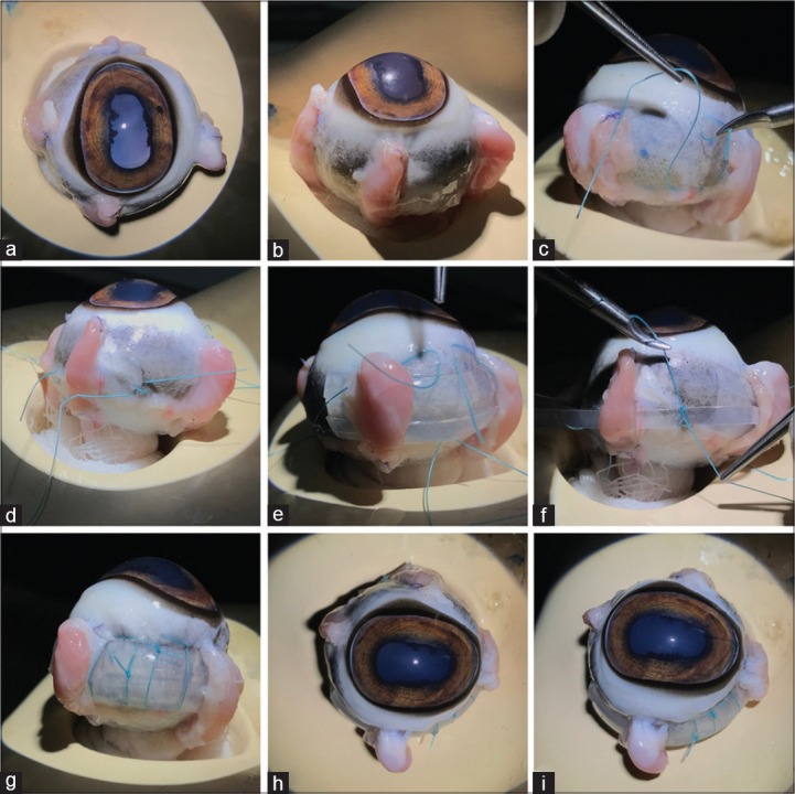
Under the surgical microscope, along the inferotemporal quadrant, marks were made at 14 mm from the limbus using gentian violet. Buckle sutures were placed at two sites along this quadrant and in rest of the quadrants, single suture for encircle was placed. Following this, buckle was passed along the inferotemporal quadrant; similarly, encirclage was passed along the 360° and buckle sutures were tied. The desired titration was achieved by manipulating the encirclage and final tie placed along the superotemporal quadrant [Fig. 1].
Figure 1.

(a) Globe mounted on the mannequin head. (b) All four rectus muscles were attached to the desired position, so as to achieve a normal anatomical orientation of rectus muscles. (c) Buckle sutures were passed along the inferior and temporal quadrant. (d) Encirclage sutures were placed in the rest of the quadrants. (e) Buckle is passed along the inferior and temporal quadrant. (f) Encirclage is passed and the sutures are tied along the respective quadrants. (g) Buckle sutures are tied and a final knot is placed. (h and i) End surgery view with perfectly placed buckle and encirclage
Discussion
Regarding training of retinal surgeries to residents, there are few laboratory surgical models regarding to teach basic vitrectomy.[1,2] However, no experimental laboratory model is available to train the residents in the field of buckling surgeries. In a report by Sagong et al., it was concluded that at least 30 buckling surgeries needed to be performed to achieve stable clinical results by a retina expert.[3] Therefore, enhancing basic buckling procedure knowledge and necessary surgical skills cannot be underestimated.
Here in this report, we would like to highlight a simple technique of performing buckling surgery on goat's eye. The goat eyes very much resemble like that of human eyes in terms of their scleral thickness and rigidity. Observations involving the anterior segment surgery training have highlighted the results after training in a limited number of residents as improvement in surgical manoeuvring and surgical confidence using questionnaire; however, here we are intended to highlight a simple technique to improve the surgical skills. Because these surgeries can be repeated on these animal eyes over several times to gather the confidence and practical experiences. Likewise, suture passage onto the sclera, buckle insertion, and encirclage passage can be practised till the perfections are achieved. Limitations of this technique are excessive tissue space for manipulation, fixed eyes with limited globe movement, and variable landmarks because of a rectangular cornea. However, the benefits of this procedure definitely overweights these limitations. The exact relation of the retinal break to the buckle position and its height cannot be demonstrated by this technique. Another limitation is that drainage procedure cannot be demonstrated using this technique. However, enhancing the suturing techniques is important to prevent possible inadvertent perforation during buckling surgery. Perforation can be a devastating complication leading to subretinal bleed and irreversible loss of sight.
To conclude, buckling surgery on goat's eye for residents under training is a simple and inexpensive technique to acquire the basic surgical skills. At the same time, the advantages of this technique are experiences of actual tissue handling while manipulating for scleral suturing, band and buckle insertion, and muscle manipulation. Thus, whenever possible, residents can be trained through these modalities to transform their surgical skills at a faster pace.
Financial support and sponsorship
Nil.
Conflicts of interest
There are no conflicts of interest.
References
- 1.Shah VA, Reddy AK, Bonham AJ, Sabates NR, Lee AG. Resident surgical practice patterns for vitreoretinal surgery in ophthalmic training programs in the United States. Ophthalmology. 2009;116:783–9. doi: 10.1016/j.ophtha.2008.11.010. [DOI] [PubMed] [Google Scholar]
- 2.Lauer A, Chan-Kai, Lauer A. Basic training module for vitreoretinal surgery and the Casey Eye Institute Vitrectomy Indices Tool for Skills Assessment. Clin Ophthalmol. 2011;5:1249–56. doi: 10.2147/OPTH.S23772. [DOI] [PMC free article] [PubMed] [Google Scholar]
- 3.Sagong M, Chang W. Learning curve of the scleral buckling operation: Lessons from the first 97 cases. Ophthalmologica. 2010;224:22–9. doi: 10.1159/000233232. [DOI] [PubMed] [Google Scholar]


