Abstract
The performance of retinal microsurgery often requires the coordinated use of both hands. During bimanual retinal surgery, dominant hand performance may be negatively impacted by poor non-dominant hand assistance. Therefore understanding bimanual latent determinants, and establishing safety criteria for bimanual manipulation is relevant to robotic development and to eventual patient care. In this paper, we present a preliminary study to quantitatively evaluate one aspect of bimanual tool use in retinal surgery. Two force sensing tools were designed and fabricated using fiber Bragg grating sensors. Tool-to-sclera contact force is measured using the developed tools and analyzed. The tool forces were recorded during five basic surgical maneuvers typical of retinal surgery. Two subjects are involved in experiments, including one clinician and one engineer. For comparison, all manipulations were replicated under robot-assisted conditions. The results indicate that the average tool-to-sclera force recorded from the dominant hand tool is significantly higher than that from the non-dominant hand tool (p = 0.004). Moreover, the average forces under robot-assisted conditions with the present steady hand robot is notably higher than freehand conditions(p = 0.01). The forces obtained from the dominant and not-dominant hand instruments indicate a weak correlation.
Keywords: tool-to-sclera force, bimanual manipulation, robot-assisted retinal surgery
I. Introduction
Retinal surgery is one of the most challenging surgical tasks due to its high precision requirements for manual dexterity. Many retinal procedures benefit from bimanual techniques. Most retinal surgery requires, at a minimum, a light source in one hand and an active tool in the other. During retinal surgery, both tools are inserted through trocars (located in the sclera) to gain access to the interior of the eyeball. Trocar to sclera contact continuously transmits the forces from the tools to the sclera. The value of the tool-to-sclera force (scleral force) varies during the operation. The latent determinants of sclera force may derive from dominant hand (DH) manipulation, non-dominant hand (NDH) manipulation, or combination of forces from both hands. Excessive scleral forces may reflect dangerous or unskilled tool use. Therefore, bimanual tool manipulation under freehand and robot-assisted conditions, is tested in order to evaluate scleral forces, safety thresholds, and force parameters during typical manipulations in retinal surgery.
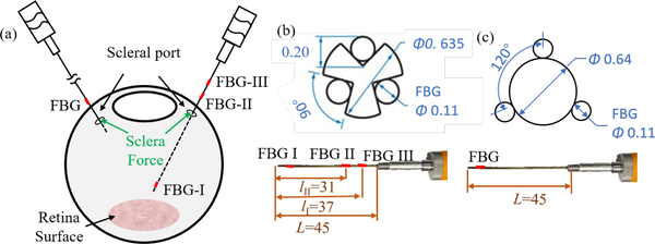
Smart or otherwise responsive instruments with force sensing capabilities are required to measure the scleral force. Therefore, our group has designed and developed a family of sensorized tools [1]–[4]. These tools are fabricated by integrating fiber Bragg grating (FBG) sensors into the contact segments of the tool shaft, thus it can independently sense forces applied in various tool shaft positions. Such multifunction tools [5] as shown in Fig.1 are capable of sensing scleral force and are further developed and applied in this paper. Similar instruments developed by other teams incorporate micro sensors in the tool handle [6], [7]. These prototypes are not, however, able to distinguish forces applied at the tool tip from the ones applied at the tool shaft, thus they are not suitable for measuring the scleral force separately.
Fig. 1.
Force sensing tools. (a) The layout of two tools inside eyeball. (b) The DH tool with its section view. (c) The NDH tool with its section view.
Robotic devices have the potential to significantly expand the capabilities of a surgeon [8], but this benefit has not been evaluated quantitatively. To better understand this potential we explore bimanual performance under robot-assisted conditions, i.e., the DH manipulates a robotic assistant to conduct surgical tasks while the NDH remains with freehand function. We did not apply the robot assistant on the NDH since the maneuver of NDH could be performed successfully by freehand due to its simple movements. In prior work we developed the steady hand eye robot (SHER) [9], [10], that is employed as the robotic assistant in this work.
In the present work, we explore the latent factors of bimanual manipulation in retinal procedures by quantitatively evaluating the forces generated by a user’s maneuvers and correlated with their concomitant performance. For the purpose of this work scleral force is considered the main outcome and primary variable to be analyzed. It is measured during five different basic retinal surgery operations performed by two subjects. Each experiment is carried out freehand, and is then repeated with the assistance of the SHER in the DH. The collected force data is analyzed independently, after which correlation analysis is performed between the scleral force exerted by the DH tool and the NDH tool.
II. Materials and Methods
Two scleral force sensing tools are designed and fabricated by the methods presented in our prior work [5]. The SHER is used as the robotic assistant for the DH. Five basic retinal surgical operations are designed to investigate the general surgical behavior.
A. Force Sensing Tool
The force sensing tool designed for the DH is shown in Fig.1 (b). It can measure the contact force between tool shaft and the sclera port. A 25-gauge stainless steel needle is machined with three grooves at an angle of 90°, and three FBGs sensors each with a diameter of 0.11 mm are glued into the grooves. The delicate FBG sensors are responsive to small strains and are able to detect the scleral forces exerted on the tool. The force sensing tool designed for the NDH to detect the contact force between tool tip and sclera port is shown in Fig. 1 (c). Three FBGs sensors are evenly glued on the outer surface of a 22-gauge tube. The tools are calibrated and validated using a precision scale with a resolution of 1 mg (Sartorius ED224S Extend Analytical Balance, Germany). The validation root mean square error for DH tool and NDH tool are 2.3 mN and 0.7 mN, respectively. During experiments, users are expected to keep the distal tip of the NDH tool around the scleral port to measure the valid force. However, the DH tool is inserted into the eyeball to conduct designed operations.
B. Basic Surgical Operations Design
To collect relevant scleral force data, we scripted five basic retinal surgery maneuvers that would be required to move the eye through a range of motion. The first three are to manipulate two tools to rotate the eyeball around each axis of the eyeball coordinate system, denoted as eyeball x-axis rotation (EXR), eyeball y-axis rotation (EYR), and eyeball zaxis rotation (EZR) shown respectively in Fig.2 (a). The fourth operation is to control the two tools to pivot around their own scleral ports while keeping the eyeball still as shown in Fig.2 (b), this is described as a tool scissor motion (TSM). During these four maneuvers, NDH tool and DH tool are performed synchronously to conduct the designated movements. The final set of maneuvers represents a combination of the aforementioned four operations, to follow the topography of the vessels (VF). VF is conducted in an eye phantom with mock vessels as shown in Fig.2 (c). During VF, the role of NDH tool is to hold the eyeball still, while the DH tool is used to follow the mock vessels. Firstly, the user is expected to move the DH tool tip above the home point, then move the DH tool to follow four simulated vessels consecutively, without touching the retina surface. Each vessel curve is tracked with one round trip. VF starts from the home position, pauses at the end of the vessel, and then returns to the home position.
Fig. 2.
The five basic operations. (a). Eyeball rotations around three axes of eyeball coordinate system. (b). Tool scissor motion. (c) Tool vessel following.
C. Experimental setup
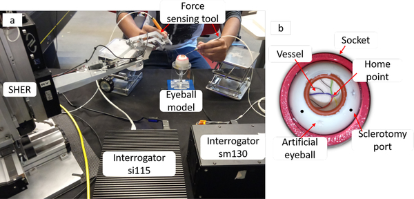
The experimental setup includes the SHER, two force sensing tools, a microscope, two FBG interrogators (SI115 and SM130, Micron Optics, Atlanta, GA), and an eye phantom, as shown Fig. 3(a). The force sensing tool is attached to the SHER with a quick release mechanism in robot-assisted experiments. The interrogators are used to monitor the FBG sensors within the spectrum from 1525 nm to 1565 nm at a 2 kHz refresh rate. The eye phantom is made of silicon rubber and placed into the 3D-printed socket. The socket is lubricated with mineral oil to produce a realistic friction coefficient analogous to the human eye. Painted lines simulate retinal vessels on the inner surface of eyeball as shown in Fig. 3 (b). Two subjects are involved in the data collection, including one engineer and one clinician. They repeated the aforementioned five maneuvers, forty times each. The first twenty trials are carried out freehand, and the last twenty are with robot-assisted.
Fig. 3.
Experimental setup of retinal surgery phantoms. (a) The force sensing tool and robotic assistant. (b) The mock eyeball model.
III. Results and Discussion
A quantitative evaluation of scleral force is performed first and this is followed by a correlation analysis between the two hand manipulations.
A. Scleral Force Analysis
The average value of scleral force is analyzed as show in Table I. The t-test and one-way analysis of variance with a value of p < 0.05 are used in the statistical analysis. The ratio value presented in the table corresponds to the ratio between the mean values of NDH tool and DH tool during the same experimental conditions. The averaged value in the table is calculated using the mean values of five operations from the same tool. The averaged scleral force of the DH hand tool is significantly higher than the value of the NDH hand tool (p = 0.004). This phenomenon is consistent for all of the experimental groups. A possible reason is that the DH tool usually leads the eyeball manipulation while the NDH tool follows it. Therefore overall, the NDH tool applied less force than the DH tool. Another notable result is that the averaged scleral force under robot-assisted conditions higher than the value under freehand conditions for all of the experimental groups (p = 0.01). This is likely the direct result of limitations on the surgeon’s dexterity, tool velocity and range of available motion resulting from limitations in present robot design. The robot’s mechanical stiffness also attenuates user tactile ability. The resultant is additional manipulation force applied to the tool handle. Moreover, the ratio decreases from the freehand condition to the robot-assisted condition when the subject is an non-clinician (such as an engineer), and the result converts to the contrary when the subject is a surgically trained clinician. The ratio value represents the utilization productivity of two hands. Therefore, it seems that the robot familiar engineer relies more heavily on the SHER, so he/she prefers to use DH hand to perform the operation. However, the clinician relies more on his/her hand, so he/she uses his/her NDH hand more often.
TABLE I.
SCLERAL FORCE ANALYSIS
| Maneuvers | Conditions | Engineer | Clinician | ||||||||
|---|---|---|---|---|---|---|---|---|---|---|---|
| NDH tool | DH tool | Ratio | NDH tool | DH tool | Ratio | ||||||
| Mean | Std | Mean | Std | Mean | Std | Mean | Std | ||||
| EXR | Freehand |
69.78 | 61.75 |
90.87 | 56.02 |
0.77 | 43.88 | 29.52 |
78.94 | 35.76 |
0.56 |
| Robot | 75.17 | 50.49 | 131.93 | 70.57 | 0.57 | 79.51 | 38.20 | 82.04 | 31.60 | 0.97 | |
| EYR | Freehand |
54.49 | 33.33 |
40.93 | 32.51 |
1.33 | 43.78 | 38.61 |
64.07 | 37.61 |
0.68 |
| Robot | 89.79 | 56.70 | 68.89 | 44.19 | 1.30 | 74.98 | 53.97 | 90.30 | 46.76 | 0.83 | |
| EZR | Freehand |
77.37 | 38.30 |
79.51 | 53.73 |
0.97 | 45.77 | 37.19 |
58.72 | 41.84 |
0.78 |
| Robot | 99.50 | 67.04 | 140.89 | 95.95 | 0.71 | 52.03 | 38.07 | 74.79 | 34.18 | 0.70 | |
| TSM | Freehand |
46.53 | 27.89 |
61.54 | 43.97 |
0.76 | 15.71 | 16.29 |
56.82 | 40.90 |
0.28 |
| Robot | 66.51 | 41.11 | 84.83 | 61.43 | 0.78 | 78.06 | 53.46 | 73.22 | 43.00 | 1.07 | |
| VF | Freehand |
38.99 | 28.87 |
64.03 | 54.54 |
0.61 | 17.70 | 11.48 |
87.08 | 59.33 |
0.20 |
| Robot | 47.46 | 28.68 | 95.08 | 79.52 | 0.50 | 37.56 | 33.11 | 69.77 | 38.50 | 0.54 | |
| Averaged value | Freehand |
57.43 | 38.03 |
67.38 | 48.15 |
0.85 | 33.37 | 26.62 |
69.13 | 43.09 |
0.48 |
| Robot | 75.69 | 48.80 | 104.32 | 70.33 | 0.73 | 64.43 | 43.36 | 78.02 | 38.81 | 0.83 | |
B. Correlations of Two Hand Manipulations
To explore the correlations of two hand manipulations, the Pearson correlation coefficient was calculated using equation (1) to compare the amount of sclera force applied between hands as shown in Table II.
| (1) |
where A and B denotes two dataset, i.e., sclera forces of two tools, μ is the mean value of the dataset, and σ is the standard deviation of the dataset.
TABLE II.
Correlations OF TWO HANDS MANIPULATIONS
| Subject | EXR | EYR | EZR | TSM | VF | |||||
|---|---|---|---|---|---|---|---|---|---|---|
| Freehand | Robot | Freehand | Robot | Freehand | Robot | Freehand | Robot | Freehand | Robot | |
| Clinician | 0.30 | 0.37 | 0.30 | 0.05 | 0.27 | 0.13 | 0.10 | 0.21 | 0.37 | 0.34 |
| Engineer | 0.23 | 0.39 | 0.21 | 0.32 | 0.33 | 0.32 | 0.24 | 0.34 | 0.47 | 0.33 |
The bidirectional limits of the correlation coefficients are −1 and 1 theoretically, and the closer to the boundary the coefficient is, the stronger linear correlations the two datasets have. The calculated correlation coefficients range from 0.05 to 0.47, therefore manipulations done with each hand are weakly correlated to each other. This result suggests that the two hands are sometimes working independently during these experiments.
IV. Conclusion
Although the results presented above are preliminary, they represent a first attempt to evaluate bimanual manipulation of tools during microsurgery. This was done by measuring the amount of scleral force applied during freehand and robot-assisted retinal surgery, as simulated by five basic maneuvers. The two force sensing tools with FBGs sensors independently measured the scleral force at the tool shaft. The SHER was used as a robotic assistant for the DH hand. Averaged scleral forces were analyzed first, and were followed by a correlation analysis. The results suggest that the two hands apply different manipulation forces and that they are weakly correlated. In the future more parameters will be considered and analyzed, the number of users will be increased as will the number of surgical skill levers of the users. Robotic assistants will be tried in both hands, to better explore and comprehend bimanual performance.
Acknowledgment
This work was supported by U.S. National Institutes of Health under grant 1R01EB023943–01 and 2R01EB000526–01. The work of C. He was supported in part by the China Scholarship Council under grant 201706020074. Research to Prevent Blindness, New York, New York, USA, and gifts by the J. Willard and Alice S. Marriott Foundation, the Gale Trust, Mr. Herb Ehlers, Mr. Bill Wilbur, Mr. and Mrs. Rajandre Shaw, Ms. Helen Nassif, Ms Mary Ellen Keck, Mr. Ronald Stiff, Donald and Maggie Feiner.
Contributor Information
Changyan He, Email: changyanhe@jhu.edu.
Iulian Iordachita, Email: iordachita@jhu.edu.
References
- [1].Balicki M, Han J-H, Iordachita I, Gehlbach P, Handa J, Taylor R, and Kang J, “Single fiber optical coherence tomography microsurgical instruments for computer and robot-assisted retinal surgery,” in International Conference on Medical Image Computing and Computer-Assisted Intervention Springer, 2009, pp. 108–115. [DOI] [PubMed] [Google Scholar]
- [2].Iordachita I, Sun Z, Balicki M, Kang JU, Phee SJ, Handa J, Gehlbach P, and Taylor R, “A sub-millimetric, 0.25 mn resolution fully integrated fiber-optic force-sensing tool for retinal microsurgery,” International journal of computer assisted radiology and surgery, vol. 4, no. 4, pp. 383–390, 2009. [DOI] [PMC free article] [PubMed] [Google Scholar]
- [3].He X, Handa J, Gehlbach P, Taylor R, and Iordachita I, “A submillimetric 3-dof force sensing instrument with integrated fiber bragg grating for retinal microsurgery,” IEEE Transactions on Biomedical Engineering, vol. 61, no. 2, pp. 522–534, 2014. [DOI] [PMC free article] [PubMed] [Google Scholar]
- [4].Gonenc B, Gehlbach P, Taylor RH, and Iordachita I, “Safe tissue manipulation in retinal microsurgery via motorized instruments with force sensing,” in SENSORS, 2017 IEEE. IEEE, 2017, pp. 1–3. [DOI] [PMC free article] [PubMed] [Google Scholar]
- [5].He X, Balicki M, Gehlbach P, Handa J, Taylor R, and Iordachita I, “A multi-function force sensing instrument for variable admittance robot control in retinal microsurgery,” in Robotics and Automation (ICRA), 2014 IEEE International Conference on IEEE, 2014, pp. 1411–1418. [DOI] [PMC free article] [PubMed] [Google Scholar]
- [6].Menciassi A, Eisinberg A, Scalari G, Anticoli C, Carrozza M, and Dario P, “Force feedback-based microinstrument for measuring tissue properties and pulse in microsurgery,” in Robotics and Automation, 2001. Proceedings 2001 ICRA. IEEE International Conference on, vol. 1 IEEE, 2001, pp. 626–631. [Google Scholar]
- [7].Seibold U, Kubler B, and Hirzinger G, “Prototype of instrument for minimally invasive surgery with 6-axis force sensing capability,” in Robotics and Automation, 2005. ICRA 2005. Proceedings of the 2005 IEEE International Conference on IEEE, 2005, pp. 496–501. [Google Scholar]
- [8].Cutler N, Balicki M, Finkelstein M, Wang J, Gehlbach P, Mc-Gready J, Iordachita I, Taylor R, and Handa JT, “Auditory force feedback substitution improves surgical precision during simulated ophthalmic surgery,” Investigative ophthalmology & visual science, vol. 54, no. 2, pp. 1316–1324, 2013. [DOI] [PMC free article] [PubMed] [Google Scholar]
- [9].Üneri A, Balicki MA, Handa J, Gehlbach P, Taylor RH, and Iordachita I, “New steady-hand eye robot with micro-force sensing for vitreoretinal surgery,” in Biomedical Robotics and Biomechatronics (BioRob), 2010 3rd IEEE RAS and EMBS International Conference on IEEE, 2010, pp. 814–819. [DOI] [PMC free article] [PubMed] [Google Scholar]
- [10].He X, Roppenecker D, Gierlach D, Balicki M, Olds K, Gehlbach P, Handa J, Taylor R, and Iordachita I, “Toward clinically applicable steady-hand eye robot for vitreoretinal surgery,” in ASME 2012 International Mechanical Engineering Congress and Exposition American Society of Mechanical Engineers, 2012, pp. 145–153. [Google Scholar]


