One of the strategies employed today to obtain new bioactive molecules with potential applications for human health (for example, antimicrobial or anticancer agents) is synthetic biology. Synthetic biology is used to biosynthesize new unnatural specialized metabolites or to force the expression of otherwise silent natural biosynthetic gene clusters. To assist the development of synthetic biology in the field of specialized metabolism, we constructed and are offering to the community a set of vectors that were intended to facilitate DNA assembly and integration in actinobacterial chromosomes. These vectors are compatible with various DNA cloning and assembling methods. They are standardized and modular, allowing the easy exchange of a module by another one of the same nature. Although designed for the assembly or the refactoring of specialized metabolite gene clusters, they have a broader potential utility, for example, for protein production or genetic complementation.
KEYWORDS: Streptomyces, synthetic biology
ABSTRACT
With the development of synthetic biology in the field of (actinobacterial) specialized metabolism, new tools are needed for the design or refactoring of biosynthetic gene clusters. If libraries of synthetic parts (such as promoters or ribosome binding sites) and DNA cloning methods have been developed, to our knowledge, not many vectors designed for the flexible cloning of biosynthetic gene clusters have been constructed. We report here the construction of a set of 12 standardized and modular vectors designed to afford the construction or the refactoring of biosynthetic gene clusters in Streptomyces species, using a large panel of cloning methods. Three different resistance cassettes and four orthogonal integration systems are proposed. In addition, FLP recombination target sites were incorporated to allow the recycling of antibiotic markers and to limit the risks of unwanted homologous recombination in Streptomyces strains when several vectors are used. The functionality and proper integration of the vectors in three commonly used Streptomyces strains, as well as the functionality of the Flp-catalyzed excision, were all confirmed. To illustrate some possible uses of our vectors, we refactored the albonoursin gene cluster from Streptomyces noursei using the BioBrick assembly method. We also used the seamless ligase chain reaction cloning method to assemble a transcription unit in one of the vectors and genetically complement a mutant strain.
IMPORTANCE One of the strategies employed today to obtain new bioactive molecules with potential applications for human health (for example, antimicrobial or anticancer agents) is synthetic biology. Synthetic biology is used to biosynthesize new unnatural specialized metabolites or to force the expression of otherwise silent natural biosynthetic gene clusters. To assist the development of synthetic biology in the field of specialized metabolism, we constructed and are offering to the community a set of vectors that were intended to facilitate DNA assembly and integration in actinobacterial chromosomes. These vectors are compatible with various DNA cloning and assembling methods. They are standardized and modular, allowing the easy exchange of a module by another one of the same nature. Although designed for the assembly or the refactoring of specialized metabolite gene clusters, they have a broader potential utility, for example, for protein production or genetic complementation.
INTRODUCTION
Synthetic biology is a domain of biotechnology that emerged at the beginning of the 21st century. It aims, for one part, at the rational engineering of biological systems to confer on them new functions. In the field of specialized metabolism, synthetic biology aims first at cloning and refactoring of silent (cryptic) biosynthetic gene clusters to afford the expression of genes and the production of metabolites that otherwise cannot be isolated and purified (1–3). Second, it is usually the method of choice for the synthesis of “unnatural natural products.” In this case, it consists either in the design and assembly of new biosynthetic gene clusters (4) or in the engineering of biosynthetic enzymes such as the modular nonribosomal peptide synthetases (NRPS) (5–7) and polyketide synthases (PKS) (8, 9). Such approaches are often referred to as combinatorial biosynthesis.
The development of synthetic biology in the field of specialized metabolism requires the development of dedicated tools and methods. In particular, it requires hosts (chassis) optimized for the production of specialized metabolites, libraries of synthetic DNA parts, such as promoters, ribosome binding sites (RBSs), or terminators, and vectors and DNA assembly methods for de novo assembly of gene clusters. Several Streptomyces strains, such as Streptomyces coelicolor (10), Streptomyces avermitilis (11), and Streptomyces albus (12, 13), have been optimized as chassis for the heterologous production of specialized metabolites. High-producing industrial strains have also been reported for the successful heterologous production of specialized metabolites (14). In parallel, efforts have been made to construct libraries of synthetic promoters (15–18) and of RBSs (15).
Many DNA assembly methods have been proposed and used so far for the assembly of DNA fragments, more specifically for the assembly of specialized metabolite biosynthetic gene clusters. These methods are mainly based on the existence of homology regions at the extremities of the fragments to be assembled, on the use of restriction enzymes, or on the use of site-specific recombinases. Examples of homology-based methods include the one-pot isothermal assembly (19), the ligase cycling reaction (LCR) (20), and direct pathway cloning (DiPaC) (3) for in vitro assembly and DNA assembler (21) based on transformation-associated recombination (TAR) in yeast or the linear plus circular homologous recombination (LCHR) method (used in the AGOS system [22]) for in vivo assembly. The first restriction enzyme-based DNA assembly method was the BioBrick assembly, based on the utilization of four restriction enzymes, two of which generate compatible cohesive ends (23). Other similar cloning methods based on the assembly of basic parts (promoter, coding sequence, terminator, etc.) into transcriptional units that can then be assembled together have since been developed (Golden Gate [24]; modular cloning, or MoClo [25]; and GoldenBraid 2.0 [26]). Finally, Olorunniji and colleagues recently established a DNA assembly method based on the use of site-specific integrases and orthogonal pairs of att sites (27).
While many DNA assembly methods have been developed, none is universal and adapted to all experimental situations. Indeed, some methods are more suitable to the assembly of (large) transcriptional units together (restriction enzyme-based methods, leaving a scar sequence but not requiring challenging PCRs of large and/or GC-rich fragments). Others are better suited to the assembly of the various elements of a transcriptional unit (homology-based methods allowing the precise positioning of the different elements without scar sequences). The size (from a few kilobases to more than 100 kb), the GC content, and the presence and number of regions presenting relatively high degrees of sequence similarities (in NRPS or PKS genes, for example) can vary a lot depending on the specialized metabolite gene cluster of interest. Thus, different experimental settings are likely to require different cloning approaches or even a combination of approaches. Therefore, the vectors used for cloning need to be flexible and adapted or easily adaptable to various assembly methods. It has been proposed that vectors built for synthetic biology should follow a standard and modular format (SEVA plasmids [28]), allowing a rapid and easy exchange of a module for another one. However, in the field of specialized metabolite synthetic biology, not many such vectors have been constructed. To our knowledge, one of the rare attempts was carried out by Phelan and colleagues (29) for the expression of genes in Streptomyces species. In their study, they describe the construction of 45 vectors based on three site-specific integration systems (φBT1, φC31, and VWB), four antibiotic resistance genes (apramycin, spectinomycin, and thiostrepton/ampicillin), and 14 promoters. These vectors were mainly designed for monocistronic gene expression, although the presence of several restriction sites could allow the assembly of a few gene cassettes.
In this study, we describe the construction of a set of 12 standardized and modular vectors designed to allow the assembly of biosynthetic gene clusters using various cloning methods in Streptomyces species, prolific producers of specialized metabolites. These vectors were designed on the model of the SEVA plasmids, although the exact architecture of these plasmids could not be used for our application. The 12 vectors were proven to be functional by the verified integration in the chromosome of three commonly used Streptomyces species. We also illustrate two possible uses of our vectors. We first refactored the albonoursin gene cluster using BioBrick assembly. Second, we genetically complemented our cgc22 mutant strain, CGCL030 (cgc22 is involved in congocidine biosynthesis [30]), by constructing a gene cassette constituted of a promoter, an RBS, cgc22, and a terminator using ligase chain reaction assembly.
RESULTS AND DISCUSSION
Design of the vectors.
The vectors were designed to meet the following specifications. It should be possible to use several vectors in the same strain (orthogonality), so different antibiotic resistance cassettes and different systems of integration at specific sites in the chromosome of Streptomyces should be used for the construction of the vectors. The vectors should be E. coli/Streptomyces shuttle vectors so that genetic constructions can be prepared in E. coli before being introduced into Streptomyces strains; thus, an E. coli origin of replication has to be included. It should be possible to introduce the vectors into Streptomyces strains by E. coli/Streptomyces intergeneric conjugation, so the presence of an origin of transfer is necessary. The vectors should be compatible with several cloning methods, including homology and restriction enzyme-based assembly methods. Finally, the vectors should be modular and flexible, so that each module can be easily replaced by an equivalent one if needed.
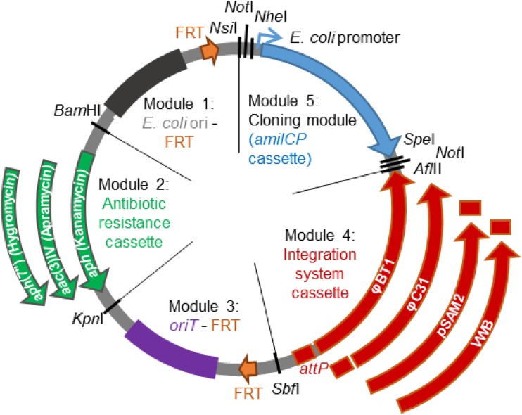
Each vector is made of five modules (Fig. 1). The first module is constituted of the E. coli origin of replication and of an Flp recombination target (FRT) recognition site for the Flp recombinase. We chose the p15A E. coli origin of replication to limit the number of plasmid copies in the cell and, thus, the metabolic burden induced by the vector, which could be important with large inserts. The second module consists of the antibiotic resistance marker. Three different resistance genes were chosen: acc(3)IV (conferring apramycin resistance), aph(7′′) (conferring hygromycin resistance), and aph (conferring kanamycin resistance). The expression of the resistance genes is under the control of a promoter that is functional in both E. coli and Streptomyces. The third module is constituted by the RP4 origin of transfer, oriT, and a second FRT site. The two FRT sites have been positioned so that the E. coli origin of replication, the antibiotic resistance cassette, and the origin of transfer can be excised once the vector is integrated in the chromosome of Streptomyces, allowing the recycling of the resistance marker and limiting the possibility of homologous recombination between two different vectors. The fourth module is the integration system cassette (integrases and their corresponding attP site) that allows site-specific integration into Streptomyces chromosomes after conjugation. Four different integration cassettes are used, derived from the integration systems of the actinophages φBT1, φC31, and VWB or of the integrative conjugative element pSAM2. Chromosomal integration sites for these systems are found in the genomes of Streptomyces species commonly used for heterologous expression (Streptomyces coelicolor, Streptomyces lividans, or Streptomyces albus J1074, for example). The construction of plasmids with four different integrase systems moreover maximizes the likelihood of being able to use at least one of them in any given strain. The last module is the cloning module. Our objective for this module was to permit the cloning and assembly of genes or gene cassettes using a variety of cloning methods (based on homology regions or on the use of restriction enzymes), as different projects may require different cloning approaches. Thus, this module was designed to allow the iterative assembly of genes (or gene cassettes) using the BioBrick assembly method (23) (see Fig. S1 in the supplemental material). We chose this assembly method rather than other methods based on the use of type IIS endonucleases (e.g., Golden Gate method [24]), as the latter enzymes cut Streptomyces genomic DNA with a high frequency (about 1 site every 1 to 1.4 kb for three of the most frequently used enzymes, BsaI, BsmBI, and BpiI, in S. coelicolor, S. avermitilis, and S. albus genomes). The BioBrick cloning system is based on the use of restriction enzymes generating compatible cohesive ends, here NheI and SpeI (Fig. S1). Once ligated, the two DNA parts are separated by a 6-bp scar sequence devoid of the NheI and SpeI restriction sites. The NheI and SpeI sites were chosen to avoid the generation of a stop codon in the scar sequence, thereby allowing the fusion of protein domains if needed, and because they are relatively rare in Streptomyces genomes. The NsiI and AflII sites that are also used in the BioBrick cloning system also are relatively scarce in Streptomyces genomes (e.g., about one site every 70 to 80 kb for NsiI and one site every 200 to 300 kb for AflII in S. coelicolor, S. avermitilis, and S. albus genomes). A NotI site is included between the NsiI and NheI sites and between the SpeI and AflII sites to facilitate the verification of the cloning. The cloning module includes an amilCP gene between the prefix and suffix sequences (31). This gene codes for a chromoprotein, giving a blue color to the cell. This cassette is meant to be replaced by the construction of interest and offers a convenient means of screening the clones containing the new construction. The five modules are separated by unique restriction sites (BamHI, KpnI, SbfI, AflII, and NsiI), so that each module (e.g., the antibiotic resistance cassette or the integration system) can easily be replaced by another one.
FIG 1.
Schematic representation of the set of modular and integrative vectors pOSV801 to pOSV812. The various antibiotic resistance cassettes and integration systems used are indicated. Each restriction enzyme site indicated is unique, except NotI (two cutting sites). E. coli ori corresponds to the E. coli p15A origin of replication. oriT is the origin of transfer. amilCP is the gene coding for an Acropora millepora chromoprotein, a protein which exhibits blue color. FRT corresponds to the sites recognized by the Flp recombinase. The promoter of module 5 is only functional in E. coli. attP sites are used by integrases to integrate the plasmid in the Streptomyces genome at a specific site.
On one side of the insert, the sequence is the same in all plasmids, and the primer on-ori (see Table 4) has been designed in the origin of replication of p15A to facilitate the verification of the insert by sequencing. On the other side of the insert, the sequence is that of the various integrase cassettes and, thus, no universal primer could be designed.
TABLE 4.
Primers used in this study
| Name | Sequence | Description |
|---|---|---|
| CEA_vec01 | ACTAGTAGCGGCCGCTTAAGCGCTCCCTGCCCGCTGTGG | Amplification integrase φBT1, suffix BioBrick (sites SpeI, NotI and AflII underlined) |
| CEA_vec02 | AATAGGAACTTCCCTGCAGGTGGCGCCGGACGGGGCTTC | Amplification integrase φBT1, site SbfI underlined |
| CEA_vec03 | CCTGCAGGGAAGTTCCTATTCTCTAGAAAGTATAGGAACTTCGTCCACGACGCCCGTGATTTTG | Amplification oriT, FRT site added in boldface, site SbfI underlined |
| CEA_vec04 | CTCACCGCGACGTGGTACCCTTTTCCGCTGCATAACCCTG | Amplification oriT, site KpnI underlined |
| CEA_vec05 | GGGTACCACGTCGCGGTGAGTTCAGG | Amplification aac(3)-IV, site KpnI underlined |
| CEA_vec06 | GGATCCGGTTCATGTGCAGCTCCATCAG | Amplification aac(3)-IV, site BamHI underlined |
| CEA_vec07 | GCTGCACATGAACCGGATCCCCTAGCGGAGTGTATACTGG | Amplification of p15A origin of replication, site BamHI underlined |
| CEA_vec08 | GCTAGCAGCGGCCGC ATGCATGAAGTTCCTATACTTTCTAGAGAATAGGAACTTCACAACTTATATCGTATGGGGCTGAC | Amplification of p15A origin of replication, FRT site added in boldface, prefix BioBrick (sites NheI, NotI, and NsiI underlined) |
| CEA_vec09 | TGCATGCGGCCGCTGCTAGCGTTTTTTGATCTCAATCAATAAAG | Amplification amilCP cassette, prefix BioBrick (sites NsiI and NotI underlined) |
| CEA_vec10 | CTTAAGCGGCCGCTACTAGTATATAAACGCAGAAAGGC | Amplification amilCP cassette, suffix BioBrick (sites AflII, NotI, and SpeI underlined) |
| CEA_vec11 | CAGTCCTGCAGGATTCCAGACGTCCCGAAGG | Amplification integrase φC31, site SbfI underlined |
| CEA_vec12 | CAGTCTTAAGCAGGCTTCCCGGGTGTCTC | Amplification integrase φC31, site AflII underlined |
| CEA_vec13 | CAGTCCTGCAGGAACGGTTCTGGCAAATATTC | Amplification integrase pSAM2, site SbfI underlined |
| CEA_vec14 | CAGTCTTAAGGTCAGTCATGCGGGCAAC | Amplification integrase pSAM2, site AflII underlined |
| CEA_vec31 | CAGTCCTGCAGGTCTCGAGCTCGCGAAAG | Amplification integrase VWB, site SbfI underlined |
| CEA_vec32 | CAGTCTTAAGGTCGACCCGTCTGACGCGTGTG | Amplification integrase VWB, site AflII underlined |
| CEA_vec17 | CTATGATCGACTGATGTCATCAGCGGTGGAGTGCAATGTCGTGACACAAGAATCCCTGTTACTTC | Amplification ORF hygromycin resistance for PCR targeting |
| CEA_vec18 | CCTTGCCCCTCCAACGTCATCTCGTTCTCCGCTCATGAGCTCAGGCGCCGGGGGCGGTGT | Amplification ORF hygromycin resistance for PCR targeting |
| CEA_vec19 | CTATGATCGACTGATGTCATCAGCGGTGGAGTGCAATGTCTCGCATGATTGAACAAGATG | Amplification ORF kanamycin resistance for PCR targeting |
| CEA_vec20 | CCTTGCCCCTCCAACGTCATCTCGTTCTCCGCTCATGAGCTCAGAAGAACTCGTCAAGAAG | Amplification ORF kanamycin resistance for PCR targeting |
| CEA_vec21 | CCAACGCACGACCGGCCGCCAGCTGTGCTTCGGTCGACACG | Site-directed mutagenesis of NheI site of φBT1 integrase, base changed underlined (T→C) |
| CEA_vec22 | CGTGTCGACCGAAGCACAGCTGGCGGCCGGTCGTGCGTTGG | Site-directed mutagenesis of NheI site of φBT1 integrase, base changed underlined (A→G) |
| CEA_vec23 | GCTGTGGTGACGAAGGAACTACTCGTTAGCCTAACTAACG | Site-directed mutagenesis of SpeI site of φBT1 integrase, base changed underlined ( A→C) |
| CEA_vec24 | CGTTAGTTAGGCTAACGAGTAGTTCCTTCGTCACCACAGC | Site-directed mutagenesis of SpeI site of φBT1 integrase, base changed underlined (T→G) |
| CEA_vec25 | CTTCCGGCGCACATGGATACCTGCAATCAAGGC | Site-directed mutagenesis of BamHI site of VWB integrase, base changed underlined (C→A) |
| CEA_vec26 | GCCTTGATTGCAGGTATCCATGTGCGCCGGAAG | Site-directed mutagenesis of BamHI site of VWB integrase, base changed underlined (G→T) |
| CEA_vec27 | CATGGAATTCGAGCTCGGTAACCGGGAATCCCCGGGTACGC | Site-directed mutagenesis of BamHI and KpnI sites of integrase pSAM2, bases changed underlined (C→A and G→A) |
| CEA_vec28 | GCGTACCCGGGGATTCCCGGTTACCGAGCTCGAATTCCATG | Site-directed mutagenesis of BamHI and KpnI sites of integrase pSAM2, bases changed underlined (C→T and G→T) |
| CEA_vec_seq_12 | TCTGGCAGCACTTTGAGGAC | Verification primer, in pSAM2 integrase, towards attP |
| CEA_vec_seq_15 | TTCGATCACGTGGGCGAAGC | Verification primer of flp excision |
| CEA_vec_seq16 | TTGCCAAAGGGTTCGTGTAG | Verification primer in oriT, towards attP of φC31 or φBT1 integrases |
| CEA_vec_seq_17 | TCAGGTCACTGTCCTGTTTC | Verification primer in φBT1 integrase, towards attP |
| CEA_vec_seq18 | AATCTTCGCCGACTTCAGC | Verification primer in φC31 integrase, towards attP |
| CEA_vec_seq_19 | GGTTTGAACTTTCCTCCCAATG | Verification primer in amilCP cassette, towards attP of pSAM2 or VWV integrases |
| CEA_vec_seq_20 | GGTGAAGAACCGGGACACC | Verification primer in VWB integrase, towards attP |
| CEA042 | GTGGTGTCGCGGAACAGACG | Verification primer in M145 and TK23, upstream of φBT1 attB site |
| CEA043 | TCCGCGACGATCCACGAC | Verification primer in M145 and TK23, downstream of φBT1 attB site |
| CEA044 | GCGTGGCGTGGACCATC | Verification primer in M145 and TK23, upstream of φC31 attB site |
| CEA045 | AATGACCTCCGGGCTTTCG | Verification primer in M145 and TK23, downstream of φC31 attB site |
| CEA046 | ACCGGCACCGCATGGCAG | Verification primer in M145 and TK23, upstream of pSAM2 attB site |
| CEA047 | ACGGCGCGTGCGGCATC | Verification primer in M145 and TK23, downstream of pSAM2 attB site |
| CEA048 | GAAAGACGGCCGACCACC | Verification primer in M145 and TK23, upstream of VWB attB site |
| CEA049 | TGCCCGCCCTCTGCATC | Verification primer in M145, downstream of VWB attB site |
| CEA050 | CTGTATGCCGCCGTCCCG | Verification primer in TK23, downstream of VWB attB site |
| CEA051 | GGTGGTGTCCCGGACCAG | Verification primer in J1074, upstream of φBT1 attB site |
| CEA052 | CCGCGACGATCCAGGACC | Verification primer in J1074, downstream of φBT1 attB site |
| CEA053 | GGCGTGGATCATGGTGATCG | Verification primer in J1074, upstream of φC31 attB site |
| CEA054 | GGTTGCGGGTGGCAAGTAG | Verification primer in J1074, downstream of φC31 attB site |
| CEA055 | CGGCCAGCTCTGCATCCC | Verification primer in J1074, upstream of pSAM2 attB site |
| CEA056 | CGGATTGTTTGCCGCCTTC | Verification primer in J1074, downstream of pSAM2 attB site |
| CEA057 | GCATGCACGGCGACCTG | Verification primer in J1074, upstream of VWB attB site |
| CEA058 | GTGACCCTGCCGGGATGG | Verification primer in J1074, upstream of VWB attB site |
| CEA_seq24 | ACCATCGCCCACGCATAAC | Verification of the loss of pUWLHFLP |
| Thio_fwd | TTGGACACCATCGCAAATC | Verification of the loss of pUWLHFLP |
| CEA036 | AAAATGCATGCGGCCGCTGCTAGCGGTGAGGCGCCACCCATCG | Amplification albonoursin cluster (sites NsiI, NotI, and NheI underlined) |
| CEA038 | AAACTTAAGGCGGCCGCTACTAGTCCGCACCATGAGCAAGTGTC | Amplification albonoursin cluster (sites AflII, NotI, and SpeI underlined) |
| F_pref_rpslp_TP | ATGCATGCGGCCGCTTCTAGAGACCGGGTCCGCGATCGGCGG | Amplification rpsl(TP)p (sites NsiI, NotI and XbaI underlined) |
| R_suff_rpslp_TP | CTTAAGGCGGCCGCTACTAGTGCTCCCTTCTCAGAAGCGCAGG | Amplification rpsl(TP)p (sites AflII, NotI and SpeI underlined) |
| onCAS001bis | GCTGCTAGCTGTTCACATTCGAACCGTCTCTG | Amplification SP22 promoter forward (truncated NotI and NheI underlined) |
| onCAS002 | ATGGACACTCCTTACTTAGAC | Amplification SP22 promoter reverse |
| onCAS003 | GTATAGGAACTTCATGCATGCGGCCGCTGCTAGCTGTTCACATTCGAACCG | Bridging oligonucleotide between plasmid pOSV812 and SP22 promoter |
| Bridge4 | ACGGTTTACAAGCATAACTAGTAGCGGCCGCTTAAGGTCGACCCGTCTG | Bridging oligonucleotide between T4 terminator and pOSV812 |
| onCAS007 | TGATCCGGTGGATGACCTTTTG | Amplification T4 terminator forward |
| onCAS008bis | GCTACTAGTTATGCTTGTAAACCGTTTTG | Amplification T4 terminator reverse (truncated NotI and SpeI underlined) |
| onCAS031 | ATGGCCACCGAGTCCGCCACC | Amplification cgc22 forward |
| onCAS032 | CTACCCGCCGTCGCCGTCGC | Amplification cgc22 reverse |
| onCAS033 | GAATACGACAGTCTAAGTAAGGAGTGTCCATATGGCCACCGAGTCCGCC | Bridging oligonucleotide between SP22 promoter and cgc22 |
| onCAS034 | GACGGCGACGGCGGGTAGTGATCCGGTGGATGACCTTTTGAATGAC | Bridging oligonucleotide between cgc22 and T4 terminator |
| on-ori | ATTTCAGTGCAATTTATCTCTTC | Universal sequencing primer in p15A origin for verification of the insert |
Construction of the vectors.
The first vector, pOSV800, was assembled by Gibson isothermal assembly (19) from five PCR-amplified DNA fragments, one for each module. The apramycin resistance gene and the φBT1 integration system were used for this first assembly. The final twelve vectors all derive from pOSV800 (Table 1 and Fig. S2). The NheI and the SpeI restriction sites present in the integration cassette of pOSV800 were removed by site-directed mutagenesis, yielding pOSV801. The vector pOSV802 was constructed by replacing the φBT1 integration cassette of pOSV800 with the φC31 integration cassette. The vectors pOSV806 (resistance to kanamycin) and pOSV810 (resistance to hygromycin) next were obtained by the replacement in pOSV802 of the aac(3)-IV gene with the aph and aph(7′′) genes by λ-Red recombination (32).
TABLE 1.
Description of the constructed vectors
| Name | Accession numbersa | Drug to which vector confers resistance | Integration system |
|---|---|---|---|
| pOSV801 | 126044/LMBP 11369 | Apramycin | φBT1 |
| pOSV802 | 126595/LMBP 11370 | Apramycin | φC31 |
| pOSV803 | 126596/LMBP 11371 | Apramycin | pSAM2 |
| pOSV804 | 126597/LMBP 11372 | Apramycin | VWB |
| pOSV805 | 126598/LMBP 11373 | Hygromycin | φBT1 |
| pOSV806 | 126606/LMBP 11374 | Hygromycin | φC31 |
| pOSV807 | 126600/LMBP 11375 | Hygromycin | pSAM2 |
| pOSV808 | 126601/LMBP 11376 | Hygromycin | VWB |
| pOSV809 | 126602/LMBP 11377 | Kanamycin | φBT1 |
| pOSV810 | 126603/LMBP 11378 | Kanamycin | φC31 |
| pOSV811 | 126604/LMBP 11379 | Kanamycin | pSAM2 |
| pOSV812 | 126605/LMBP 11380 | Kanamycin | VWB |
Accession numbers before slashes are from the Addgene plasmid repository, and those after the slashes are from the BCCM/GeneCorner Plasmid Collection.
The vector pOSV803 was constructed by replacing the φBT1 integration cassette of pOSV800 with the pSAM2 integration cassette after the removal of the BamHI and KpnI sites from this cassette by site-directed mutagenesis. The vectors pOSV807 (resistance to hygromycin) and pOSV811 (resistance to kanamycin) were next obtained by the replacement in pOSV803 of the apramycin resistance cassette by the hygromycin (from pOSV806) and kanamycin (from pOSV810) resistance cassettes, respectively.
Similarly, pOSV804 was constructed by replacing the φBT1 integration cassette of pOSV800 by the VWB integration cassette after the removal of the BamHI site from the VWB integration cassette by site-directed mutagenesis. The vectors pOSV808 (resistance to hygromycin) and pOSV812 (resistance to kanamycin) were next obtained by the replacement in pOSV804 of the apramycin resistance cassette with the hygromycin and kanamycin resistance cassettes, respectively.
Finally, pOSV805 (resistance to hygromycin) and pOSV809 (resistance to kanamycin) were obtained by the replacement in pOSV801 of the apramycin resistance cassette with the hygromycin and kanamycin resistance cassettes, respectively.
Verification of the functionality of the vectors: integration into Streptomyces chromosome.
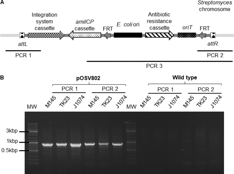
To verify that the 12 vectors we constructed all were functional, we integrated them in the chromosome of three Streptomyces strains commonly used for heterologous expression: Streptomyces coelicolor M145, Streptomyces lividans TK23, and Streptomyces albus J1074. The vectors were introduced into the Streptomyces strains by intergeneric conjugation from E. coli. The exconjugants were selected for using the appropriate antibiotics, and resistant clones were verified by PCR on extracted genomic DNA. The general principle for the PCR verification of the correct integration of the vectors at the expected chromosomal site is presented in Fig. 2A. Briefly, two DNA fragments encompassing the attL and attR sites were amplified by PCR (PCR 1 and PCR 2). The results of these PCR verifications for the integration of pOSV802 are presented in Fig. 2B. DNA fragments with a size of roughly 900 bp were amplified as expected when using the genomic DNA of the Streptomyces strains bearing the pOSV802 plasmid as the matrix. The sequences surrounding the attL and attR sites were verified. No PCR amplification was observed when the genomic DNAs of the wild-type strains were used as the matrix. Thus, these results confirmed the integration of pOSV802 at the expected site in the chromosome of the three Streptomyces species.
FIG 2.
Verification of the integration of pOSV802 in S. coelicolor M145, S. lividans TK23, and S. albus J1074 chromosomes. (A) Principle of the PCR verification of the integration of the pOSV801 to pOSV812 vectors in the Streptomyces chromosomes (PCR 1 and PCR 2) (PCR 3, PCR verification before excision of modules 1 to 3). (B) PCR fragments obtained by PCR 1 (attL region; expected sizes, 913 bp for M145 and TK23, 888 bp for J1074) and by PCR 2 (attR region; expected sizes, 911 bp for M145 and TK23, 907 bp for J1074) on the three Streptomyces strains bearing pOSV802. No PCR amplification is expected when the genomic DNA of the wild-type Streptomyces strains is used as the matrix. MW corresponds to the molecular weight ladder (Thermo Scientific GeneRuler DNA ladder mix).
Results of the PCR verification of the correct integration of the eleven other vectors are presented in the supplemental material (Fig. S3 to S9). All PCR products had the expected size, indicating that the vectors integrated at the expected location in the Streptomyces chromosomes. Altogether, these experiments demonstrate that the 12 plasmids (i) are replicative in E. coli, (ii) can be transferred by intergeneric conjugation into Streptomyces, (iii) confer the expected resistance, and (iv) integrate at the expected location in the chromosome of Streptomyces.
Excision of modules 1, 2, and 3 using the flp recombinase.
One potential difficulty when multiple genetic constructions need to be integrated in Streptomyces chromosomes is the limited number of antibiotic resistance markers that are functional in a given strain. To allow the recycling of resistance markers, we included in our vectors FRT sites surrounding module 1 (E. coli origin of replication), module 2 (antibiotic resistance cassette), and module 3 (origin of transfer). Thus, once a vector has been integrated in a Streptomyces chromosome, these three modules, which are no longer necessary, can be excised using the Flp recombinase brought in trans by a replicative plasmid, leaving a scar of 34 bp (33).
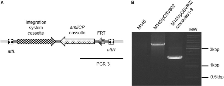
To verify that modules 1, 2, and 3 could be excised using the Flp recombinase, we used the pUWLHFLP plasmid constructed by A. Luzhetskyy's group (34) and followed the protocol described in reference 33 to excise modules 1 to 3 in S. coelicolor M145/pOSV802 as an example. The pUWLHFLP plasmid is a replicative plasmid that allows the constitutive expression of an flp gene with codon usage optimized for Streptomyces species. Approximately one apramycin-sensitive clone was obtained for each 100 clones screened, which is roughly ten times less than what was previously described (33). One sensitive clone was chosen for PCR verification of the excision of the modules 1 to 3 (Fig. 3). As expected, a smaller (1.6-kb) fragment was amplified with the genomic DNA of the sensitive clone M145 containing pOSV802 from which modules 1 to 3 had been deleted compared to the 4.2-kb fragment obtained with S. coelicolor M145/pOSV802 genomic DNA. The sequencing of the 1.6-kb fragment confirmed the correct excision of modules 1 to 3.
FIG 3.
Verification of the excision of modules 1, 2, and 3 by Flp recombinase. (A) Principle of the PCR verification of the Flp-catalyzed excision of modules 1 to 3 (PCR 3; Fig. 2A shows PCR 3 on nonexcised pOSV802). (B) PCR fragments obtained by PCR 3; expected sizes, 4,192 bp for M145/pOSV802 and 1,637 bp for M145 containing pOSV802 after excision of modules 1 to 3 by the Flp recombinase.
This experiment demonstrated the feasibility of the excision of modules 1 to 3 after the integration of one of our vectors in the chromosome of a Streptomyces species. As the pUWLHFLP plasmid is relatively unstable, it can be lost after two rounds of growth on the solid medium soya flour mannitol (SFM) without selection pressure, allowing the integration of a second vector bearing the same resistance marker. It should be noted that it will not be possible to use the pUWLHFLP plasmid, which bears a hygromycin resistance gene when pOSV805 to pOSV808 (bearing a hygromycin resistance gene) are used. However, other plasmids for the expression of Flp in Streptomyces have been constructed harboring different resistance markers, e.g., thiostrepton resistance (33).
Refactoring the albonoursin gene cluster.
The pOSV801 to pOSV812 vectors were mainly designed for the assembly of gene cassettes to form new gene clusters or to refactor silent gene clusters, although their use may not be limited to these applications. To illustrate one of the possible uses of our vectors, we decided to refactor the albonoursin gene cluster. Albonoursin [cyclo(ΔPhe-ΔLeu)], produced by Streptomyces noursei, belongs to the family of diketopiperazine metabolites studied in our group. Its biosynthetic gene cluster consists of three genes, albA, albB, and albC (35). We chose to express the alb gene under the control of the rpsL(TP) constitutive promoter (2) and to assemble the required elements using the BioBrick assembly method.
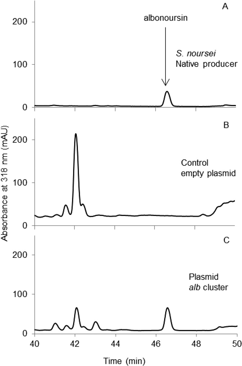
The rpsL(TP) promoter followed by the ribosome binding site (RBS) sequence of tipA (36) was first cloned into pOSV802, yielding pCEA005. Similarly, the alb gene cluster was cloned in pOSV802, yielding pCEA006. Finally, the NheI/AflII fragment of pCEA006 containing the alb gene cluster was cloned into SpeI/AflII-digested pCEA005, and the resulting pCEA007 plasmid was introduced into S. coelicolor M145 by intergeneric conjugation. To verify that S. coelicolor M145/pCEA007 produced albonoursin, the culture supernatant of this strain, together with the culture supernatants of S. noursei (positive control) and of S. coelicolor M145/pOSV802 (negative control), were analyzed by liquid chromatography-mass spectrometry (LC-MS). The chromatograms (Fig. 4) and the MS spectra and fragmentation patterns (Fig. S10) (37) confirmed that M145/pCEA007 produces albonoursin.
FIG 4.
HPLC analysis of albonoursin production. Chromatograms of the analysis of the culture supernatants of the native albonoursin producer S. noursei (A), the control S. coelicolor M145/pOSV802 (B), and S. coelicolor M145/pCEA007 (C).
Genetic complementation of mutant strain: assembly of a gene cassette using LCR in pOSV812.
Cloning methods based on the use of restriction enzymes necessitate the presence or introduction of restriction sites in the sequence, which may sometimes be problematic (for example, for the fusion of protein domains or for the cloning of an RBS sequence in front of a coding sequence). In these cases, the use of seamless cloning methods is preferable. To demonstrate that gene cassettes could be assembled in our vectors using such seamless cloning methods, we undertook the genetic complementation of a mutant constructed previously during the study of the congocidine biosynthetic gene cluster (mutant strain CGCL030) (30). Congocidine is a pyrrolamide antibiotic assembled by an atypical NRPS. The gene cgc22, deleted in the strain CGCL030, encodes an acyl-coenzyme A synthetase that activates the pyrrole precursor during congocidine assembly. To construct the plasmid for genetic complementation, we assembled three DNA fragments in pOSV802 by LCR (20): the SP22 constitutive promoter with the RBS of the capsid φC31 gene (15), the cgc22 gene, and the T4 terminator (38). The LCR method is based on the ligation of DNA fragments using bridging oligonucleotides whose sequences are complementary to the sequences of the extremities of the DNA fragments to be assembled (Fig. S11). The assembly is achieved through multiple cycles of denaturation-annealing-ligation using a thermostable ligase. This method has the advantages of working for the assembly of very short fragments (<100 bp) and does not necessitate the existence of homology regions at the extremities of the DNA fragments that will be assembled.
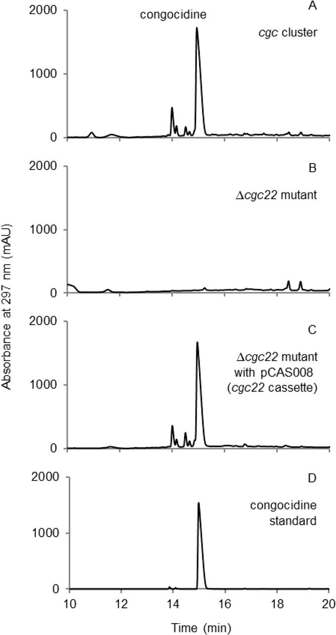
Each DNA fragment was amplified by PCR. The oligonucleotides used for the amplification of the promoter and RBS fragment and of the T4 terminator fragment were designed to reconstitute the prefix and the suffix sequences once all the fragments have been assembled in the vector. All PCR fragments were phosphorylated and assembled in one step with the NotI/Klenow-digested vector pOSV812. To verify that the constructed gene cassette was functional, the pCAS008 plasmid was introduced by intergeneric conjugation in the S. lividans CGCL030 strain expressing the whole cgc gene cluster except for cgc22 (30). The supernatants of 4-day cultures of CGCL030/pCAS008, CGCL030, and CGCL006 expressing the complete cgc gene cluster were then analyzed by high-performance liquid chromatography (HPLC). Figure 5 shows that production of congocidine is restored in CGCL030/pCAS008, demonstrating the functionality of the constructed gene cassette.
FIG 5.
HPLC analysis of the genetic complementation of the Δcgc22 mutant. Chromatograms of the analysis of the culture supernatant of the CGCL006 strain expressing the complete cgc cluster (A), the culture supernatant of the CGCL030 mutant strain expressing the cgc cluster except for cgc22 (B), the culture supernatant of the CGCL083 strain (CGCL030 genetically complemented with pCAS008) (C), and the congocidine standard (D).
In conclusion, we constructed a set of plasmids dedicated to DNA assembly and integration in Streptomyces chromosomes. We aimed at offering a modular and flexible platform that can be used in various experimental settings, from the assembly of small gene cassettes to the assembly of larger DNA fragments, and that will be compatible with a large variety of cloning methods. Varying the nature of the resistance cassette (resistance to three different antibiotics) and of the integration system (four different systems), we constructed a total of 12 plasmids. To increase our plasmid collection, we plan in the future to add new resistance cassettes (e.g., erythromycin) and integration systems (e.g., integration systems from TG1, φJoe, or SV1 [39–41]) but also to include new modules, such as the CEN-ARS module (1) for DNA cloning and assembly in yeast. All of our plasmids will be made available to the community through deposition in plasmid collections such as Addgene or the BCCM/GeneCorner Plasmid Collection.
MATERIALS AND METHODS
Bacterial strains, plasmids, and growth conditions.
Strains and plasmids used in this study are listed in Tables 2 and 3. E. coli strains were grown at 37°C in LB or Super Optimal Broth (SOB) medium complemented with MgSO4 (20 mM final), supplemented with appropriate antibiotics as needed. Soya flour mannitol (SFM) medium (42) was used for genetic manipulations of Streptomyces strains and spore stock preparations. Streptomyces strains were grown at 28°C in MP5 (43) for congocidine or albonoursin production.
TABLE 2.
Strains used during the study
| Strain | Description | Reference or source |
|---|---|---|
| Escherichia coli DH5α | General cloning host | Promega |
| Escherichia coli ET12567/pUZ8002 | Host strain for conjugation from E. coli to Streptomyces | 55 |
| Escherichia coli ET12567/pUZ8003 | Host strain for conjugation from E. coli to Streptomyces when using vectors containing the kanamycin resistance cassette (pUZ8003 is modified pUZ8002 with aph replaced by bla) | Our unpublished data |
| Escherichia coli S17-1 | Host strain for conjugation from E. coli to Streptomyces when using vectors containing the kanamycin resistance cassette | 56 |
| Escherichia coli BW25113/pIJ790 | Host strain for PCR targeting | 32 |
| S. coelicolor M145 | Streptomyces host strain for heterologous expression | 42 |
| S. lividans TK23 | Streptomyces host strain for heterologous expression | 42 |
| S. albus J1074 | Streptomyces host strain for heterologous expression | 42 |
| S. noursei ATCC11455 | Albonoursin native producer | ATCC |
| S. coelicolor M145/pOSV801 | M145 containing pOSV801 | This work |
| S. coelicolor M145/pOSV802 | M145 containing pOSV802 | This work |
| S. coelicolor M145/pOSV803 | M145 containing pOSV803 | This work |
| S. coelicolor M145/pOSV804 | M145 containing pOSV804 | This work |
| S. coelicolor M145/pOSV805 | M145 containing pOSV805 | This work |
| S. coelicolor M145/pOSV806 | M145 containing pOSV806 | This work |
| S. coelicolor M145/pOSV807 | M145 containing pOSV807 | This work |
| S. coelicolor M145/pOSV808 | M145 containing pOSV808 | This work |
| S. coelicolor M145/pOSV809 | M145 containing pOSV809 | This work |
| S. coelicolor M145/pOSV810 | M145 containing pOSV810 | This work |
| S. coelicolor M145/pOSV811 | M145 containing pOSV811 | This work |
| S. coelicolor M145/pOSV812 | M145 containing pOSV812 | This work |
| S. lividans TK23/pOSV801 | TK23 containing pOSV801 | This work |
| S. lividans TK23/pOSV802 | TK23 containing pOSV802 | This work |
| S. lividans TK23/pOSV803 | TK23 containing pOSV803 | This work |
| S. lividans TK23/pOSV804 | TK23 containing pOSV804 | This work |
| S. lividans TK23/pOSV805 | TK23 containing pOSV805 | This work |
| S. lividans TK23/pOSV806 | TK23 containing pOSV806 | This work |
| S. lividans TK23/pOSV807 | TK23 containing pOSV807 | This work |
| S. lividans TK23/pOSV808 | TK23 containing pOSV808 | This work |
| S. lividans TK23/pOSV809 | TK23 containing pOSV809 | This work |
| S. lividans TK23/pOSV810 | TK23 containing pOSV810 | This work |
| S. lividans TK23/pOSV811 | TK23 containing pOSV811 | This work |
| S. lividans TK23/pOSV812 | TK23 containing pOSV812 | This work |
| S. albus J1074/pOSV801 | J1074 containing pOSV801 | This work |
| S. albus J1074/pOSV802 | J1074 containing pOSV802 | This work |
| S. albus J1074/pOSV803 | J1074 containing pOSV803 | This work |
| S. albus J1074/pOSV804 | J1074 containing pOSV804 | This work |
| S. albus J1074/pOSV805 | J1074 containing pOSV805 | This work |
| S. albus J1074/pOSV806 | J1074 containing pOSV806 | This work |
| S. albus J1074/pOSV807 | J1074 containing pOSV807 | This work |
| S. albus J1074/pOSV808 | J1074 containing pOSV808 | This work |
| S. albus J1074/pOSV809 | J1074 containing pOSV809 | This work |
| S. albus J1074/pOSV810 | J1074 containing pOSV810 | This work |
| S. albus J1074/pOSV811 | J1074 containing pOSV811 | This work |
| S. albus J1074/pOSV812 | J1074 containing pOSV812 | This work |
| S. coelicolor M145 containing pOSV802 from which modules 1 to 3 had been deleted | M145 containing pOSV802 after excision with Flp | This work |
| S. coelicolor M145/pCEA007 | M145 containing pCEA007 | This work |
| CGCL006 | TK23 containing pCGC002 (cgc cluster) | 30 |
| CGCL030 | TK23 containing pCGC221 (cgc cluster with cgc22 deleted) | 30 |
| CGCL083 | CGCL030 containing pCAS008 | This work |
TABLE 3.
Plasmids used in this study
| Plasmid | Description | Reference or source |
|---|---|---|
| pCR-Blunt | E. coli cloning vector | Invitrogen |
| pRT801 | Source of the φBT1 integrase fragment | 45 |
| pAC-BETA | Source of the origin of replication p15A | 48 |
| pOSV408 | Source of the origin of transfer | 46 |
| pSET152 | Source of the apramycin resistance cassette and of the φC31 integrase fragment | 47 |
| psB1C3-BBa_K1155003 | Source of the amilCP cassette | iGEM registry of standard biological parts |
| pKT02 | Source of the VWB integrase fragment | 52 |
| pOSV215 | Source of the T4 terminator | 54 |
| pOSV554 | Source of the integrase pSAM2 fragment | Our unpublished data |
| pOSV400 | Source of the ORF of hygromycin resistance gene | Our unpublished data |
| pOSV401 | Source of the ORF of kanamycin resistance gene | Our unpublished data |
| pSL128 | Source of the albonoursin cluster (albA, albB, and albC) | 35 |
| pCEA001 | pUC57 containing rpsl(TP)p and tipA RBS | Genecust |
| pCEA002 | pGEM-T easy containing rpsl(TP)p and tipA RBS with the last 6 nucleotides replaced by the SpeI site | This work |
| pCEA003 | Plasmid pCR-Blunt containing pSAM2 integrase, used for site-directed mutagenesis | This work |
| pCEA004 | Plasmid pCR-Blunt containing VWB integrase, used for site-directed mutagenesis | This work |
| pCEA005 | pOSV802 containing rpsl(TP)p and tipA RBS with the last 6 nucleotides replaced by the SpeI site | This work |
| pCEA006 | pOSV802 containing the genes albA, albB, and albC instead of the amilCP cassette | This work |
| pCEA007 | pOSV802 containing rpsl(TP)p and the albonoursin cluster instead of amilCP | This work |
| pOSV800 | Plasmid constructed containing apramycin resistance and φBT1 integrase with two BioBrick sites NheI and SpeI in φBT1 integrase | This work |
| pOSV801 | Plasmid constructed containing apramycin resistance and φBT1 integrase | This work |
| pOSV802 | Plasmid constructed containing apramycin resistance and φC31 integrase | This work |
| pOSV803 | Plasmid constructed containing apramycin resistance and pSAM2 integrase | This work |
| pOSV804 | Plasmid constructed containing apramycin resistance and VWB integrase | This work |
| pOSV805 | Plasmid constructed containing hygromycin resistance and φBT1 integrase | This work |
| pOSV806 | Plasmid constructed containing hygromycin resistance and φC31 integrase | This work |
| pOSV807 | Plasmid constructed containing hygromycin resistance and pSAM2 integrase | This work |
| pOSV808 | Plasmid constructed containing hygromycin resistance and VWB integrase | This work |
| pOSV809 | Plasmid constructed containing kanamycin resistance and φBT1 integrase | This work |
| pOSV810 | Plasmid constructed containing kanamycin resistance and φC31 integrase | This work |
| pOSV811 | Plasmid constructed containing kanamycin resistance and pSAM2 integrase | This work |
| pOSV812 | Plasmid constructed containing kanamycin resistance and VWB integrase | This work |
| pCAS008 | pOSV812 with cassette SP22p-cgc22-T4 terminator instead of amilCP | This work |
DNA preparation and manipulations.
All oligonucleotides used in this study were purchased from Eurofins and are listed in Table 4. The high-fidelity DNA polymerase Phusion (Thermo Fisher Scientific) was used to amplify the fragments used for the construction of the vectors. DreamTaq polymerase (Thermo Fisher Scientific) was used for PCR verification of plasmid integration in Streptomyces strains. DNA fragments were purified from agarose gels using the NucleoSpin gel and PCR cleanup kit from Macherey-Nagel. DNA extractions and manipulations, Escherichia coli transformations, and E. coli/Streptomyces conjugations were performed according to standard procedures (42, 44).
Construction of pOSV800.
pOSV800 was constructed by assembling five fragments, coming from five different vectors, using the one-pot isothermal assembly developed by Gibson et al. (19). The first fragment (φBT1 integrase gene and attP site) was amplified from pRT801 (45) using the CEA_vec01 and CEA_vec02 primers. The second fragment (oriT origin of transfer) was amplified from pOSV408 (46) using the CEA_vec03 and CEA_vec04 primers. The third fragment [apramycin resistance cassette aac(3)-IV] was amplified from pSET152 (47) using CEA_vec05 and CEA_vec06 primers. The fourth fragment (p15A origin of replication) was amplified from pAC-BETA (48) using CEA_vec07 and CEA_vec08 primers. The fifth and last fragment (amilCP cassette surrounded by a BioBrick-like prefix [NsiI, NotI, and NheI sites] and suffix [SpeI, NotI, and AflII]) was amplified from pSB1C3-BBa-K1155003 (iGEM registry of standard biological parts) using CEA_vec09 and CEA_vec10 primers. Two FRT sites were introduced in the primer sequences of CEA_vec03 and CEA_vec08. The PCR products were purified and diluted to 100 ng/μl. One microliter of each of the PCR products was used for the assembly. A mix containing T5 exonuclease (New England Biolabs [NEB]), Taq ligase (NEB), and Phusion high-fidelity polymerase (Thermo Fisher Scientific) in the appropriate buffer was prepared by following the protocol described by Gibson (49). The reaction was carried out by adding 5 μl of DNA to 15 μl of the mix and incubating at 50°C for 1 h. Five microliters was used for a standard transformation of E. coli DH5α. The amilCP cassette, coding for a blue protein, allowed the easy screening of potential correct clones. Plasmid DNA was extracted from a blue clone, and the sequence of the plasmid was confirmed by sequencing.
Construction of pOSV801.
The φBT1 integrase gene in pOSV800 contains NheI and SpeI restriction sites that were chosen for the BioBrick type of cloning. To remove these sites, one base was modified by site-directed mutagenesis by following a protocol described previously (50). CEA_vec21 and CEA_vec22 were used to remove the NheI site by replacing an A with a G at position 123 in the integrase gene sequence (position 38926 of the φBT1 bacteriophage genome sequence), conserving the amino acid leucine (CTA becoming CTG) in the protein. Similarly, CEA_vec23 and CEA_vec24 were used to remove the SpeI site in the terminator downstream of the φBT1 integrase gene at position 40663 in the φBT1 bacteriophage genome sequence, replacing a T with a G.
Briefly, the plasmid was amplified using the first pair of oligonucleotides with the Phusion polymerase. One microliter of DpnI was added to the reaction mixture to digest the original vector for 2 h at 37°C, and competent E. coli DH5α cells were transformed with 5 μl of the mixture. The second site-directed mutagenesis was performed by following the same protocol. The sequence of the resulting plasmid was verified by sequencing, and the plasmid was named pOSV801.
Construction of pOSV802 to pOSV812.
The pOSV802 to pOSV812 vectors all were derived from pOSV800, except for pOSV805 and pOSV809, which were derived from pSV801 (see Fig. S2 in the supplemental material). The eleven vectors were confirmed by restriction analyses and by sequencing each fragment obtained by PCR. The φBT1 integration cassette was replaced either by the φC31, VWB, or pSAM2 integration cassettes, and the aac(3)-IV gene was replaced by either the aph or the aph(7′′) genes. The use of the pSAM2 (from pOSV554 [51]) and VWB integration (from pKT02 [52]) cassette necessitated the removal of KpnI and BamHI sites and of a BamHI site, respectively. Thus, these cassettes were first cloned into pCR-Blunt by following the procedure advised by Invitrogen, yielding pCEA003 and pCEA004, respectively. The BamHI site from the VWB integrase was removed by site-directed mutagenesis using the oligonucleotides CEA_025 and CEA_026 by changing base 1008 of the integrase gene sequence from C to A, thereby keeping the amino acid unchanged (ATC becoming ATA, isoleucine). The mutation in the resulting plasmid pCEA004 was verified by sequencing. The KpnI and BamHI sites, located upstream of the integrase pSAM2 coding sequence and only 3 bp apart, were removed in a single round of site-directed mutagenesis using the oligonucleotides CEA_027 and CEA_028. The mutations in the resulting plasmid pCEA003 were verified by sequencing.
To replace the φBT1 integration cassette with the φC31 integration cassette in pOSV800, the φC31 integration cassette was amplified by PCR from pSET152 (47) using the oligonucleotides CEA_vec11 and CEA_vec12. The PCR product was digested by SbfI and AflII and cloned into the SbfI- and AflII-digested pOSV800, yielding pOSV802. The replacement of the φBT1 integration cassette by the pSAM2 integration cassette in pOSV800 was executed likewise, cloning the 1.6-kb SbfI/AflII fragment from pCEA003 into the SbfI- and AflII-digested pOSV800, yielding pOSV803. The same protocol was used to replace the φBT1 integration cassette by the VWB integration cassette in pOSV800, yielding pOSV804.
The replacement of the aac(3)-IV gene (apramycin resistance) with the aph(7′′) gene (hygromycin resistance) or the aph gene (kanamycin resistance) in pOSV802 was carried out by λ-Red recombination as described by Gust and colleagues (32). The aph(7′′) and aph genes were amplified by PCR using the oligonucleotides CEA_vec_017 and CEA_vec_018 for aph(7′′) and CEA_vec_019 and CEA_vec_020 for aph, and the PCR products were used to replace the aac(3)-IV gene in pOSV802, yielding pOSV806 and pOSV810, respectively. The joining sequences were confirmed by sequencing. Sequencing showed that the sequences of aph and aph(7′′) were as predicted, except for base 188 of aph(7′′), in which A was replaced by G, leading to the replacement of Asp (GAC) by Gly (GGC). However, no functional difference has been observed, and the plasmid confers full resistance to hygromycin.
To replace the aac(3)-IV gene cassette in pOSV801, pOSV803, and pOSV804 with the aph(7′′) gene cassette, the 1.4-kb KpnI/BamHI fragment of pOSV806 was cloned into KpnI/BamHI-digested pOSV801, pOSV803, and pOSV804, yielding pOSV805, pOSV807, and pOSV808, respectively. Using the same protocol, the aac(3)-IV gene was replaced in pOSV801, pOSV803, and pOSV804 by the aph gene cassette, yielding pOSV809, pOSV811, and pOSV812, respectively. The vectors obtained were verified by restriction analyses.
Verification of the integration of the vectors in Streptomyces species.
The 12 vectors constructed were introduced in three Streptomyces species (Streptomyces coelicolor M145, Streptomyces lividans TK23, and Streptomyces albus J1074) by intergeneric conjugation by following the standard procedure (42). E. coli ET12567/pUZ8002 was used as a donor strain for pOSV801 to pOSV808. For pOSV809 to pSOV812, which confer resistance to kanamycin, we used E. coli S17-1 as a donor strain to perform conjugation with S. lividans TK23 and S. albus J1074 and E. coli ET12567/pUZ8003 as a donor strain to perform conjugation with S. coelicolor M145. Genomic DNA was extracted from the exconjugants obtained. To confirm that the vectors had been integrated into the host chromosomal DNA at the expected sites, PCR 1 and PCR 2 were performed as shown in Fig. 2, using the primers CEA_vec_seq12, CEA_vec_seq_16–20, and CEA_42–58. These PCRs amplify a fragment of about 900 bp only if the plasmid is integrated at the expected chromosomal attB site.
Excision mediated by the flp recombinase.
We used M145/pOSV802 to verify that modules 1, 2, and 3 could be excised using the Flp recombinase once integrated into the chromosome of Streptomyces. For this purpose, we used the plasmid pUWLHFLP and followed the protocol described previously (33). pUWLHFLP is similar to pUWLFLP, but the thiostrepton resistance cassette was replaced by a hygromycin resistance cassette (34). Briefly, pUWLHFLP was introduced by intergeneric conjugation into the strain M145/pOSV802, and exconjugants were replicated on SFM plates containing 100 μg/ml hygromycin. After one round of liquid culture in tryptic soy broth, stocks of spores were made. Spore dilutions were plated on SFM supplemented with nalidixic acid, and the clones were screened for loss of apramycin resistance by replica plating. The loss of the fragment of the vector was subsequently confirmed by amplifying the fragment around both FRT sites (PCR 3, primers CEA_vec_seq15 and CEA_045 [Fig. 3]), which was then sequenced. Stocks of spores of the confirmed clones were prepared on SFM supplemented with nalidixic acid, and the loss of the helper vector pUWLHFLP was confirmed by PCR (primers thio-fwd and CEA_seq24).
Construction of pCEA007.
The albonoursin gene cluster, constituted of the three genes albA, albB, and albC, was cloned into pOSV802 and placed under the control of the rpsL(TP) promoter (2) by following the BioBrick assembly procedure (Fig. S1). The pCEA001 plasmid was used to amplify the rpsL(TP) promoter sequence followed by the tipA RBS sequence using the primers F_pref_rpslp_TP and R_suff_rpslp_TP. The PCR product was cloned into pGEM-T Easy, and the resulting plasmid was named pCEA002. The 0.4-kb NsiI/SpeI-digested fragment of pCEA002 was ligated into NsiI/SpeI-digested pOSV802, yielding pCEA005. The insert sequence of pCEA005 was confirmed by sequencing. The albonoursin gene cluster was amplified from pSL128 (35) using the primers CEA036 and CEA038. The PCR product was digested by NsiI and SpeI and ligated into NsiI/AflII-digested pOSV802, yielding pCEA006. The sequence of the insert was confirmed by sequencing. pCEA006 was then digested by AflII and NheI and the 1.8-kb fragment was ligated into the SpeI/AflII-digested pCEA005, yielding pCEA007. The resulting plasmid, pCEA007, was confirmed by digestion by NotI and by EcoRI/HindIII. This plasmid was introduced in S. coelicolor M145 by intergeneric conjugation.
Construction of the pCAS008 plasmid.
The pCAS008 plasmid, expressing the cgc22 gene under the control the SP22 promoter (15), was assembled using the ligase cycling reaction as previously described (53). pOSV812 was digested by NotI, and Klenow was added to the mix in order to obtain blunt ends. The 5-kb fragment was purified on agarose gel. The gene cgc22 was amplified from the cosmid pCGC002 (30) with the primers onCAS031 and onCAS032.The promoter SP22 was ordered from Eurofins Genomics as a synthetic gene fragment and amplified with the primers onCAS001bis and onCAS002. The T4 terminator was amplified from the plasmid pOSV215 (54) with the primers onCAS007 and onCAS008bis. The primers upstream of the promoter SP22 and downstream of the terminator were designed in order to recreate the BioBrick prefix and suffix (NsiI, NotI, and NheI and SpeI, NotI, and AflII, respectively). All fragments were then phosphorylated and ligated via ligase cycling reaction. The sequence of the resulting plasmid pCAS008 was confirmed by sequencing. The pCAS008 plasmid was introduced in S. lividans CGCL030 by intergeneric conjugation.
LC and LC-MS analyses.
For albonoursin production, S. coelicolor M145/pCEA006, M145/pOSV802, and S. noursei strains were cultivated for 5 days in MP5 medium at 30°C. Supernatants were filtered using the Mini-UniPrep syringeless filter devices (0.2 μm; Whatman). The samples were analyzed on an Atlantis C18 T3 column (250 mm by 4.6 mm, 5 μm, column temperature of 30°C) using an Agilent 1200 HPLC instrument equipped with a quaternary pump. The filtrates were eluted using a 0% to 45% linear gradient of solvent B (solvent A, 0.1% HCOOH in H2O; solvent B, 0.1% HCOOH in CH3CN) for 45 min (flow rate, 1 ml/min). Albonoursin was detected by monitoring absorbance at 318 nm (35). A Bruker Daltonics Esquire HCT ion trap mass spectrometer equipped with an orthogonal atmospheric pressure interface-electrospray ionization (AP-ESI) source was used for LC-MS analyses. The LC flow was split 1/10 to the mass spectrometer and 9/10 to a diode array detector. The ESI source was operated in positive mode with the nebulizing gas set to a pressure of 241 kPa. The drying gas was set to 8 liters·min−1, and the drying temperature was set to 340°C. Nitrogen served as the drying and nebulizing gas, and helium gas was introduced into the ion trap both for efficient trapping and cooling of the ions and for fragmentation processes. Ionization and mass analysis conditions (capillary high voltage, skimmer and capillary exit voltages, and ion transfer parameters) were optimized for detection of compounds in the m/z range of 50 to 600. For structural characterization by fragmentation, an isolation width of 1 mass unit was used. A fragmentation energy ramp was used for automatically varying the fragmentation amplitude to optimize the MS/MS process. For LC-MS analyses, filtrates were eluted using a slightly modified gradient: after 5 min of an isocratic run at 100% buffer A, the concentration of buffer B was linearly increased over 50 min to reach 50%.
For congocidine production, S. lividans CGCL083, CGCL030, and CGCL006 strains were cultivated in MP5 medium for 4 days at 30°C. Supernatants were filtered using Mini-UniPrep syringeless filter devices (0.2 μm; Whatman). The samples were analyzed on an Atlantis C18 T3 column (250 mm by 4.6 mm, 5 μm, column temperature of 30°C) using an Agilent 1200 HPLC instrument with a quaternary pump. Samples were eluted under isocratic conditions of 0.1% HCOOH in H2O (solvent A)-0.1% HCOOH in CH3CN (solvent B) (95:5) at 1 ml/min for 7 min, followed by a gradient to 40:60 A:B over 23 min. Congocidine was detected by monitoring absorbance at 297 nm (30).
Supplementary Material
ACKNOWLEDGMENTS
We thank Hervé Leh for critical reading of the manuscript.
The research received funding from ANR-14-CE16-0003-01. The funders had no role in study design, data collection and interpretation, or the decision to submit the work for publication.
Footnotes
Supplemental material for this article may be found at https://doi.org/10.1128/AEM.00485-19.
REFERENCES
- 1.Yamanaka K, Reynolds KA, Kersten RD, Ryan KS, Gonzalez DJ, Nizet V, Dorrestein PC, Moore BS. 2014. Direct cloning and refactoring of a silent lipopeptide biosynthetic gene cluster yields the antibiotic taromycin A. Proc Natl Acad Sci U S A 111:1957–1962. doi: 10.1073/pnas.1319584111. [DOI] [PMC free article] [PubMed] [Google Scholar]
- 2.Shao Z, Rao G, Li C, Abil Z, Luo Y, Zhao H. 2013. Refactoring the silent spectinabilin gene cluster using a plug-and-play scaffold. ACS Synth Biol 2:662–669. doi: 10.1021/sb400058n. [DOI] [PMC free article] [PubMed] [Google Scholar]
- 3.Greunke C, Duell ER, D’Agostino PM, Glöckle A, Lamm K, Gulder T. 2018. Direct pathway cloning (DiPaC) to unlock natural product biosynthetic potential. Metab Eng 47:334–345. doi: 10.1016/j.ymben.2018.03.010. [DOI] [PubMed] [Google Scholar]
- 4.Bozhüyük KAJ, Fleischhacker F, Linck A, Wesche F, Tietze A, Niesert C-P, Bode HB. 2018. De novo design and engineering of non-ribosomal peptide synthetases. Nat Chem 10:275–281. doi: 10.1038/nchem.2890. [DOI] [PubMed] [Google Scholar]
- 5.Awakawa T, Fujioka T, Zhang L, Hoshino S, Hu Z, Hashimoto J, Kozone I, Ikeda H, Shin-Ya K, Liu W, Abe I. 2018. Reprogramming of the antimycin NRPS-PKS assembly lines inspired by gene evolution. Nat Commun 9:3534. doi: 10.1038/s41467-018-05877-z. [DOI] [PMC free article] [PubMed] [Google Scholar]
- 6.Baltz RH. 2018. Synthetic biology, genome mining, and combinatorial biosynthesis of NRPS-derived antibiotics: a perspective. J Ind Microbiol Biotechnol 45:635–649. doi: 10.1007/s10295-017-1999-8. [DOI] [PubMed] [Google Scholar]
- 7.Baltz RH. 2014. Combinatorial biosynthesis of cyclic lipopeptide antibiotics: a model for synthetic biology to accelerate the evolution of secondary metabolite biosynthetic pathways. ACS Synth Biol 3:748–758. doi: 10.1021/sb3000673. [DOI] [PubMed] [Google Scholar]
- 8.Yuzawa S, Mirsiaghi M, Jocic R, Fujii T, Masson F, Benites VT, Baidoo EEK, Sundstrom E, Tanjore D, Pray TR, George A, Davis RW, Gladden JM, Simmons BA, Katz L, Keasling JD. 2018. Short-chain ketone production by engineered polyketide synthases in Streptomyces albus. Nat Commun 9:4569. doi: 10.1038/s41467-018-07040-0. [DOI] [PMC free article] [PubMed] [Google Scholar]
- 9.Yuzawa S, Backman TWH, Keasling JD, Katz L. 2018. Synthetic biology of polyketide synthases. J Ind Microbiol Biotechnol 45:621–633. doi: 10.1007/s10295-018-2021-9. [DOI] [PubMed] [Google Scholar]
- 10.Gomez-Escribano JP, Bibb MJ. 2011. Engineering Streptomyces coelicolor for heterologous expression of secondary metabolite gene clusters. Microb Biotechnol 4:207–215. doi: 10.1111/j.1751-7915.2010.00219.x. [DOI] [PMC free article] [PubMed] [Google Scholar]
- 11.Komatsu M, Komatsu K, Koiwai H, Yamada Y, Kozone I, Izumikawa M, Hashimoto J, Takagi M, Omura S, Shin-Ya K, Cane DE, Ikeda H. 2013. Engineered Streptomyces avermitilis host for heterologous expression of biosynthetic gene cluster for secondary metabolites. ACS Synth Biol 2:384–396. doi: 10.1021/sb3001003. [DOI] [PMC free article] [PubMed] [Google Scholar]
- 12.Kallifidas D, Jiang G, Ding Y, Luesch H. 2018. Rational engineering of Streptomyces albus J1074 for the overexpression of secondary metabolite gene clusters. Microb Cell Factories 17:25. doi: 10.1186/s12934-018-0874-2.. [DOI] [PMC free article] [PubMed] [Google Scholar]
- 13.Myronovskyi M, Rosenkränzer B, Nadmid S, Pujic P, Normand P, Luzhetskyy A. 2018. Generation of a cluster-free Streptomyces albus chassis strains for improved heterologous expression of secondary metabolite clusters. Metab Eng 49:316–324. doi: 10.1016/j.ymben.2018.09.004. [DOI] [PubMed] [Google Scholar]
- 14.Baltz RH. 2016. Genetic manipulation of secondary metabolite biosynthesis for improved production in Streptomyces and other actinomycetes. J Ind Microbiol Biotechnol 43:343–370. doi: 10.1007/s10295-015-1682-x. [DOI] [PubMed] [Google Scholar]
- 15.Bai C, Zhang Y, Zhao X, Hu Y, Xiang S, Miao J, Lou C, Zhang L. 2015. Exploiting a precise design of universal synthetic modular regulatory elements to unlock the microbial natural products in Streptomyces. Proc Natl Acad Sci U S A 112:12181–12186. doi: 10.1073/pnas.1511027112. [DOI] [PMC free article] [PubMed] [Google Scholar]
- 16.Seghezzi N, Amar P, Koebmann B, Jensen PR, Virolle M-J. 2011. The construction of a library of synthetic promoters revealed some specific features of strong Streptomyces promoters. Appl Microbiol Biotechnol 90:615–623. doi: 10.1007/s00253-010-3018-0. [DOI] [PubMed] [Google Scholar]
- 17.Siegl T, Tokovenko B, Myronovskyi M, Luzhetskyy A. 2013. Design, construction and characterisation of a synthetic promoter library for fine-tuned gene expression in actinomycetes. Metab Eng 19:98–106. doi: 10.1016/j.ymben.2013.07.006. [DOI] [PubMed] [Google Scholar]
- 18.Sohoni SV, Fazio A, Workman CT, Mijakovic I, Lantz AE. 2014. Synthetic promoter library for modulation of actinorhodin production in Streptomyces coelicolor A3(2). PLoS One 9:e99701. doi: 10.1371/journal.pone.0099701. [DOI] [PMC free article] [PubMed] [Google Scholar]
- 19.Gibson DG, Young L, Chuang R-Y, Venter JC, Hutchison CA, Smith HO. 2009. Enzymatic assembly of DNA molecules up to several hundred kilobases. Nat Methods 6:343–345. doi: 10.1038/nmeth.1318. [DOI] [PubMed] [Google Scholar]
- 20.de Kok S, Stanton LH, Slaby T, Durot M, Holmes VF, Patel KG, Platt D, Shapland EB, Serber Z, Dean J, Newman JD, Chandran SS. 2014. Rapid and reliable DNA assembly via ligase cycling reaction. ACS Synth Biol 3:97–106. doi: 10.1021/sb4001992. [DOI] [PubMed] [Google Scholar]
- 21.Shao Z, Zhao H, Zhao H. 2009. DNA assembler, an in vivo genetic method for rapid construction of biochemical pathways. Nucleic Acids Res 37:e16. doi: 10.1093/nar/gkn991. [DOI] [PMC free article] [PubMed] [Google Scholar]
- 22.Basitta P, Westrich L, Rösch M, Kulik A, Gust B, Apel AK. 2017. AGOS: a plug-and-play method for the assembly of artificial gene operons into functional biosynthetic gene clusters. ACS Synth Biol 6:817–825. doi: 10.1021/acssynbio.6b00319. [DOI] [PubMed] [Google Scholar]
- 23.Shetty RP, Endy D, Knight T. 2008. Engineering BioBrick vectors from BioBrick parts. J Biol Eng 2:5. doi: 10.1186/1754-1611-2-5. [DOI] [PMC free article] [PubMed] [Google Scholar]
- 24.Engler C, Kandzia R, Marillonnet S. 2008. A one pot, one step, precision cloning method with high throughput capability. PLoS One 3:e3647. doi: 10.1371/journal.pone.0003647. [DOI] [PMC free article] [PubMed] [Google Scholar]
- 25.Weber E, Engler C, Gruetzner R, Werner S, Marillonnet S. 2011. A modular cloning system for standardized assembly of multigene constructs. PLoS One 6:e16765. doi: 10.1371/journal.pone.0016765. [DOI] [PMC free article] [PubMed] [Google Scholar]
- 26.Sarrion-Perdigones A, Vazquez-Vilar M, Palací J, Castelijns B, Forment J, Ziarsolo P, Blanca J, Granell A, Orzaez D. 2013. GoldenBraid 2.0: a comprehensive DNA assembly framework for plant synthetic biology. Plant Physiol 162:1618–1631. doi: 10.1104/pp.113.217661. [DOI] [PMC free article] [PubMed] [Google Scholar]
- 27.Olorunniji FJ, Merrick C, Rosser SJ, Smith MCM, Stark WM, Colloms SD. 2017. Multipart DNA assembly using site-specific recombinases from the large serine integrase family. Methods Mol Biol 1642:303–323. doi: 10.1007/978-1-4939-7169-5_19. [DOI] [PubMed] [Google Scholar]
- 28.Silva-Rocha R, Martínez-García E, Calles B, Chavarría M, Arce-Rodríguez A, de Las Heras A, Páez-Espino AD, Durante-Rodríguez G, Kim J, Nikel PI, Platero R, de Lorenzo V. 2013. The standard European vector architecture (SEVA): a coherent platform for the analysis and deployment of complex prokaryotic phenotypes. Nucleic Acids Res 41:D666–D675. doi: 10.1093/nar/gks1119. [DOI] [PMC free article] [PubMed] [Google Scholar]
- 29.Phelan RM, Sachs D, Petkiewicz SJ, Barajas JF, Blake-Hedges JM, Thompson MG, Reider Apel A, Rasor BJ, Katz L, Keasling JD. 2017. Development of next generation synthetic biology tools for use in Streptomyces venezuelae. ACS Synth Biol 6:159–166. doi: 10.1021/acssynbio.6b00202. [DOI] [PubMed] [Google Scholar]
- 30.Juguet M, Lautru S, Francou F-X, Nezbedová S, Leblond P, Gondry M, Pernodet J-L. 2009. An iterative nonribosomal peptide synthetase assembles the pyrrole-amide antibiotic congocidine in Streptomyces ambofaciens. Chem Biol 16:421–431. doi: 10.1016/j.chembiol.2009.03.010. [DOI] [PubMed] [Google Scholar]
- 31.Alieva NO, Konzen KA, Field SF, Meleshkevitch EA, Hunt ME, Beltran-Ramirez V, Miller DJ, Wiedenmann J, Salih A, Matz MV. 2008. Diversity and evolution of coral fluorescent proteins. PLoS One 3:e2680. doi: 10.1371/journal.pone.0002680. [DOI] [PMC free article] [PubMed] [Google Scholar]
- 32.Gust B, Chandra G, Jakimowicz D, Yuqing T, Bruton CJ, Chater KF. 2004. Lambda red-mediated genetic manipulation of antibiotic-producing Streptomyces. Adv Appl Microbiol 54:107–128. doi: 10.1016/S0065-2164(04)54004-2. [DOI] [PubMed] [Google Scholar]
- 33.Fedoryshyn M, Petzke L, Welle E, Bechthold A, Luzhetskyy A. 2008. Marker removal from actinomycetes genome using Flp recombinase. Gene 419:43–47. doi: 10.1016/j.gene.2008.04.011. [DOI] [PubMed] [Google Scholar]
- 34.Siegl T, Luzhetskyy A. 2012. Actinomycetes genome engineering approaches. Antonie Van Leeuwenhoek 102:503–516. doi: 10.1007/s10482-012-9795-y. [DOI] [PubMed] [Google Scholar]
- 35.Lautru S, Gondry M, Genet R, Pernodet JL. 2002. The albonoursin gene cluster of S. noursei: biosynthesis of diketopiperazine metabolites independent of nonribosomal peptide synthetases. Chem Biol 9:1355–1364. doi: 10.1016/S1074-5521(02)00285-5. [DOI] [PubMed] [Google Scholar]
- 36.Holmes DJ, Caso JL, Thompson CJ. 1993. Autogenous transcriptional activation of a thiostrepton-induced gene in Streptomyces lividans. EMBO J 12:3183–3191. doi: 10.1002/j.1460-2075.1993.tb05987.x. [DOI] [PMC free article] [PubMed] [Google Scholar]
- 37.Li Y, Lai Y-M, Lu Y, Yang Y-L, Chen S. 2014. Analysis of the biosynthesis of antibacterial cyclic dipeptides in Nocardiopsis alba. Arch Microbiol 196:765–774. doi: 10.1007/s00203-014-1015-x. [DOI] [PubMed] [Google Scholar]
- 38.Prentki P, Krisch HM. 1984. In vitro insertional mutagenesis with a selectable DNA fragment. Gene 29:303–313. doi: 10.1016/0378-1119(84)90059-3. [DOI] [PubMed] [Google Scholar]
- 39.Morita K, Yamamoto T, Fusada N, Komatsu M, Ikeda H, Hirano N, Takahashi H. 2009. The site-specific recombination system of actinophage TG1. FEMS Microbiol Lett 297:234–240. doi: 10.1111/j.1574-6968.2009.01683.x. [DOI] [PubMed] [Google Scholar]
- 40.Fayed B, Younger E, Taylor G, Smith M. 2014. A novel Streptomyces spp. integration vector derived from the S. venezuelae phage, SV1. BMC Biotechnol 14:51. doi: 10.1186/1472-6750-14-51. [DOI] [PMC free article] [PubMed] [Google Scholar]
- 41.Fogg PCM, Haley JA, Stark WM, Smith M. 2017. Genome integration and excision by a new Streptomyces bacteriophage, ϕJoe. Appl Environ Microbiol 83:e02767-16. doi: 10.1128/AEM.02767-16. [DOI] [PMC free article] [PubMed] [Google Scholar]
- 42.Kieser T, Bibb M, Buttner M, Hopwood DA. 2000. Practical Streptomyces genetics. John Innes Foundation, Norwich, UK. [Google Scholar]
- 43.Pernodet JL, Alegre MT, Blondelet-Rouault MH, Guérineau M. 1993. Resistance to spiramycin in Streptomyces ambofaciens, the producer organism, involves at least two different mechanisms. J Gen Microbiol 139:1003–1011. doi: 10.1099/00221287-139-5-1003. [DOI] [PubMed] [Google Scholar]
- 44.Sambrook J, Russell DW. 2001. Molecular cloning: a laboratory manual, 3rd ed CSHL Press, Cold Spring Harbor, NY. [Google Scholar]
- 45.Gregory MA, Till R, Smith M. 2003. Integration site for Streptomyces phage φBT1 and development of site-specific integrating vectors. J Bacteriol 185:5320–5323. doi: 10.1128/jb.185.17.5320-5323.2003. [DOI] [PMC free article] [PubMed] [Google Scholar]
- 46.Vingadassalon A, Lorieux F, Juguet M, Le Goff G, Gerbaud C, Pernodet J-L, Lautru S. 2015. Natural combinatorial biosynthesis involving two clusters for the synthesis of three pyrrolamides in Streptomyces netropsis. ACS Chem Biol 10:601–610. doi: 10.1021/cb500652n. [DOI] [PubMed] [Google Scholar]
- 47.Bierman M, Logan R, O'Brien K, Seno ET, Rao RN, Schoner BE. 1992. Plasmid cloning vectors for the conjugal transfer of DNA from Escherichia coli to Streptomyces spp. Gene 116:43–49. doi: 10.1016/0378-1119(92)90627-2. [DOI] [PubMed] [Google Scholar]
- 48.Cunningham FX, Pogson B, Sun Z, McDonald KA, DellaPenna D, Gantt E. 1996. Functional analysis of the beta and epsilon lycopene cyclase enzymes of Arabidopsis reveals a mechanism for control of cyclic carotenoid formation. Plant Cell 8:1613–1626. doi: 10.1105/tpc.8.9.1613. [DOI] [PMC free article] [PubMed] [Google Scholar]
- 49.Gibson DG. 2011. Enzymatic assembly of overlapping DNA fragments. Methods Enzymol 498:349–361. doi: 10.1016/B978-0-12-385120-8.00015-2. [DOI] [PMC free article] [PubMed] [Google Scholar]
- 50.Laible M, Boonrod K. 2009. Homemade site directed mutagenesis of whole plasmids. J Vis Exp 27:1135. doi: 10.3791/1135. [DOI] [PMC free article] [PubMed] [Google Scholar]
- 51.Nguyen HC, Karray F, Lautru S, Gagnat J, Lebrihi A, Ho Huynh TD, Pernodet J-L. 2010. Glycosylation steps during spiramycin biosynthesis in Streptomyces ambofaciens: involvement of three glycosyltransferases and their interplay with two auxiliary proteins. Antimicrob Agents Chemother 54:2830–2839. doi: 10.1128/AAC.01602-09. [DOI] [PMC free article] [PubMed] [Google Scholar]
- 52.Van Mellaert L, Mei L, Lammertyn E, Schacht S, Anne J. 1998. Site-specific integration of bacteriophage VWB genome into Streptomyces venezuelae and construction of a VWB-based integrative vector. Microbiology 144:3351–3358. doi: 10.1099/00221287-144-12-3351. [DOI] [PubMed] [Google Scholar]
- 53.Chandran S. 2017. Rapid assembly of DNA via ligase cycling reaction (LCR). Methods Mol Biol 1472:105–110. doi: 10.1007/978-1-4939-6343-0_8. [DOI] [PubMed] [Google Scholar]
- 54.Raynal A, Karray F, Tuphile K, Darbon-Rongère E, Pernodet J-L. 2006. Excisable cassettes: new tools for functional analysis of Streptomyces genomes. Appl Environ Microbiol 72:4839–4844. doi: 10.1128/AEM.00167-06. [DOI] [PMC free article] [PubMed] [Google Scholar]
- 55.Flett F, Mersinias V, Smith CP. 1997. High efficiency intergeneric conjugal transfer of plasmid DNA from Escherichia coli to methyl DNA-restricting streptomycetes. FEMS Microbiol Lett 155:223–229. doi: 10.1111/j.1574-6968.1997.tb13882.x. [DOI] [PubMed] [Google Scholar]
- 56.Simon R, Priefer U, Pühler A. 1983. A broad host range mobilization system for in vivo genetic engineering: transposon mutagenesis in gram negative bacteria. Nat Biotechnol 1:784–791. doi: 10.1038/nbt1183-784. [DOI] [Google Scholar]
Associated Data
This section collects any data citations, data availability statements, or supplementary materials included in this article.


