Fig. 1.
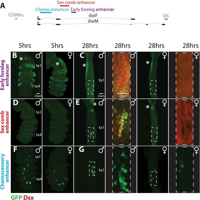
Three modular enhancers drive doublesex expression in the foreleg. (A) A map of the doublesex locus with the positions of chemosensory (blue), sex comb (red) and foreleg (purple) dsx enhancers. Thick gray lines represent the flanking genes lds and CD98hc, thick black lines are the exons of dsx, and thin black lines are the dsx introns. (B-G) Foreleg expression patterns of the three dsx enhancers, with enhancer-driven GFP in green and magnifications of 28 h samples of the early foreleg and sex comb enhancers showing Dsx antibody staining in red. hrs, h APF. The early foreleg and sex comb enhancers constructs (B-E) are directly adjacent to GFP, whereas the chemosensory enhancer (42C06; Pfeiffer et al., 2008) construct (F,G) is a GAL4 driver crossed to a UAS-GFP.nls line. (B) At 5 h APF, the early foreleg enhancer recapitulates the Dsx expression pattern in the leg epithelium (Tanaka et al., 2011) and shows a similar expression pattern in males (left) and females (right). Both sexes show ectopic expression in the proximal first tarsal segment (ta1) (asterisks in B-E). (C) At 28 h APF, the early foreleg enhancer shows clear sexual dimorphism in the first tarsal segment. Males show strong expression in the epithelial cells surrounding the sex comb and weak to no expression in the sex comb bristle cells. Females show no expression in the distal ta1, consistent with the loss of Dsx expression by this stage (red channel shows non-specific cytoplasmic background in an over-exposed image). Both males and females show ectopic expression in the proximal ta1 (asterisks), in the same region as at 5 h APF (B), that is not seen by Dsx antibody staining (Tanaka et al., 2011). (D) The sex comb enhancer is not active in either sex at 5 h APF, except for a proximal patch of ectopic expression seen near the joint in some individuals. (E) At 28 h APF, the sex comb enhancer is active in the bristle cells of the male sex comb (large nuclei), and weakly in the epithelial cells ventral to the sex comb. Females show weak expression in the distal portion of the segment. Both sexes show ectopic expression in the joint between the tibia and ta1. (F) At 5 h APF, the chemosensory enhancer is active in small clusters of cells in ta1-ta5 in both sexes, with more GFP-positive cells in males than in females. (G) At 28 h APF, both sexes show small clusters of expression in ta1-ta5; only ta1 is shown. (No Dsx antibody staining was performed in F,G.)

