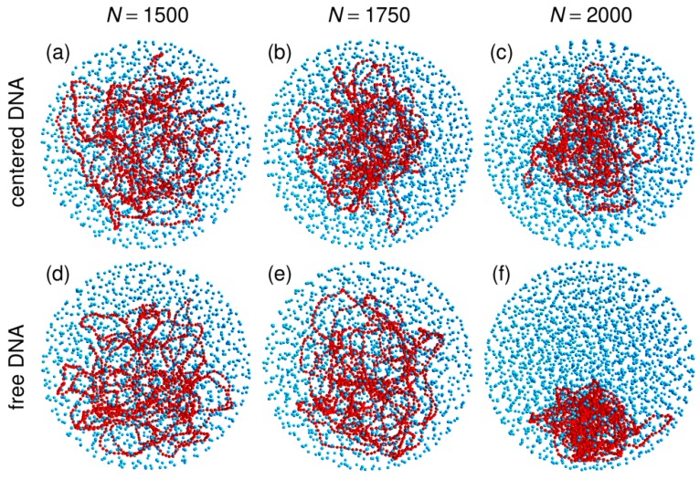
Figure 1.
Representative snapshots of simulations performed with the spherical confinement chamber and (panels (a) and (d)), 1750 (panels (b) and (e)), or 2000 (panels (c) and (f)) crowders. For panels (a), (b), and (c), the center of the confinement sphere was repositioned on top of the center of mass of the DNA chain after each integration time step, while the centering step was omitted for panels (d), (e), and (f). DNA beads are colored in red and spherical crowders in cyan. Crowders are represented at ¼ of their actual radius, in order that the DNA chain may be seen through the layers of crowders. The confinement sphere is not shown.

