The survival rate of acral lentiginous melanoma is poorer than that of other cutaneous melanoma types, largely owing to difficulty in diagnosis and more advanced stages at presentation.1 A single-center retrospective study of 53 acral melanomas found that at least 34% (n = 18) were initially misdiagnosed; of the misdiagnosed cases, 50% (n = 9) were amelanotic.2 The amelanotic variant of acral volar melanoma is scarcely reported, and its clinical and dermoscopic characteristics are unknown. Özdemir et al3 described a dermoscopic feature on the periphery of pigmented acral lentiginous melanomas as a “vascularized parallel ridge pattern,” defined as erythema and dotted vessels filling the ridges and sparing the furrows. However, volar angiomas have similarly been observed to harbor a vascularized parallel ridge pattern on dermoscopy.4,5 Herein, we describe the dermoscopic features of a subungual melanoma with an amelanotic volar component and compare these findings with the dermoscopic features of volar hemangiomas.
Report of a Case |
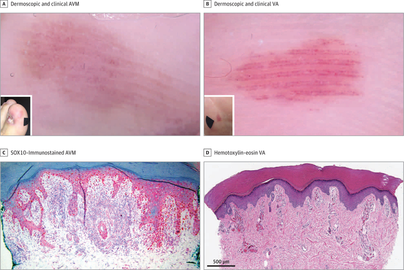
An adult man presented with a new diagnosis of melanoma of the left great toe. He reported a gradual worsening dystrophy of the left great toenail over the past 5 to 6 years. A biopsy of the nail bed confirmed a diagnosis of melanoma, at least 0.57 mm in Breslow depth. Clinical inspection of the left hallux revealed irregular brown pigmentation on the distal aspect of the dorsum of the hallux and a prominent pink tumor on the medial aspect. Dermoscopic examination of the pink plaque revealed chaotically distributed red dotted vessels on the ridges, sparing the furrows (Figure, A). A punch biopsy of the red plaque was interpreted as melanoma in situ, with both superficial acrosyringeal/eccrine duct and deep eccrine gland involvement (Figure, C). The patient underwent amputation of the left great toe at the distal interphalangeal join, and the final Breslow depth was 4.6 mm. Findings of a sentinel lymph node biopsy of the left groin were negative.
Figure. Clinical, Dermoscopic, and Histopathological Presentation of Amelanotic Volar Melanoma (AVM) and Volar Angioma (VA).
A and B, Polarized light dermoscopy, original magnification ×10. A, Dermoscopic and clinical (inset) appearance of AVMwith parallel ridge presentation of chaotically distributed red dots. B, Dermoscopic and clinical (inset) appearance of VA with parallel ridge presentation of red dots aligned at the edges of the ridges with sparing of the eccrine pores. C, Immunohistochemical analysis of AVMin situ using SOX10 immunostain, showing atypical melanocytes in the epidermis and involving the eccrine ducts, with a slight increase in the density of small vascular channels in the superficial dermis associated with inflammation (original magnification ×10). D, Histopathologic analysis of VA showing capillary vascular proliferations that extend up into the dermal papillae that surround the crista profunda intermedia and spare the eccrine structures (original magnification ×4).
We also examined 3 patients with volar hemangiomas. They all presented dermoscopically with a parallel ridge pattern composed of red-to-purple dots. In contrast to the amelanotic volar melanoma, the dots were regularly aligned at the edges of the ridges, sparing the eccrine pores (Figure, B). Histopathological examination of a biopsy specimen of a volar hemangioma revealed capillary vascular proliferations that extended up into the dermal papillae, sparing the eccrine structures (Figure, D).
Discussion |
The presence of a pigmented parallel ridge pattern has been shown in numerous studies to be associated with acral lentiginous melanoma and is particularly helpful for recognizing acral melanoma in situ.6 Herein we describe a case of acral lentiginous melanoma with prominent amelanotic volar involvement that displayed a vascular parallel ridge pattern composed of chaotically distributed red dots. Additionally, we and others have found that volar hemangioma, which is an important differential diagnosis for amelanotic acral melanoma, has a distinct dermoscopic presentation of a parallel ridge vascular pattern composed of red or purple dots aligned at the edges of the ridges and sparing the eccrine pores (the linear,double-dotted ridge pattern).4,5 Our observations require validation in future studies but may aid in distinguishing volar amelanotic melanoma from volar angioma.
Acknowledgments
Funding/Support: This work was funded in part through the National Institutes of Health/National Cancer Institute Cancer Center Support Grant P30 CA008748.
Role of the Funder/Sponsor: The funder had no role in the design and conduct of the study; collection, management, analysis, and interpretation of the data; preparation, review, or approval of the manuscript; and decision to submit the manuscript for publication.
Footnotes
Conflict of Interest Disclosures: None reported.
References
- 1.Bradford PT, Goldstein AM, McMaster ML, Tucker MA. Acral lentiginous melanoma: incidence and survival patterns in the United States, 1986–2005. Arch Dermatol 2009;145(4):427–434. doi: 10.1001/archdermatol.2008.609 [DOI] [PMC free article] [PubMed] [Google Scholar]
- 2.Soon SL, Solomon ARJ Jr, Papadopoulos D, Murray DR, McAlpine B, Washington CV. Acral lentiginous melanoma mimicking benign disease: the Emory experience. JAm Acad Dermatol 2003;48(2):183–188. doi: 10.1067/mjd.2003.63 [DOI] [PubMed] [Google Scholar]
- 3.Özdemir F, Errico MA, Yaman B, Karaarslan I. Acral lentiginous melanoma in the Turkish population and a new dermoscopic clue for the diagnosis. Dermatol Pract Concept 2018;8(2):140–148. doi: 10.5826/dpc.0802a14 [DOI] [PMC free article] [PubMed] [Google Scholar]
- 4.Phan A, Dalle S, Marcilly M-C, Bergues J-P, Thomas L. Benign dermoscopic parallel ridge pattern variants. Arch Dermatol 2011;147(5):634. doi: 10.1001/archdermatol.2011.47 [DOI] [PubMed] [Google Scholar]
- 5.Freites-Martinez A, Moreno-Torres A, Núñez AH, Martinez-Sanchez D, Huerta-Brogeras M, Borbujo J. Angioma serpiginosum: report of an unusual acral case and review of the literature. An Bras Dermatol 2015;90(3)(suppl 1): 26–28. doi: 10.1590/abd1806-4841.20153794 [DOI] [PMC free article] [PubMed] [Google Scholar]
- 6.Saida T, Miyazaki A, Oguchi S, et al. Significance of dermoscopic patterns in detecting malignant melanoma on acral volar skin: results of a multicenter study in Japan. Arch Dermatol 2004;140(10):1233–1238. doi: 10.1001/archderm.140.10.1233 [DOI] [PubMed] [Google Scholar]


