Abstract
BACKGROUND
Identification of the morphology of the genial tubercles (GTs) is valuable for different dental applications. The morphological pattern of the GTs is still controversial, and therefore, the study of its morphology using cone beam computed tomography (CBCT) plays a valuable role in resolving the controversy.
AIM
To assess the morphological pattern, dimensions and position of the GTs using CBCT among a selected Saudi population.
METHODS
CBCT records of 155 Saudi subjects (49 female and 106 male) were used to assess the pattern and size of the GTs and to determine the distance from the apices of the lower central incisors to the superior border of the GTs (I-SGT) and the distance from the inferior border of the GTs to the menton (IGT-M).
RESULTS
The results of this study showed that the most common morphological pattern was of two superior GTs and a rough impression below them (36.8%), followed by two superior GTs and a median ridge representing fused inferior GTs below them (22.6%) and a single median eminence or projection (20%). The classically described pattern, of two superior and two inferior GTs placed one above the other, was found in only 14.2% of cases, while 6.4% of the studied cases had no GTs. The mean width and height were 6.23 ± 1.93 mm and 6.67 ± 3.04 mm, respectively, while the mean I-SGT and IGT-M measurements were 8.26 ± 2.7 mm and 8.13 ± 3.07 mm, respectively.
CONCLUSION
The GTs are a controversial anatomical landmark with wide variation in their morphological pattern. The most common pattern among the studied Saudi sample was of two superior GTs and a rough impression below them, and there were no significant differences between males and females.
Keywords: Genial tubercles, Cone beam computed tomography, Morphological analysis, Mandible, Anatomical landmark
Core tip: The morphological pattern of the genial tubercles (GTs) is controversial. Classically, they are described as four elevations equidistant between the upper and lower edges of the mandible that are arranged in pairs and surround the lingual foramina bilaterally; however, several osteological and radiological studies proved that there is wide variation in their morphology. This retrospective study was conducted to determine the morphological pattern, size and position of the GTs using cone beam computed tomography among a selected Saudi population.
INTRODUCTION
Genial tubercles (GTs), also known as spinae mentalis, genial apophysis and mental spines GTs are small eminences of bone found on the lingual side of the mandible at the midline and are important landmarks for maxillofacial surgeons, radiologists, prosthodontists and general dentists[1,2].
The GTs serve as the insertion of the geniohyoid muscles in the lower and the genioglossus muscles in the upper portions of the tubercles. The action of these muscles is related to tongue mobility and swallowing, which are important for speech and feeding[3].
Although the genial tubercles are classically described in different anatomy textbooks as four mental spines on the lingual surface of the symphysis menti arranged in two pairs placed one above the other, they show different patterns in their positions and shapes[4,5].
Cone beam computed tomography (CBCT) is an accurate method to evaluate the morphology, size and position of the GTs[6,7].
Accurate identification of the GTs morphology, size and/or position using three-dimensional (3D) imaging is valuable for different applications, such as preparation for genioglossus advancement in the treatment of obstructive sleep apnea[8,9], estimation of the safe zone before implant surgery in the interforaminal region of the mandible[6] and evaluation of mandibular asymmetry on CBCT images[10].
In cases of extreme atrophy of the aged edentulous mandibles, when the GTs remain as bony projections in the floor of the mouth, they can pose a great prosthodontic challenge[11-13].
In this context, it is very important to identify the morphology of the GTs and their relation to the mandibular anterior teeth and to the margins of the mandible.
In the literature, there is no study describing the morphology of the GTs among the Saudi population; thus, the aim of this study is to determine the morphological pattern, size and position of the GTs using CBCT among a selected Saudi population.
MATERIAL AND METHODS
Study design
CBCT images used in this study were obtained from the Radiology Department Archive of the Dental Clinics Center, Qassim University, and the study protocol was approved by the Ethical Committee of the Dental Research Center, College of Dentistry, Qassim University, Saudi Arabia.
The inclusion criteria included patients from both sexes, above the age of 17 years and with full anterior dentition. The exclusion criteria included completely edentulous patients; patients with missing anterior teeth or those with mandibular asymmetry, congenital or developmental deformities; patients with traumatic injury or pathologic changes in the mandible; and patients with blurred or distorted CBCT images.
CBCT images were acquired using the Galileos® Comfort plus System (Sirona 3D, Germany) with settings as follows: X-ray generator, 98 kV and 3-6 mA. Focal spot size according to IEC 60336 was 0.5 mm, and the total filtration according to IEC 60522 was > 2.5 mm. The detector was an Image Intensifier by Siemens with the following settings: Pixels: 1000; FPS: 15-30; dynamics: 12 Bits; image volume: 15.4 cm, spherical volume – collimated 15 cm × 8.5 cm; voxel size 0.25/0.125 mm; scan time/exposure time 14 s/2-5 s. Galileos software was used, which allowed linear measurements of images and detection of the GT pattern.
CBCT scans were oriented to standardize the measurements so that the bilateral zygomatic structures were at the same level in the axial view. The infra orbital foramina of the right and left sides were parallel to the horizontal line in the coronal view. In the sagittal view, the Frankfort plane represented the true horizontal axis.
To reduce measurement error, all measurements were repeated on 2 separate occasions in 1-wk intervals, and the average values were recorded. Differences between the 2 readings were used to assess intra examiner variation with a paired t-test.
GTs pattern assessment
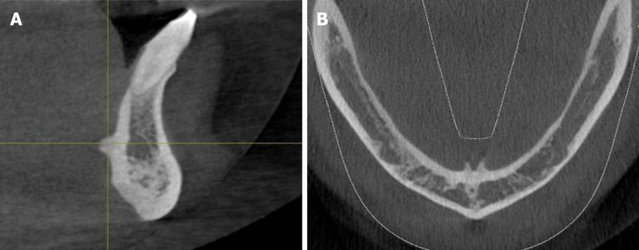
The morphological patterns of the GTs were studied and grouped into five patterns as follows: The classic description of four spines, two superior and two inferior tubercles (Type I); two superior GTs and a median ridge representing fused inferior GTs below them (Type II); two superior GTs and a rough impression below them (Type III); a single median eminence or projection (Type IV); and absence of the GTs (Type V). The pattern of the GTs was evaluated in the axial view, together with the sagittal view. (Figure 1).
Figure 1.
Detection of the genial tubercles pattern in the sagittal and axial views.
GTs position and size assessment
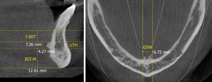
The following parameters were measured in millimeters: GTs height (GTH), GTs width (GTW), distance from the superior border of the GTs to the apex of the lower central incisors (I-SGT) and the distance from the inferior border of the GTs to the menton (IGT-M).
GTW was measured in the axial view at the level of the widest level of the GTs, while GTH was measured in the sagittal view as the vertical distance between the level of the most superior and the most inferior borders of the GTs.
I-SGT and IGT-M were measured by drawing tangential lines from the apex of the lower central incisors to the superior border of the GTs and from the inferior border of the GTs to the inferior border of the mandible (menton), respectively (Figure 2).
Figure 2.
Measurements of the genial tubercle height, distance from the superior border of the genial tubercles to the apex of the lower central incisors and the distance from the inferior border of the genial tubercles to the menton in the sagittal view and genial tubercles width in the axial view. GTH: Genial tubercles height; GTW: Genial tubercles width; I-SGT: GTs to the lower central incisors; IGT-M: Genial tubercles to the menton.
Statistical analysis
The collected data were tabulated and analyzed using SPSS 20.0 (Statistical Package for Scientific Studies) for Windows. A paired t-test was used for intra examiner calibration, and a chi square test was used for comparison between genial tubercle patterns in both sexes. All statistical tests were adjusted at a significance level of P < 0.05.
RESULTS
The data were collected from 155 Saudi patients aged 17 to 63 years of both sexes, who were treated at the Dental Clinics Center of Qassim University and fulfilled the inclusion and exclusion criteria. The sex distribution was 106 (69.4%) males and 49 females (31.6%). The intraexaminer reliability showed no statistically significant difference between the 2 image readings by using a paired t-test (P > 0.05) and had almost perfect agreement (P = 0.92 for the measurements and P = 0.94 for the pattern distribution).
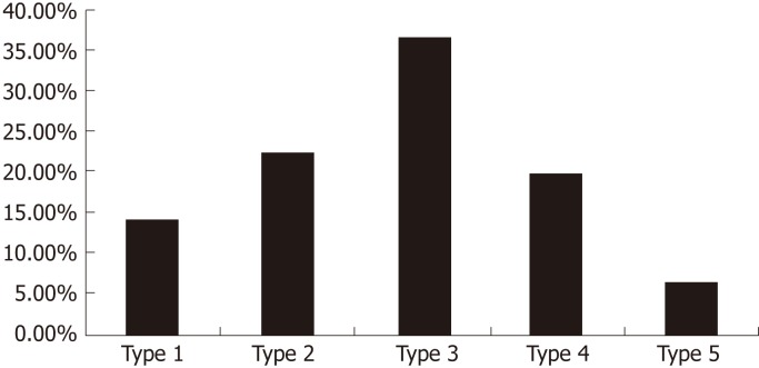
Regarding the pattern distribution of the GTs among the selected sample (Table 1 and Figure 3), Type III (36.8%) was the most common pattern, followed by Type II (22.6%), Type IV (20%) and Type I (14.2%), while Type V was the least common among the pattern types (6.4%). Regarding the correlation of the pattern of the GTs with sex, there was no statistically significant difference between sexes at P ≤ 0.05 using the chi-square test.
Table 1.
Genial tubercles pattern distribution as a function of sex
| Type |
Gender |
Total |
||||
|
Male (n = 106) (% = 32%) |
Female (n = 49) (% = 68%) |
|||||
| No | % | No | % | No | % | |
| I | 14 | 13.20% | 8 | 16.30% | 22 | 14.20% |
| II | 24 | 22.60% | 11 | 22.40% | 35 | 22.60% |
| III | 39 | 36.80% | 18 | 36.70% | 57 | 36.80% |
| IV | 23 | 21.70% | 8 | 16.30% | 31 | 20% |
| V | 6 | 5.70% | 4 | 8.20% | 10 | 6.40% |
χ2 = 1.0391, P = 0.9
Figure 3.
Genial tubercles pattern distribution.
The GTH and GTW as well as the distance from the GTs superior border to the apices of the lower central incisors (I-SGT) and the distance from the inferior border of the GTs to the menton (IGT-M) are tabulated in Table 2.
Table 2.
Dimensions of the genial tubercles
| Dimensions | Mean ± SD | Maximum | Minimum |
| GTH (mm) | 6.67 ± 3.04 | 16.08 | 2.34 |
| GTW (mm) | 6.23 ± 1.93 | 11.77 | 1.8 |
| I-SGT (mm) | 8.26 ± 2.7 | 15.82 | 1.57 |
| IGT-M (mm) | 8.13 ± 3.07 | 14.9 | 1.31 |
GTH: Genial tubercles height; GTW: Genial tubercles width; I-SGT: GTs to the lower central incisors; IGT-M: Genial tubercles to the menton.
The mean GTH measured was 6.67 ± 3.04 mm, and the mean GTW was 6.23 ± 1.93 mm. The average I-SGT was 8.26 ± 2.7 mm, and the average IGT-M was 8.13 ± 3.07 mm.
DISCUSSION
The main advantages of CBCT are accessibility, ease of handling, and availability of in-office imaging, and it offers a real-size dataset with multiplanar cross-sectional and 3-dimensional reconstructions from a single scan with a low radiation dose and relatively low cost compared with conventional computed tomography[14].
Several studies demonstrated an acceptable accuracy of linear measurements of alveolar bone and mandibular anatomy in CBCT[15]. The results of a study conducted by Hueman et al[9] showed the accuracy of CBCT in identifying the anatomic location of the GTs. Therefore, we used CBCT to explore the morphological pattern, dimensions and position of this important landmark.
The morphological pattern of the GTs is controversial and debated; classically, it is described as four elevations equidistant between the upper and lower edges of the mandible that are arranged in pairs and surround the lingual foramina bilaterally. However, several osteological and radiological studies proved that there is wide variation in their morphology.
Regarding the GTs pattern distribution among our selected sample, Type III was the most common type, and type V was the least common type, indicating that type I (the classic pattern) is not the most common type. This observation was also previously described in 2 osteological studies, one conducted among the Indian population by Singh et al[4] and one conducted among the Brazilian population by Oda et al[5].
The results of the width and height measurements of our study were 6.23 ± 1.93 mm and 6.67 ± 3.04 mm, respectively, which is in line with the results of the radiological study conducted by Wang et al[7], who examined the CBCT records of ninety Taiwanese patients and stated that the ranges of GTH measurements were 6.5-7.9 mm and the ranges of the GTW measurements were 7.1-8.2 mm. This is also in line with the study conducted by Yin et al[16], who studied the morphometry of GTs in the Chinese population with both anatomical and imaging techniques and correlated them. He concluded that the height and width of the GTs, which were measured by spiral computed tomography, were 6.17 ± 0.71 mm and 7.01 ± 1.13, respectively.
The IGT-M measurements in our study (8.13 ± 3.07 mm) were in line with the measurements made by Hueman et al[9] (11.2 ± 3.6 mm), who measured the distance from the middle part of the tubercle to the menton.
The mean I-SGT measurement in our study was 8.26 ± 2.7 mm, and a similar result was discussed by Kolsuz et al[17], who studied the anatomy of the genial tubercle using CBCT among a Turkish sample population and found that the mean I-SGT was 8.1 ± 1.7 mm in males and 7.7 ± 1.8 in females with class I occlusion.
In conclusion, the morphological pattern of the GTs is controversial; the GTs are classically described as four elevations equidistant between the upper and lower edges of the mandible that are arranged in pairs, but this is not the most common pattern. The most common pattern among the studied Saudi sample population is of two superior GTs and a rough impression below them with no significant difference between males and females. Further studies should be conducted with larger sample sizes to obtain an accurate morphological analysis of the GTs among different ethnic groups.
ARTICLE HIGHLIGHTS
Research background
The genial tubercles (GTs) are an important anatomical landmark that is located in the midline of the lingual side of the mandible and is important for multiple clinical and surgical interventions. For years, it was described as four spines arranged in two pairs, one above the other, and few osteological and radiological studies demonstrated the wide variation in its morphology. cone beam computed tomography (CBCT) is an effective and simple method to use in the assessment of GTs morphology among different ethnic groups.
Research motivation
To the best of our knowledge, no previous studies have assessed the pattern of GTs using CBCT, and no previous studies have assessed the dimensions and position of the GTs among the Saudi population or any other Arab population using either osteological or radiological methods.
Research objectives
The aim of this study was to assess the pattern, size and position of the GTs using CBCT among a selected Saudi population.
Research methods
We used CBCT images of 155 male and female Saudi subjects who fulfilled the inclusion and exclusion criteria for this study. Galileos software was used to assess the GTs pattern and to collect all the linear measurements required to determine its dimensions and position in relation to the menton and the apices of the mandibular central incisors.
Research results
Of the 155 studied subjects, 106 were males, and 49 were females; ages ranged from 17 to 63 years. According to the analysis of the pattern of the GTs , we found that the prevalence of Type III was 36.8%, followed by Type II (22.6%) and Type IV (20%), while the classically described pattern (Type I) was 14.2%. Type V was the least common among the pattern types (6.4%). There was no statistically significant difference between the sexes. Regarding its dimensions and position, we found that the mean GTs height was 6.67 ± 3.04 mm, the mean width was 6.23 ± 1.93 mm, and the average distance from the apices of the mandibular central incisors to its superior border was 8.26 ± 2.7 mm. the average distance between the GTs inferior border and the menton was 8.13 ± 3.07 mm.
Research conclusions
The morphological pattern of the GTs is controversial; the classically described GTs pattern of four elevations, equidistant between the upper and lower edges of the mandible, that are arranged in pairs, is not the most common pattern. The most common pattern among the studied Saudi sample was of two superior GTs and a rough impression below them with no significant difference between the sexes.
Research perspectives
Our results suggest that CBCT might be a simple, valuable and effective tool for conducting an accurate morphological analysis of the GTs among different ethnic groups and resolving the controversy about their morphology.
ACKNOWLEDGEMENTS
The authors would like to thank the College of Dentistry, Qassim University, Saudi Arabia, for providing the radiographic records and the approval of this study.
Footnotes
Institutional review board statement: The study protocol was reviewed and conducted with approval by the Ethical Committee, Dental Research Center, College of Dentistry, Qassim University, Saudi Arabia.
Informed consent statement: Patients were not required to give informed consent to the study because the analysis used anonymous data that were obtained from the Radiology Department Archive, Dental Clinics Center, Qassim University, KSA after each patient agreed to treatment by written consent.
Conflict-of-interest statement: All authors declare that they have no conflicts of interest.
Peer-review started: May 8, 2019
First decision: June 17, 2019
Article in press: July 25, 2019
Specialty type: Radiology, Dentistry, Oral Medicine and medical imaging
Country of origin: Saudi Arabia
Peer-review report classification
Grade A (Excellent): 0
Grade B (Very good): 0
Grade C (Good): C
Grade D (Fair): 0
Grade E (Poor): 0
P-Reviewer: Gao BL S-Editor: Cui LJ L-Editor: A E-Editor: Ma YJ
Contributor Information
Yasser A Araby, Department of Prosthetic Dental Sciences, College of Dentistry, Qassim University, Qassim 51452, Saudi Arabia.
Ahmed A Alhirabi, Dental Intern, College of Dentistry, Qassim University, Qassim 51452, Saudi Arabia. dr.yasser.araby@qudent.org.
Abdelaleem H Santawy, Department of Maxillofacial Surgery and Diagnostic Sciences, College of Dentistry, Qassim University, Qassim 51452, Saudi Arabia.
References
- 1.Thomson A. On the Presence of Genial Tubercles on the Mandible of Man, and their Suggested Association with the Faculty of Speech. J Anat Physiol. 1915;50:43–74. [PMC free article] [PubMed] [Google Scholar]
- 2.Mintz SM, Ettinger AC, Geist JR, Geist RY. Anatomic relationship of the genial tubercles to the dentition as determined by cross-sectional tomography. J Oral Maxillofac Surg. 1995;53:1324–1326. doi: 10.1016/0278-2391(95)90594-4. [DOI] [PubMed] [Google Scholar]
- 3.Ryan JM, Ross D, Obeid G. Genial tubercle fracture: a case report and review of the literature. J Oral Maxillofac Surg. 2010;68:2338–2341. doi: 10.1016/j.joms.2010.02.032. [DOI] [PubMed] [Google Scholar]
- 4.Singh V, Anand MK, Dinesh K. Variations in the pattern of mental spines and spinous mental foramina in dry adult human mandibles. Surg Radiol Anat. 2000;22:169–173. doi: 10.1007/s00276-000-0169-1. [DOI] [PubMed] [Google Scholar]
- 5.Oda LS, Iyomasa MM, Watanabe IS. Morphologic analysis of the "spina mentalis" in adult mandibles of Brazilian whites and negroes. Rev Bras Pesqui Med Biol. 1977;10:357–360. [PubMed] [Google Scholar]
- 6.Voon YS, Patil PG. Safe zone in anterior mandible related to the genial tubercle for implant osteotomy in a Chinese-Malaysian population: A CBCT study. J Prosthet Dent. 2018;119:568–573. doi: 10.1016/j.prosdent.2017.05.011. [DOI] [PubMed] [Google Scholar]
- 7.Wang YC, Liao YF, Li HY, Chen YR. Genial tubercle position and dimensions by cone-beam computerized tomography in a Taiwanese sample. Oral Surg Oral Med Oral Pathol Oral Radiol. 2012;113:e46–e50. doi: 10.1016/j.oooo.2011.11.021. [DOI] [PubMed] [Google Scholar]
- 8.Barbick MB, Dolwick MF. Genial tubercle advancement for obstructive sleep apnea syndrome: a modification of design. J Oral Maxillofac Surg. 2009;67:1767–1770. doi: 10.1016/j.joms.2009.03.051. [DOI] [PubMed] [Google Scholar]
- 9.Hueman EM, Noujeim ME, Langlais RP, Prihoda TJ, Miller FR. Accuracy of cone beam computed tomography in determining the location of the genial tubercle. Otolaryngol Head Neck Surg. 2007;137:115–118. doi: 10.1016/j.otohns.2007.02.035. [DOI] [PubMed] [Google Scholar]
- 10.Lee SY, Choi DS, Jang I, Song GS, Cha BK. The genial tubercle: A prospective novel landmark for the diagnosis of mandibular asymmetry. Korean J Orthod. 2017;47:50–58. doi: 10.4041/kjod.2017.47.1.50. [DOI] [PMC free article] [PubMed] [Google Scholar]
- 11.Păuna MR, Babiuc I, Farcaşiu AT. Prosthodontic management of an extreme atrophy of the mandible correlated with a prominent genial tubercle - a clinical report. Rom J Morphol Embryol. 2015;56:867–870. [PubMed] [Google Scholar]
- 12.Solomon EG. A critical analysis of complete denture impression procedures: contribution of early prosthodontists in India-part I. J Indian Prosthodont Soc. 2011;11:172–182. doi: 10.1007/s13191-011-0089-2. [DOI] [PMC free article] [PubMed] [Google Scholar]
- 13.Shohat I, Shoshani Y, Taicher S. Fracture of the genial tubercles associated with a mandibular denture: a clinical report. J Prosthet Dent. 2003;89:232–233. doi: 10.1067/mpr.2003.46. [DOI] [PubMed] [Google Scholar]
- 14.Fatemitabar SA, Nikgoo A. Multichannel computed tomography versus cone-beam computed tomography: linear accuracy of in vitro measurements of the maxilla for implant placement. Int J Oral Maxillofac Implants. 2010;25:499–505. [PubMed] [Google Scholar]
- 15.Ludlow JB, Laster WS, See M, Bailey LJ, Hershey HG. Accuracy of measurements of mandibular anatomy in cone beam computed tomography images. Oral Surg Oral Med Oral Pathol Oral Radiol Endod. 2007;103:534–542. doi: 10.1016/j.tripleo.2006.04.008. [DOI] [PMC free article] [PubMed] [Google Scholar]
- 16.Yin SK, Yi HL, Lu WY, Guan J, Wu HM, Cao ZY, Yu DZ, Huang YY, Wu CG. Anatomic and spiral computed tomographic study of the genial tubercles for genioglossus advancement. Otolaryngol Head Neck Surg. 2007;136:632–637. doi: 10.1016/j.otohns.2006.10.034. [DOI] [PubMed] [Google Scholar]
- 17.Kolsuz ME, Orhan K, Bilecenoglu B, Sakul BU, Ozturk A. Evaluation of genial tubercle anatomy using cone beam computed tomography. J Oral Sci. 2015;57:151–156. doi: 10.2334/josnusd.57.151. [DOI] [PubMed] [Google Scholar]


