Abstract
Carcinoid heart disease is a devastating paraneoplastic consequence of unchecked hormone production from neuroendocrine tumors (NET) and often results in right-sided heart failure. While it occurs frequently in NET patients with carcinoid syndrome, cardiac metastases occur much less often and are usually only incidentally found. Gallium-68 dotatate (ga-68) is an imaging tracer which binds to somatostatin receptor 2 with greater avidity than Indium-111, the tracer used commonly in octreotide scans. Ga-68 PET/CT is the most sensitive study for detecting occult NET metastases and has emerged as the current imaging gold standard. We describe two cases from Vanderbilt University Medical Center and Stanford University Medical Center where asymptomatic patients with well-differentiated midgut NET were diagnosed with intra-cardiac metastases using ga-68 PET/CT. Management of these patients was altered based on the findings as they underwent extensive cardiac evaluation and initiation of therapy with octreotide. Fortunately, they have not suffered life-threatening cardiac complications seen in some NET patients, from other published series, such as bradycardia, heart block, syncope and arrhythmias. These possibilities suggest early cardiology evaluation and consideration of other therapies beyond octreotide, such as surgery or PRRT, may be essential for all NET patients found to have intra-cardiac metastases.
Keywords: Well differentiated neuroendocrine tumor, Asymptomatic cardiac metastases, gallium-68 dotatate PET/CT, Carcinoid heart disease
INTRODUCTION
Carcinoid heart disease (CHD) is a major cause of morbidity and mortality in metastatic neuroendocrine tumor (NET) patients. It typically develops in the setting of a paraneoplastic process where excess NET-produced hormone byproducts escape metabolism by the liver and generate fibrous plaques that deposit on heart valves (predominantly right-sided). In patients who develop CHD, 3-year overall survival (OS) is 31%, compared to nearly double that OS rate in patients who do not develop the phenomenon [1]. While CHD develops in 50% of NET patients with carcinoid syndrome, cardiac metastases are much rarer and have been reported in <5% of patients [2,3]. In contrast to CHD, patients with these occult metastases (generally found incidentally on systemic imaging or autopsy reports), are usually asymptomatic. Cardiac metastases can arise in the myocardium of any heart chamber, septa, or pericardium. 40 cases of cardiac NET metastases from midgut origin (the most common primary site for NET), have been reported to-date and this number will likely increase as more NET patients undergo routine imaging with gallium-68 dotatate (ga-68) PET/CT [3,4,5]. The Ga-68 tracer binds to somatostatin receptor 2 (SSTR2) with greater avidity than the 111In pentetreotide tracer (used for octreotide scans) and is more sensitive for detecting bony and visceral lesions [6].
We describe here two well-differentiated midgut NET patients from Vanderbilt University Medical Center (VUMC) and Stanford University Medical Center (SUMC) with asymptomatic cardiac metastases detected by ga-68 PET/CT.
Case 1
A 54-year-old man with previously resected (2006) Stage III (T4N2) well-differentiated jejunal NET underwent a ga-68 PET/CT after establishing care with a new oncologist. Although he was completely asymptomatic, and his lab work suggested normal plasma and urine levels of 5-HIAA and chromogranin A, his oncologist ordered the scan as he had not been imaged since 2011.
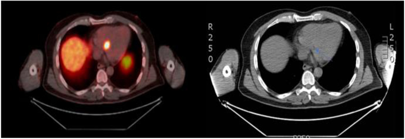
The ga-68 PET/CT revealed hypermetabolic foci in his interventricular septum and deep pelvic lymph nodes. He was referred to cardiology and a TTE confirmed a soft tissue mass in the interventricular septum. No valvular deficits or outflow tract obstruction were appreciated on the study, ECG was negative for arrhythmia and he began octreotide in early April 2018. He continues to receive octreotide without issue.
Case 2
A 75-year-old man who was initially thought to have localized well-differentiated ileal NET underwent a preoperative ga-68 PET/CT scan indicating a lesion in the interatrial septal region. He subsequently underwent resection of his primary lesion in February 2017 and began octreotide shortly thereafter. Patient was then referred to cardiology, where a cardiac MRI confirmed the interatrial septum mass. A TTE demonstrated normal ejection fraction with no evidence of valvular defects or abnormalities in his outflow tract. His most recent ga-68 PET/CT from November 2017 suggests an unchanged appearance of his interatrial septal mass. The patient remains asymptomatic, has not developed any evidence of carcinoid heart disease, and continues to receive octreotide.
DISCUSSION
Both cases represent instances where ga-68 PET/CT was able to identify occult intra-cardiac metastases in otherwise asymptomatic NET patients. Patient management was altered based on the findings from the scans, which prompted rigorous diagnostic cardiac evaluation (including cardiology evaluation, ECGs, echocardiograms and cardiac MRIs) and somatostatin analog (SSA) therapy initiation. Although these patients have not required surgical intervention based on the size, location, and symptoms from their intra-cardiac lesions, there have been some life-threatening complications (heart failure, arrhythmias, ischemic symptoms, tamponade) from NET cardiac metastases described in the literature [7,8,9,10]. In a recent case reported by Caldiera et al., a patient with small bowel NET developed two syncopal episodes from right ventricular outflow (RV) tract obstruction along with bradycardia from impaired AV conduction due to the presence of a 4.9 × 3.6 cm metastasis along the right ventricular free wall [8]. His symptoms resolved post resection of the RV tumor. Patel et al. published the experience of a small bowel NET patient with previously normal cardiac function who died of sudden cardiac arrest from a left bundle branch block; autopsy revealed interruption of bundle of His fibers by metastatic infiltration [9].Shehata et al. presented the case of a small bowel NET patient who died from complete heart block; autopsy revealed significant infiltration of the AV node by tumor [10]. Other treatment approaches in patients with more rapidly growing or symptomatic somatostatin-avid cardiac metastases include surgical resection, peptide receptor radionucleotide therapy (PRRT) or in rare cases, chemotherapy with regimens such as capecitabine and temozolomide [11].
The largest existing series about cardiac metastases from gastrointestinal NET detected by ga-68 PET/CT was published by Calissendorff et al. [3]. Among 92 NET patients in this series, 4 had cardiac metastases. Three out of the 4 patients had small bowel primaries and overt evidence of carcinoid syndrome. Two of these 3 patients had cardiac metastases at the time of diagnosis; however, only 1 patient had interventricular septal involvement. Beyond their series, the authors examined patient characteristics from previously published cases (35) of cardiac metastases from well-differentiated midgut NET. From these cases, only 5 patients had interventricular septum involvement, making it the rarest intra-cardiac metastatic site. The most common intra-cardiac metastatic sites were the left and right ventricle. Included within these cases was data from Jann et al. who reported on 28 patients [2]. 21 of 28 patients had evidence of carcinoid syndrome by biochemical evaluation, while 10 of these patients also demonstrated CHD.
CONCLUSION
Neither of our two described patients had evidence of carcinoid syndrome, CHD, or symptoms from their cardiac lesions. The first patient had a NET metastasis in his interventricular septum while the second had it in his interatrial septum. Although no direct cardiac intervention resulted from the detection of their metastases, diagnostic evaluation and SSA therapy were initiated in both these patients. The ability of ga-68 PET/CT to detect occult disease in precarious locations speaks to the power of the imaging modality and why it is being accepted as the current gold standard for imaging well differentiated NET patients. In our opinion, any NET patient with a confirmed intra-cardiac metastasis warrants, at-minimum, a cardiology evaluation to rule out life threatening complications.
Figure 1:
Cross-sectional imaging from Patient 1. On left, ga-68 PET/CT demonstrates avidity in the inter-ventricular septum. This corresponds to anatomic lesion (blue arrow) on CT imaging portion of the study seen on the right.
Figure 2:
Cross-sectional imaging from Patient 2. On left, ga-68 PET/CT demonstrates avidity in the interatrial septum. This corresponds to anatomic lesion (blue arrow) on CT imaging portion of the study seen on the right.
FUNDING
This research received no specific grant from any funding agency in the public, commercial, or not-for-profit organization.
Footnotes
DECLARATION OF CONFLICTING INTEREST
The authors have no conflicts of interest (political, personal, religious, ideological, academic, intellectual, commercial or any other) to declare in relation to this manuscript.
REFERENCES
- 1.Fox D, Khattar R. Carcinoid heart disease: presentation, diagnosis, and management. Heart 2004;90(10):1224–1228. [DOI] [PMC free article] [PubMed] [Google Scholar]
- 2.Jann H, Wertenbruch T, Pape U et al. A matter of the heart: myocardial metastases in neuroendocrine tumors. Horm Metab Res. 2010;42(13):967–76. [DOI] [PubMed] [Google Scholar]
- 3.Calissendorff J, Sundin A, Falhammar H et al. 68Ga-DOTA-TOC-PET/CT detects heart metastases from ileal neuroendocrine tumors. Endocrine 2014;47(1):169–176. [DOI] [PubMed] [Google Scholar]
- 4.Bonsen LR, Aalbersberg EA, Tesselaar M et al. Cardiac neuroendocrine tumour metastases: case reports and review of the literature. Nucl Med Commun. 2016;37(5):461–465. [DOI] [PubMed] [Google Scholar]
- 5.Coulier B, Gielen I, Colin GC et al. Multiple ileal neuroendocrine tumors (NETs) with cardiac metastasis and ectopic ileal pancreas. Diagnostic and Interventional Imaging 2018; 10.1016/j.diii.2018.04.010. [DOI] [PubMed] [Google Scholar]
- 6.Hofman M, Lau WF, Hicks R. Somatostatin Receptor Imaging with 68Ga DOTATATE PET/CT: Clinical Utility, Normal Patterns, Pearls, and Pitfalls in Interpretation. Nuclear Medicine 2015; 35(2):500–516. [DOI] [PubMed] [Google Scholar]
- 7.Vaduganathan M, Patel N, Lubitz S et al. A “Malignant” Arrhythmia: Cardiac Metastasis and Ventricular Tachycardia. Tex Heart Inst J 2016;43(6):558–559. [DOI] [PMC free article] [PubMed] [Google Scholar]
- 8.Caldiera C, Sayad D, Strosberg J et al. Surgical Treatment of an Isolated Metastatic Myocardial Neuroendocrine Tumor. Ann Thorac Surg. 2016;101(2):747–749. [DOI] [PubMed] [Google Scholar]
- 9.Patel S, Heetun M, Gurjar SV et al. A rare case of intra-cardiac metastasis from an appendiceal carcinoid tumour without liver metastases. Int J Colorectal Dis 2009;24(8):993–994. [DOI] [PubMed] [Google Scholar]
- 10.Shehata B, Thomas J, Doudenko-Rufforny I. Metastatic Carcinoid to the Conducting System—Is It a Rare or Merely Unrecognized Manifestation of Carcinoid Cardiopathy? Archives of Pathology & Laboratory Medicine 2002;126(12):1538–1540. [DOI] [PubMed] [Google Scholar]
- 11.Makis M, McCann K, Bryanton M et al. Cardiac Metastases of Neuroendocrine Tumors Treated With 177Lu DOTATATE Peptide Receptor Radionuclide Therapy or 131I-MIBG Therapy. Clinical Nuclear Medicine 2015;40(12):962–964. [DOI] [PubMed] [Google Scholar]


