Abstract
Extracts of 35 samples of European propolis were tested against wild type and resistant strains of the protozoal pathogens Trypanosoma brucei, Trypanosoma congolense and Leishmania mexicana. The extracts were also tested against Crithidia fasciculata a close relative of Crithidia mellificae, a parasite of bees. Crithidia, Trypanosoma and Leishmania are all members of the order Kinetoplastida. High levels of activity were obtained for all the samples with the levels of activity varying across the sample set. The highest levels of activity were found against L. mexicana. The propolis samples were profiled by using liquid chromatography with high resolution mass spectrometry (LC-MS) and principal components analysis (PCA) of the data obtained indicated there was a wide variation in the composition of the propolis samples. Orthogonal partial least squares (OPLS) associated a butyrate ester of pinobanksin with high activity against T. brucei whereas in the case of T. congolense high activity was associated with methyl ethers of chrysin and pinobanksin. In the case of C. fasciculata highest activity was associated with methyl ethers of galangin and pinobanksin. OPLS modelling of the activities against L. mexicana using the mass spectrometry produced a less successful model suggesting a wider range of active components.
Subject terms: Mass spectrometry, Antiparasitic agents
Introduction
Propolis is a resinous substance collected by bees, generally from plant buds. Its composition varies widely according to the vegetation surrounding the bee hive1. It is collected on the hindlegs of the bee and is removed with the help of other bees upon return to the hive and layered onto surfaces and used to fill any gaps within the hive, helping to maintain a sterile environment within the hive. In Northern Europe and other temperate regions such as Northern China and North America propolis is generally collected from the buds of poplar species1,2, whereas in Southern Europe the predominant sources are various Cypress species and in tropical regions several different plant sources may be utilised1,3. Propolis almost always displays high activity against Trypanosoma brucei and other protozoa, particularly those from the order Kinetoplastida, and we have found this to be the case regardless of the region of origin. Antiprotozoal activity has been found in propolis from Libya, Nigeria, Cameroon, Saudi Arabia and Brazil3–10. Although propolis is also antibacterial this activity is often only moderate in most samples and absent in others; generally, the strongest antibacterial activity is found in tropical propolis samples11,12. It has recently become clear that protozoal infection in bees is widespread, this was originally thought to be caused by Crithidia mellificae, which has been found to be associated with a higher incidence of winter colony collapse in Belgian bee colonies13, but it is now thought that the protozoal species Lotmaria passim14,15 is the main infecting organism. It has been found that DNA from L. passim is the most abundant DNA from a pathogenic organism within the DNA profile for the microbiome of Scottish bees16. Recently, L. passim has also been found in Africanised bees from Argentina, Uruguay and Chile and in this report a heavy burden of infection was found to be associated with a higher incidence of Varroa mite infestation17. Thus far there is no evidence that bees ingest propolis but since the spread of the protozoal infection occurs via faeces, coating the surfaces in the hive with propolis that is active against trypanosomatids could prevent transmission18. It remains an unanswered question just how important propolis is to the bee, and what its exact mechanism is in keeping down infections within the hive. European propolis has been extensively characterised and is composed of a complex mixture of >300 flavonoids and cinnamic acid derivatives19,20 and even though it has been worked on for many years there still remain components in it that have not been completely chemically or biologically characterised20, especially with regards to their antimicrobial properties. In this paper we report the activity of 35 European propolis samples against Trypanosoma brucei, Trypanosoma congolense, Leishmania mexicana and Crithidia fasciculata.
Results
Figure 1 shows the spread of the compositions of the propolis samples in a PCA model. We have previously characterised most of the major components in propolis from the UK by using accurate mass measurement with LC-MSn20. Although the samples have broadly similar compositions, there are some quite marked variations in individual components. For instance, Fig. 2 shows extracted ion chromatograms for a major component, pinobanksin acetate, across three samples from different positions in the PCA plot. Pinobanksin acetate is most abundant in the Bulgarian samples, which contain ~3.5-fold more of the compound than a sample from Northern Ireland. In contrast, Fig. 3 shows extracted ion traces for a component putatively identified as trimethyl dihydrokaempferol, which is abundant in the Northern Ireland sample but only present at low levels in the Bulgarian sample. Table 1 shows the results obtained in testing the 35 samples of European propolis against Trypanosoma brucei, Trypanosoma congolense and the multidrug resistant strain Trypanosoma brucei B48. Of these, 4 samples displayed high activity, i.e. EC50 values < 5 µg/mL, and 21 displayed intermediate activity between 5 and 10 µg/mL for the standard drug-sensitive strain Lister 427WT. The propolis samples from Norfolk displayed the highest activity, followed by the adjoining county of Suffolk and nearby Northamptonshire. The EC50 values for the multidrug resistant stain B48 were within ~1.5-fold of the control (Resistance Index (RI) 0.63–1.56; average 0.83 ± 0.04) although the RI for pentamidine was 222 (P < 0.001, Student’s unpaired t-test; Table 1).
Figure 1.
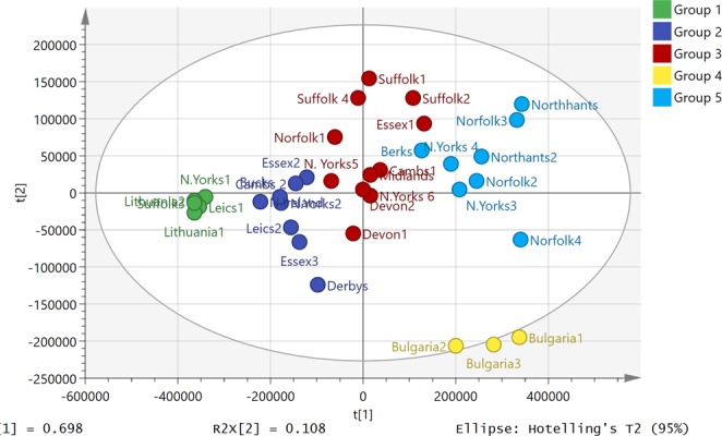
PCA plot showing the variation of propolis composition across 35 European propolis samples (Pareto scaled based on 233 components).
Figure 2.
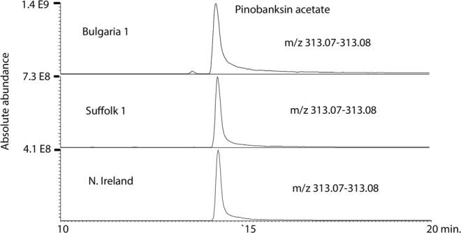
Extracted ion trace showing variation in the levels of pinobanksin acetate across 3 European propolis samples.
Figure 3.
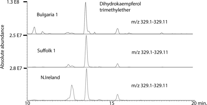
Extracted ion trace showing variation in trimethyl dihydrokaempferol across 3 European propolis samples.
Table 1.
The activity (µg/ml) of 35 European propolis samples against the standard drug-sensitive T. brucei 427WT and multi-drug resistant strain T. brucei B48, and T. congolense.
| Propolis sample | T. brucei | T. congolense | |||
|---|---|---|---|---|---|
| 427WT EC50 | B48 EC50 | R.I. | P value | IL3000 EC50 | |
| Suffolk 4, UK | 7.42 ± 0.37 | 5.7 ± 0.17 | 0.77 | 0.013 | 8.46 ± 1.47 |
| Bulgaria 1 | 5.20 ± 0.18 | 3.6 ± 0.52 | 0.69 | 0.043 | 3.69 ± 0.79 |
| Suffolk 2, UK | 6.69 ± 0.36 | 7.7 ± 1.1 | 1.15 | 0.423 | 5.66 ± 1.55 |
| North Yorkshire 1, UK | 13.5 ± 0.61 | 11.0 ± 0.70 | 0.82 | 0.058 | 18.9 ± 1.1 |
| Northamptonshire 1, UK | 4.49 ± 0.22 | 3.0 ± 0.20 | 0.67 | 0.007 | 5.69 ± 1.10 |
| Essex 1, UK | 5.97 ± 0.17 | 4.6 ± 0.26 | 0.77 | 0.013 | 4.40 ± 0.47 |
| Essex 2, UK | 14.0 ± 0.13 | 10.6 ± 1.6 | 0.75 | 0.102 | 17.3 ± 2.4 |
| Norfolk 1, UK | 5.23 ± 0.49 | 3.3 ± 0.31 | 0.63 | 0.029 | 3.08 ± 0.90 |
| Devon 1, UK | 8.57 ± 0.26 | 10.8 ± 1.2 | 1.26 | 0.144 | 11.4 ± 1.8 |
| Leicestershire 1, UK | 13.7 ± 1.18 | 11.6 ± 2.3 | 0.85 | 0.448 | 15.3 ± 3.0 |
| Leicestershire 2, UK | 17.8 ± 2.16 | 22.1 ± 1.4 | 1.24 | 0.169 | 27.6 ± 5.3 |
| Derbyshire, UK | 11.8 ± 0.57 | 9.5 ± 1.49 | 0.81 | 0.228 | 26.4 ± 4.5 |
| Lithuania 1 | 18.4 ± 1.30 | 22.1 ± 0.24 | 1.20 | 0.049 | 30.9 ± 2.8 |
| Lithuania 2 | 16.1 ± 0.93 | 25.0 ± 1.0 | 1.56 | 0.003 | 23.4 ± 1.4 |
| Suffolk 1, UK | 6.82 ± 0.87 | 4.5 ± 0.23 | 0.66 | 0.058 | 5.12 ± 0.68 |
| Suffolk 3, UK | 4.37 ± 0.18 | 2.9 ± 0.15 | 0.66 | 0.003 | 3.26 ± 1.03 |
| Bulgaria 2 | 5.80 ± 0.36 | 4.1 ± 0.41 | 0.71 | 0.036 | 2.06 ± 1.12 |
| Bulgaria 3 | 6.28 ± 0.69 | 5.3 ± 0.14 | 0.84 | 0.249 | 1.96 ± 1.01 |
| Cambridgeshire 1, UK | 9.79 ± 0.37 | 8.2 ± 0.32 | 0.84 | 0.034 | 5.65 ± 1.95 |
| Norfolk 2, UK | 6.18 ± 0.27 | 4.2 ± 0.41 | 0.68 | 0.015 | 2.13 ± 0.38 |
| Northamptonshire 2, UK | 5.24 ± 0.42 | 3.4 ± 0.39 | 0.65 | 0.030 | 4.83 ± 1.67 |
| Cambridgeshire 2, UK | 12.7 ± 0.09 | 10.3 ± 1.22 | 0.81 | 0.116 | 7.78 ± 2.15 |
| North Yorkshire 2, UK | 18.5 ± 0.48 | 14.9 ± 0.31 | 0.81 | 0.003 | 16.5 ± 3.1 |
| Northern Ireland, UK | 6.30 ± 0.33 | 6.7 ± 0.34 | 1.06 | 0.476 | 15.2 ± 4.2 |
| North Yorkshire 3, UK | 6.97 ± 0.60 | 5.4 ± 0.72 | 0.77 | 0.174 | 4.90 ± 1.53 |
| North Yorkshire 4, UK | 6.79 ± 0.45 | 4.7 ± 0.31 | 0.69 | 0.019 | 4.99 ± 2.06 |
| North Yorkshire 5, UK | 10.0 ± 0.06 | 9.0 ± 1.3 | 0.90 | 0.477 | 7.41 ± 1.25 |
| North Yorkshire 6, UK | 8.75 ± 0.34 | 7.3 ± 0.41 | 0.83 | 0.055 | 13.6 ± 3.1 |
| Essex 3, UK | 6.86 ± 0.71 | 5.4 ± 0.18 | 0.79 | 0.122 | 35.7 ± 6.5 |
| Berkshire, UK | 6.23 ± 0.12 | 4.2 ± 0.30 | 0.67 | 0.003 | 4.07 ± 1.10 |
| Midlands, UK | 5.28 ± 0.51 | 4.7 ± 0.31 | 0.89 | 0.395 | 6.12 ± 1.82 |
| Devon 2, UK | 8.68 ± 0.43 | 5.6 ± 0.23 | 0.65 | 0.003 | 7.52 ± 1.62 |
| Buckinghamshire, UK | 17.4 ± 0.96 | 13.1 ± 1.5 | 0.75 | 0.071 | 28.4 ± 6.0 |
| Norfolk 3, UK | 3.67 ± 0.30 | 2.5 ± 0.14 | 0.68 | 0.028 | 3.47 ± 0.92 |
| Norfolk 4, UK | 4.19 ± 0.21 | 2.9 ± 0.04 | 0.69 | 0.004 | 3.60 ± 0.99 |
| Pentamidine (µM) | 0.0027 ± 3.90E-04 | 0.6 ± 0.01 | 222 | <0.0001 | N.D. |
| Diminazene (µM) | N.D. | N.D. | 0.37 ± 0.12 | ||
Effective Concentration 50% (EC50) values (µg/ml) are given as averages and SEM of 3 independent experiments for T. brucei and 3–4 experiments for T. congolense. P value is based on a Student’s unpaired t-test, comparing T. brucei WT and B48. R. I. is the resistance index, being the ratio of the EC50 values for T. brucei WT and B48. N.D., not determined.
OPLS was used to model the activity of the different propolis samples against T. brucei B48 in relation to their composition. It was possible to produce a model for 33 of the samples based on 5 components, including a butyl ester of pinobanksin, which produced a reasonable fit of predicted against observed activity shown in Fig. 4 (the corresponding loadings plot is shown in Fig. S1). The highest activity was associated with a butyl ester of pinobanksin and a propionyl ester of pinobanksin. Table S1 includes MSn data used to further characterise the compounds associated with high activity. It can be seen from the extracted ion trace shown in Fig. 5 that the highest activity sample from Norfolk contains about 4 times the concentration of pinobanksin butyrate present in the lowest activity sample from Leicestershire. The wild type strain of T. brucei 427 gave similar results. Figure S2 shows an OPLS plot of predicted against measured activity with the corresponding loadings plot shown in Fig. S3. The highest activity is again associated with a butyl ester of pinobanksin and two propionyl esters of pinobanksin.
Figure 4.
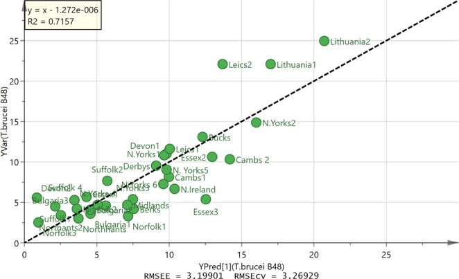
OPLS plot of observed against predicted activity against T. brucei B48 for 33 propolis samples based on five components.
Figure 5.
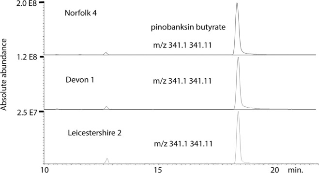
Extracted ion traces pinobanksin butyrate in samples with high, moderate and low activity against T. brucei.
The same propolis samples were also tested against the veterinary trypanosome species T. congolense (Table 1) with very similar results, as the average of the ratio of EC50 (Tbb427WT)/EC50(T. congolense) was 1.21 ± 0.11. Interestingly, the two Bulgarian samples were ~3-fold more active against T. congolense than against either of the T. brucei clones, as was one sample from Norfolk, UK. Figure 6 shows the OPLS plot obtained for the activity against T. congolense. The correlation between composition and activity was based on seven components. Figure S4 shows the corresponding loadings plot. There was a stronger fit for this plot than for the activity against T. brucei B48 and all 35 samples could be included in the model. Most active components against T. congolense are different from the most active against T. brucei and thus the OPLS plot highlights, galangin, an isomer of kaempferol, and a methylether of chrysin as the most active components (Table S1).
Figure 6.
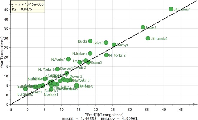
OPLS plot of observed against predicted activity against T. congolense for 35 propolis samples based on seven components.
Table 2 shows the data obtained from testing propolis against C. fasciculata which is a closer relative to the trypanosomatids that infect bees than T. brucei is. A wide range of activities were obtained. In many cases the samples were less active against C. fasciculata than against T. brucei. The OPLS model did not give as strong a correlation with the components in the sample as for T. congolense (Fig. 7) although it was possible to reduce the number of variables supporting the plot to thus giving a better indication of which components might be associated with high activity. The corresponding loadings plot is shown in Fig. S5. Galangin methyl ether is associated with high activity and this can be seen in Fig. 8 where one of the most active samples from Essex has about four times the amount of this component in comparison to a sample from Leicestershire.
Table 2.
EC50 values (µg/ml) for European propolis against C. fasciculata (n = 3).
| Propolis | C. fasciculate EC50 AVG ± SEM | Ratio EC50(Tbb)/EC50(Cf) | P value |
|---|---|---|---|
| Suffolk 4, UK | 6.41 ± 0.22 | 1.16 | 0.0798 |
| Bulgaria 1 | 3.78 ± 0.65 | 1.37 | 0.1048 |
| Suffolk 2, UK | 2.80 ± 0.47 | 2.39 | 0.0029 |
| North Yorkshire 1, UK | 8.56 ± 1.19 | 1.57 | 0.0215 |
| Northamptonshire 1, UK | 3.54 ± 0.20 | 1.27 | 0.0324 |
| Essex 1, UK | 2.72 ± 0.23 | 2.20 | 0.0004 |
| Essex 2, UK | 13.4 ± 0.94 | 1.05 | 0.5182 |
| Norfolk 1, UK | 3.05 ± 0.48 | 1.71 | 0.0340 |
| Devon 1, UK | 8.11 ± 1.43 | 1.06 | 0.7664 |
| Leicestershire 1, UK | 9.58 ± 0.25 | 1.43 | 0.0269 |
| Leicestershire 2, UK | 23.8 ± 1.85 | 0.75 | 0.1030 |
| Derbyshire, UK | 5.64 ± 0.68 | 2.09 | 0.0022 |
| Lithuania 1 | 5.92 ± 0.03 | 3.10 | 0.0007 |
| Lithuania 2 | 10.1 ± 1.56 | 1.59 | 0.0310 |
| Suffolk 1, UK | 9.46 ± 1.03 | 0.72 | 0.1213 |
| Suffolk 3, UK | 7.94 ± 0.70 | 0.55 | 0.0077 |
| Bulgaria 2 | 6.11 ± 0.66 | 0.95 | 0.6931 |
| Bulgaria 3 | 5.55 ± 0.57 | 1.13 | 0.4633 |
| Cambridgeshire 1, UK | 8.44 ± 0.69 | 1.16 | 0.1597 |
| Norfolk 2, UK | 5.64 ± 0.93 | 1.10 | 0.6068 |
| Northamptonshire 2, UK | 4.62 ± 0.56 | 1.13 | 0.4258 |
| Cambridgeshire 2, UK | 22.7 ± 1.06 | 0.56 | 0.0007 |
| North Yorkshire 2, UK | 13.7 ± 1.15 | 1.35 | 0.0187 |
| Northern Ireland, UK | 11.6 ± 0.77 | 0.54 | 0.0032 |
| North Yorkshire 3, UK | 5.04 ± 0.71 | 1.38 | 0.1062 |
| North Yorkshire 4, UK | 2.95 ± 0.25 | 2.30 | 0.0018 |
| North Yorkshire 5, UK | 7.46 ± 1.00 | 1.34 | 0.0647 |
| North Yorkshire 6, UK | 3.98 ± 0.15 | 2.20 | 0.0002 |
| Essex 3, UK | 14.0 ± 0.99 | 0.49 | 0.0043 |
| Berkshire, UK | 5.56 ± 0.70 | 1.12 | 0.4015 |
| Midlands, UK | 3.27 ± 0.54 | 1.62 | 0.0540 |
| Devon 2, UK | 2.58 ± 0.43 | 3.36 | 0.0006 |
| Buckinghamshire, UK | 21.4 ± 1.34 | 0.81 | 0.0716 |
| Norfolk 3, UK | 4.34 ± 0.35 | 0.84 | 0.2208 |
| Norfolk 4, UK | 4.21 ± 0.49 | 1.00 | 0.9715 |
| PAOa (µM) | 5.35 ± 4.72 | 5.44 | 5.17 |
aPAO = phenylarsine oxide.
Figure 7.
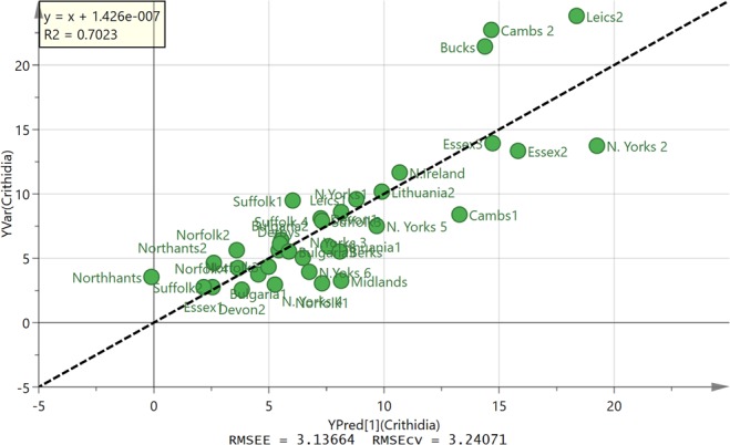
OPLS model of predicted against observed activity for propolis against C. fasciculata based on 4 components.
Figure 8.
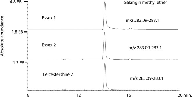
Extracted ion traces for galangin methyl ether in samples with high, moderate and low activity against C. fasciculata.
Table 3 shows the activity obtained for 25 of the propolis samples against L. mexicana. The activity of the propolis samples against L. mexicana was higher than that obtained against T. brucei, with average EC50 values below 1 µg/mL for 52% of samples, and all EC50 values were under 5 µg/mL. The highest activity was obtained for one of the Bulgarian samples, at 0.35 ± 0.03 µg/mL. In most cases activity was equal or superior against the miltefosine APC12-resistant cell line, giving an average Resistance Index of 0.74 ± 0.09, but it was not possible to fit a strong an OPLS model for the data obtained for L. mexicana as for the T. brucei data, probably because the range of activities obtained across the samples is lower than for T. brucei and the number of samples tested was smaller. The activities obtained against Leishmania were an order of magnitude higher than those obtained for C. fasciculata and T. brucei, as shown in Fig. 9.
Table 3.
The activity (µg/ml) of propolis against wild type and miltefosine-APC12 resistant L. mexicana (C12Rx).
| Propolis ID | L. mexicana wild type | L. mexicana C12Rx | Resistance Index | ttest |
|---|---|---|---|---|
| (µg/mL) | (µg/mL) | |||
| Suffolk 4, UK | 1.04 ± 0.19 | 0.81 ± 0.15 | 0.78 | 0.40 |
| Bulgaria 1 | 0.35 ± 0.03 | 0.29 ± 0.04 | 0.85 | 0.33 |
| Suffolk 2, UK | 0.85 ± 0.14 | 0.45 ± 0.03 | 0.53 | 0.048 |
| North Yorkshire 1, UK | 0.90 ± 0.17 | 0.94 ± 0.15 | 0.96 | 0.87 |
| Northamptonshire 1, UK | 0.59 ± 0.05 | 0.28 ± 0.08 | 0.48 | 0.029 |
| Essex 1, UK | 0.62 ± 0.07 | 0.37 ± 0.07 | 0.60 | 0.073 |
| Essex 2, UK | 0.89 ± 0.10 | 0.42 ± 0.09 | 0.47 | 0.027 |
| Norfolk 1, UK | 1.94 ± 0.44 | 0.61 ± 0.003 | 0.31 | 0.027 |
| Devon 1, UK | 4.97 ± 0.23 | 0.95 ± 0.16 | 0.25 | 0.00014 |
| Leicestershire 1, UK | 5.67 ± 0.43 | 1.33 ± 0.09 | 0.23 | 0.00058 |
| Leicestershire 2, UK | 4.71 ± 0.33 | 1.06 ± 0.02 | 0.23 | 0.00041 |
| Derbyshire, UK | 1.23 ± 0.08 | 0.50 ± 0.17 | 0.41 | 0.016 |
| Lithuania 1 | 1.51 ± 0.06 | 1.35 ± 0.02 | 0.89 | 0.064 |
| Lithuania 2 | 0.65 ± 0.12 | 1.55 ± 0.01 | 2.38 | 0.0018 |
| Suffolk 1, UK | 0.67 ± 0.05 | 0.79 ± 0.09 | 1.17 | 0.32 |
| Suffolk 3 UK | 1.02 ± 0.18 | 0.50 ± 0.04 | 0.49 | 0.048 |
| Bulgaria 2 | 1.13 ± 0.17 | 0.69 ± 0.22 | 0.61 | 0.19 |
| Bulgaria 3 | 1.17 ± 0.18 | 0.78 ± 0.11 | 0.67 | 0.14 |
| Cambridgeshire 1, UK | 2.38 ± 0.40 | 1.53 ± 0.21 | 0.64 | 0.13 |
| Norfolk 2, UK | 0.93 ± 0.06 | 0.60 ± 0.05 | 0.65 | 0.020 |
| Northamptonshire 2, UK | 0.65 ± 0.05 | 0.49 ± 0.002 | 0.78 | 0.018 |
| North Yorkshire 2 | 2.68 ± 0.15 | 1.36 ± 0.08 | 0.51 | 0.003 |
| Northern Ireland | 0.61 ± 0.05 | 0.78 ± 0.17 | 1.27 | 0.17 |
| North Yorkshire 4, UK | 0.72 ± 0.22 | 0.67 ± 0.06 | 0.94 | 0.75 |
| North Yorkshire 5, UK | 0.42 ± 0.12 | 0.58 ± 0.07 | 1.38 | 0.12 |
| Miltefosine APC 12 | 0.1 ± 0.03 | 67.0 ± 12.6 | 670 | <0.001 |
| Miltefosine APC 16 | 2.0 ± 0.20 | 56 ± 9.7 | 28 | <0.001 |
All EC50 values are given as average ± SEM (n = 3). Statistical difference between EC50 values of the same sample against two strains was analysed using Student’s unpaired t-test.
Figure 9.
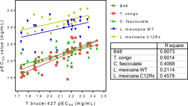
Correlation between the EC50 values of propolis samples against T. brucei 427WT and the other parasite strains and species.
Discussion
The importance of propolis to bees is not entirely clear, in so far as some strains of bee do not collect much of it. However, experimental work has been carried out in order to establish the role of propolis in protecting the hive against infection21–29. There is evidence that bees that collect greater amounts of propolis are healthier and produce more viable broods than bees which are selected for reduced propolis collection21. Bees that collected propolis were found to exhibit superior hygienic behaviour in comparison with those that collected less22. It was found that a parasite challenge encouraged bees to collect more propolis and that the propolis envelop improved the immunity of colonies against infection23–27. As in the current study, regional variations in the antimicrobial properties of propolis have been found to exist28. Several acyl esters of flavonoids were recently isolated from temperate propolis and were fully characterised by spectroscopic methods. The isolated compounds were tested against honey bee pathogens Paenibacillus larvae (American foulbrood) and Ascosphaera apis (chalkbrood). The most active compound against A. apis was pinobanksin 3-butyrate while the most active compound against Paenibacillus larvae was pinobanksin 3-octanoate29. The OPLS model for activity against T. brucei reflects this with two butyrate esters of pinobanksin being associated with the highest activity samples. In the current case the EC50 values against T. brucei in µg/ml terms are similar to those obtained for purified pinobanksin butyrate tested against A. apis. Interestingly the most active acyl flavonoid tested against Paenibacillus larvae was different from the most active against A. apis and this would seem to be the same in the current case, particularly for C. fasciculata, where the most active components were a methyl ether of galangin, a methyl ether of pinobanksin and pinobanksin. Thus propolis would appear to have broad spectrum activity with individual components in the mixture having activity against different organisms. Figure 9 shows a good overall correlation between the effects of the various samples against each of the kinetoplastid species. Especially between T. brucei and T. congolense the correlation is very close, which is important as African animal trypanosomiasis is caused by multiple Trypanosoma species including T. congolense, T. b. brucei and, in Eastern Africa, T. b. rhodesiense30 and the disease has now spread far beyond Africa for T. vivax and T. evansi31. Even more important is that the correlation between the drug-resistant and the sensitive strains was very good, with activity against the resistant strains on average better than against the parental strains. This is in line with previous observations that cross-resistance with current drugs will not be a problem for propolis-derived phytochemicals3,6, although it cannot be denied that resistance to any new, propolis-derived compound is likely to arise at some point unless a suitable combination therapy can be devised32.
The consistent high levels of activity obtained for propolis extracts against protozoa coupled with the prevalence of protozoal DNA amongst the DNA of pathogenic species in the metagenome16 of the bee suggests that these organisms may exert a greater pressure than may be currently appreciated on bee health. There remains much to understand about the role of propolis in bee health and also with regard to its potential in treating human infections, and the broad anti-kinetoplastid activity of propolis components reported here gives ample scope for further investigations.
Materials and Methods
Chemicals and materials
Absolute ethanol, HPLC grade acetonitrile, methanol, formic acid, water and Acrodisc syringe filters were obtained from Fisher Scientific (Loughborough, UK). 36 raw propolis samples were collected from different areas of the UK and Europe following a request by Mr James Fearnley for people to submit samples for testing. Miltefosine analogues APC12 and APC16 were obtained from Anatrace (Ohio, USA).
Extraction of propolis samples
A sample of each propolis sample (500 mg) was extracted with 10 ml of ethanol by sonication for 1 h. The solvent was evaporated under a stream of nitrogen and the extracts were weighed and then redissolved in 5 ml of ethanol and then aliquoted into volumes containing 10 mg which were then blown to dryness under a stream of nitrogen.
LC-MS Conditions
LC-MS was carried out by using an Accela pump connected to an Orbitrap Exactive mass spectrometer operated in positive/negative switching mode. The sheath gas and auxiliary gas were set at 50 and 17 arbitrary units, respectively. The needle voltage was 4.5 kV in positive mode and 4.0 kV in negative mode. The heated capillary temperature was 320 °C. The HPLC was fitted with an ACE C18 column 150 × 4.6 mm, 3 µM particle size (Hichrom, Reading, UK). Solvent A was 0.1% formic acid in water and solvent B was 0.1% formic acid in acetonitrile. The flow rate was 0.3 ml/min and the solvent gradient was as follows: 0 min 30% B, 30 min 100% B, 40 min 100% B, 41 min 30% B, 50 min 30% B. The files were processed by using m/z Mine 20.1 and then the masses were searched against an in-house database. The extracted data was then processed by using Simca P 14.1 (Umetrics, Umea, Sweden). To produce PCA and OPLS models33,34. MSn experiments for characterisation of the activity marker compounds were carried out on an LTQ Orbitrap with a collision energy of 35 V and used the chromatographic and mass spectrometry conditions given above.
Strains and cultures
Bloodstream forms of T. b. brucei were grown in standard HMI-9 medium with 10% fetal bovine serum at 37 °C/5% CO2, in vented culture flasks, exactly as described)35. The standard laboratory strain Lister 427WT36 was used as drug sensitive standard and the multi-drug resistant clone B4837 was used to assess the potential for cross-resistance with the diamidine and melaminophenyl arsenical classes of trypanocides. T. congolense strain IL3000 (Savannah-type) was cultured as described previously in Minimal Essential Medium (MEM) base with 10% goat serum, supplemented with 14 µL/L β-mercapto-ethanol, glutamine and antibiotics as described38.
Transgenic Leishmania mexicana promastigotes (5 × 10⁶ cells.ml) of strain MYNC/BZ/62/M379 expressing the firefly luciferase gene and sensitive to the miltefosine APC12 with 12 alkyl carbon chain called APC1239 were designated WT; a related strain, C12Rx, resistant to 80 µg/mL APC12, was selected under controlled conditions by a stepwise progressive increase of APC12 (Fig. S6), with surviving stationary phase cells at each dose, used to inoculate subsequent cultures. Cells able to grow in the presence of the drug were cloned under drug pressure by limiting dilution to 1 cell/ml in 20 ml of growth medium and plated out into 96-well plates. Both were cultured in complete Modified Eagle’s Medium (M199 supplemented with 10% (v/v) heat inactivated foetal calf serum) at 25 °C. The transgenic line cultures were further supplemented with Hygromycin B in order to retain the luciferase gene.
A standard wild-type C. fasciculata (strain HS6, kind gift of Professor Terry K. Smith, University of St-Andrews, UK) was grown at 27 °C in axenic serum-free defined media containing yeast extract (5 mg/mL), tryptone (4 mg/mL), sucrose (15 mg/mL), triethanolamine (4.4 mg/mL) and Tween 80 (0.5%) and supplemented with 10 μg/mL of haemin, exactly as described by Kipandula et al.40.
Testing against T. brucei, T. congolense and C. fasciculata
The extracts were tested against T. brucei as described previously3,6, using our standard Alamar blue® (resazurin) method in white opaque 96 well plates (Greiner Bio-One, Frickenhausen, Germany), with 23 doubling dilutions and a no-drug control for each sample, using 2 × 10⁴ T. brucei or 5 × 10⁴ T. congolense per well and incubating 48 h with test compound prior to the addition of resazurin sodium salt (Sigma) and a further incubation of 24 h. The method is based on live but not cells metabolizing blue, non-fluorescent resazurin to pink, fluorescent resorufin, with fluorescence intensity being proportional to cell numbers41. Stock solutions of each compound or mixture prepared in DMSO for each concentration so that there was a constant percentage of DMSO per well (1% v/v).
Testing against C. fasciculata involved a very similar procedure, using 5 × 10³ cells/well and incubations of 48 h and 24 h (27 °C, 5% CO2) before and after the addition of resazurin, respectively. Cell densities were determined using a haemocytometer after adding 1% v/v glycerol to the culture sample to immobilize the parasites. Cell density was then adjusted to 5 × 10⁴ cells/mL with fresh medium, of which 100 µL was added to each well of a pre-prepared 96-well plate with the doubling dilution of test compound/sample.
Fluorescence was determined using a FLUOstar Optima (BMG Labtech, Durham, NC, USA) plate reader (λex = 544 nm; λem = 590 nm) and the output was plotted to a sigmoid curve with variable slope (Prism 5.0, GraphPad software) to obtain 50% effective concentrations (EC50 values).
Testing against L. mexicana
A miltefosine APC12-resistant L. mexicana was strain was selected as shown in Fig. S6. Both cell lines were screened with propolis samples at a starting concentration of 0.125 mg/ml, doubly diluted eleven times across a 96 well plate in triplicate and incubated for 72 h at 25 °C. Wells with no propolis added were used in control experiments. After, luciferin solution (1 µg/ml) was added and the light emitted was measured using a luminometer (Biotek Synergy HT) at a wavelength of 440/40 nm. Viability was taken to be proportional to light emitted from for each drug-treated well, and was expressed as a faction of emission from the ‘no drug’ control. IC50 values were determined using Prism 5.0, GraphPad software.
Supplementary information
Acknowledgements
GUE was supported by a studentship from the Nigerian Tertiary Education Trust Fund. AA was supported by a Saudi Government studentship.
Author Contributions
A.A., S.A., A.M.D., S.G., G.U.E., D.G.W. and R.W. carried out the experimental work and data processing and interpretation. W.S., D.G.W., J.O.I., H.P.d.K. and J.F. contributed to the authorship of the manuscript. J.F. collected the propolis samples.
Data Availability
The datasets generated during and/or analysed during the current study are available from the corresponding author on reasonable request.
Competing Interests
The authors declare no competing interests.
Footnotes
Publisher’s note: Springer Nature remains neutral with regard to jurisdictional claims in published maps and institutional affiliations.
Supplementary information
Supplementary information accompanies this paper at 10.1038/s41598-019-47840-y.
References
- 1.Bankova V, Popova M, Trusheva B. The phytochemistry of the honeybee. Phytochemistry. 2018;155:1–11. doi: 10.1016/j.phytochem.2018.07.007. [DOI] [PubMed] [Google Scholar]
- 2.Wilson MB, Spivak M, Hegeman AD, Rendahl A, Cohen JD. Metabolomics Reveals the Origins of Antimicrobial Plant Resins Collected by Honey Bees. PloS one. 2013;8:e77512. doi: 10.1371/journal.pone.0077512. [DOI] [PMC free article] [PubMed] [Google Scholar]
- 3.Omar RM, et al. Chemical characterisation of Nigerian red propolis and its biological activity against Trypanosoma brucei. Phytochemical Analysis. 2016;27:107–115. doi: 10.1002/pca.2605. [DOI] [PubMed] [Google Scholar]
- 4.Siheri W, et al. Chemical and antimicrobial profiling of propolis from different regions within Libya. PLoS One. 2016;11:e0155355. doi: 10.1371/journal.pone.0155355. [DOI] [PMC free article] [PubMed] [Google Scholar]
- 5.Siheri W, et al. The isolation of antiprotozoal compounds from Libyan propolis. Phytotherapy research. 2014;28:1756–1760. doi: 10.1002/ptr.5194. [DOI] [PubMed] [Google Scholar]
- 6.Omar R, et al. The chemical characterization of Nigerian propolis samples and their activity against Trypanosoma brucei. Scientific reports. 2017;7:923. doi: 10.1038/s41598-017-01038-2. [DOI] [PMC free article] [PubMed] [Google Scholar]
- 7.Nweze NE, et al. Effects of Nigerian red propolis in rats infected with Trypanosoma brucei brucei. Comparative Clinical Pathology. 2017;26:1129–1133. doi: 10.1007/s00580-017-2497-0. [DOI] [Google Scholar]
- 8.Almutairi S, et al. New anti-trypanosomal active prenylated compounds from African propolis. Phytochemistry Letters. 2014;10:35–39. doi: 10.1016/j.phytol.2014.07.002. [DOI] [Google Scholar]
- 9.Almutairi S, et al. Isolation of diterpenes and flavonoids from a new type of propolis from Saudi Arabia. Phytochemistry letters. 2014;10:160–163. doi: 10.1016/j.phytol.2014.08.022. [DOI] [Google Scholar]
- 10.Do Nascimento TG, et al. Polymeric Nanoparticles of Brazilian red propolis extract: preparation, characterization, antioxidant and leishmanicidal activity. Nanoscale research letters. 2016;11:301. doi: 10.1186/s11671-016-1517-3. [DOI] [PMC free article] [PubMed] [Google Scholar]
- 11.Seidel V, Peyfoon E, Watson DG, Fearnley J. Comparative study of the antibacterial activity of propolis from different geographical and climatic zones. Phytotherapy Research. 2008;22:1256–1263. doi: 10.1002/ptr.2480. [DOI] [PubMed] [Google Scholar]
- 12.Raghukumar R, Vali L, Watson D, Fearnley J, Seidel V. Antimethicillin‐resistant Staphylococcus aureus (MRSA) activity of ‘pacific propolis’ and isolated prenylflavanones. Phytotherapy research. 2010;24:1181–1187. doi: 10.1002/ptr.3096. [DOI] [PubMed] [Google Scholar]
- 13.Ravoet J, et al. Comprehensive bee pathogen screening in Belgium reveals Crithidia mellificae as a new contributory factor to winter mortality. PLoS One. 2013;8:e72443. doi: 10.1371/journal.pone.0072443. [DOI] [PMC free article] [PubMed] [Google Scholar]
- 14.Schwarz RS, et al. Characterization of two species of trypanosomatidae from the honey bee Apis mellifera: Crithidia mellificae Langridge and McGhee, and Lotmaria passim n. gen., n. sp. Journal of Eukaryotic Microbiology. 2015;62:567–583. doi: 10.1111/jeu.12209. [DOI] [PubMed] [Google Scholar]
- 15.Ravoet J, et al. Differential diagnosis of the honey bee trypanosomatids Crithidia mellificae and Lotmaria passim. Journal of invertebrate pathology. 2015;130:21–27. doi: 10.1016/j.jip.2015.06.007. [DOI] [PubMed] [Google Scholar]
- 16.Regan T, et al. Characterisation of the British honey bee metagenome. Nature. Communications. 2018;9:4995. doi: 10.1038/s41467-018-07426-0. [DOI] [PMC free article] [PubMed] [Google Scholar]
- 17.Castelli L, et al. Detection of Lotmaria passim in Africanized and European honey bees from Uruguay, Argentina and Chile. Journal of invertebrate pathology. 2018;160:95–97. doi: 10.1016/j.jip.2018.11.004. [DOI] [PubMed] [Google Scholar]
- 18.Ruiz‐Gonzalez MX, Brown MJ. Honey bee and bumblebee trypanosomatids: specificity and potential for transmission. Ecological Entomology. 2006;31:616–622. doi: 10.1111/j.1365-2311.2006.00823.x. [DOI] [Google Scholar]
- 19.De Groot, A. C., Popova, M. P. & Bankova, V. S. An update on the constituents of poplar-type propolis. Wapserveen, The Netherlands: Acdegroot publishing, ISBN 978-90-813233-0-7 (2014).
- 20.Saleh K, Zhang T, Fearnley J, Watson DG. A comparison of the constituents of propolis from different regions of the United Kingdom by liquid chromatography-high resolution mass spectrometry using a metabolomics approach. Current Metabolomics. 2015;3:42–53. doi: 10.2174/2213235X03666150328000505. [DOI] [Google Scholar]
- 21.Simone-Finstrom M, Spivak M. Propolis and bee health: the natural history and significance of resin use by honey bees. Apidologie. 2010;41:295–311. doi: 10.1051/apido/2010016. [DOI] [Google Scholar]
- 22.Nicodemo D, Malheiros EB, De Jong D, Couto RHN. Increased brood viability and longer lifespan of honeybees selected for propolis production. Apidologie. 2014;45:269–275. doi: 10.1007/s13592-013-0249-y. [DOI] [Google Scholar]
- 23.Nicodemo D, De Jong D, Couto RHN, Malheiros B. Honey bee lines selected for high propolis production also have superior hygienic behavior and increased honey and pollen stores. Genetics and Molecular Research. 2013;12:6931–6938. doi: 10.4238/2013.December.19.12. [DOI] [PubMed] [Google Scholar]
- 24.Simone-Finstrom MD, Spivak M. Increased resin collection after parasite challenge: a case of self-medication in honey bees? PloS one. 2012;7:e34601. doi: 10.1371/journal.pone.0034601. [DOI] [PMC free article] [PubMed] [Google Scholar]
- 25.Simone-Finstrom M, Borba R, Wilson M, Spivak M. Propolis counteracts some threats to honey bee health. Insects. 2017;8:46. doi: 10.3390/insects8020046. [DOI] [PMC free article] [PubMed] [Google Scholar]
- 26.Borba, R. S., Klyczek, K. K., Mogen, K. L. & Spivak, M. Seasonal benefits of a natural propolis envelope to honey bee immunity and colony health. Journal of Experimental Biology, jeb. 127324 (2015). [DOI] [PubMed]
- 27.Borba RS, Spivak M. Propolis envelope in Apis mellifera colonies supports honey bees against the pathogen, Paenibacillus larvae. Scientific Reports. 2017;7:11429. doi: 10.1038/s41598-017-11689-w. [DOI] [PMC free article] [PubMed] [Google Scholar]
- 28.Wilson M, Brinkman D, Spivak M, Gardner G, Cohen JD. Regional variation in composition and antimicrobial activity of US propolis against Paenibacillus larvae and Ascosphaera apis. Journal of invertebrate pathology. 2015;124:44–50. doi: 10.1016/j.jip.2014.10.005. [DOI] [PubMed] [Google Scholar]
- 29.Wilson MB, et al. 3-Acyl dihydroflavonols from poplar resins collected by honey bees are active against the bee pathogens Paenibacillus larvae and Ascosphaera apis. Phytochemistry. 2017;138:83–92. doi: 10.1016/j.phytochem.2017.02.020. [DOI] [PubMed] [Google Scholar]
- 30.Giordani F, Morrison LJ, Rowan TG, De Koning HP, Barrett MP. The animal trypanosomiases and their chemotherapy: a review. Parasitology. 2016;143:1862–1889. doi: 10.1017/S0031182016001268. [DOI] [PMC free article] [PubMed] [Google Scholar]
- 31.Aregawi WG, Agga GE, Abdi RD, Büscher P. Systematic review and meta-analysis on the global distribution, host range, and prevalence of Trypanosoma evansi. Parasites & Vectors. 2019;12:67. doi: 10.1186/s13071-019-3311-4. [DOI] [PMC free article] [PubMed] [Google Scholar]
- 32.de Koning HP. Drug resistance in protozoan parasites. Emerging Topics in Life Sciences. 2017;1:627–632. doi: 10.1042/ETLS20170113. [DOI] [PMC free article] [PubMed] [Google Scholar]
- 33.Eriksson, L., Byrne, T., Johansson, E., Trygg, J. & Vikstrom, C. In Multi- and Megavariate Data Analysis: Basic Principles and Application Ch. 503, 455–456 (MKS Umetrics AB, 2013).
- 34.Eriksson L, Trygg J, Wold S. CV-ANOVA for significance testing of PLS and OPLS (R) models. J Chemometr. 2008;22:594–600. doi: 10.1002/cem.1187. [DOI] [Google Scholar]
- 35.Gudin S, et al. Trypanosoma brucei: a survey of pyrimidine transport activities. Experimental parasitology. 2006;114:118–125. doi: 10.1016/j.exppara.2006.02.018. [DOI] [PubMed] [Google Scholar]
- 36.de Koning HP, MacLeod A, Barrett MP, Cover B, Jarvis SM. Further evidence for a link between melarsoprol resistance and P2 transporter function in African trypanosomes. Molecular and biochemical parasitology. 2000;106:181–185. doi: 10.1016/S0166-6851(99)00206-6. [DOI] [PubMed] [Google Scholar]
- 37.Bridges DJ, et al. Loss of the high affinity pentamidine transporter is responsible for high levels of cross-resistance between arsenical and diamidine drugs in African trypanosomes. Molecular pharmacology. 2007;71:1098–1108. doi: 10.1124/mol.106.031351. [DOI] [PubMed] [Google Scholar]
- 38.Cerone Michela, Uliassi Elisa, Prati Federica, Ebiloma Godwin U., Lemgruber Leandro, Bergamini Christian, Watson David G., de A. M. Ferreira Thais, Roth Cardoso Gabriella Simões Heyn, Soares Romeiro Luiz A., de Koning Harry P., Bolognesi Maria Laura. Discovery of Sustainable Drugs for Neglected Tropical Diseases: Cashew Nut Shell Liquid (CNSL)‐Based Hybrids Target Mitochondrial Function and ATP Production in Trypanosoma brucei. ChemMedChem. 2019;14(6):621–635. doi: 10.1002/cmdc.201800790. [DOI] [PMC free article] [PubMed] [Google Scholar]
- 39.Hurrell BP, et al. Rapid sequestration of Leishmania mexicana by neutrophils contributes to the development of chronic lesion. PLoS pathogens. 2015;11:e1004929. doi: 10.1371/journal.ppat.1004929. [DOI] [PMC free article] [PubMed] [Google Scholar]
- 40.Kipandula W, Young SA, MacNeill SA, Smith TK. Screening of the MMV and GSK open access chemical boxes using a viability assay developed against the kinetoplastid Crithidia fasciculata. Molecular and biochemical parasitology. 2018;222:61–69. doi: 10.1016/j.molbiopara.2018.05.001. [DOI] [PubMed] [Google Scholar]
- 41.Gould MK, Vu XL, Seebeck T, de Koning HP. Propidium iodide-based methods for monitoring drug action in the kinetoplastidae: comparison with the Alamar Blue assay. Analytical biochemistry. 2008;382:87–93. doi: 10.1016/j.ab.2008.07.036. [DOI] [PubMed] [Google Scholar]
Associated Data
This section collects any data citations, data availability statements, or supplementary materials included in this article.
Supplementary Materials
Data Availability Statement
The datasets generated during and/or analysed during the current study are available from the corresponding author on reasonable request.


