Abstract
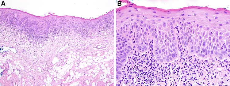
Leukoplakia and erythroplakia are two entities under the moniker of “oral potentially malignant disorders” that are highly associated with the presence of oral epithelial dysplasia (OED) at first biopsy, while lesions of submucous fibrosis develop OED after being present for years. Importantly, traumatic/frictional keratoses are often mistaken clinically for leukoplakia and it is important for the pathologist to recognize and report them as such. The features of OED have been well-described and other architectural features will be discussed here, in particular verrucous and papillary architecture, bulky epithelial proliferation and epithelial atrophy. Proliferative leukoplakia, verrucous or otherwise, often show only hyperkeratosis in early lesions, with development of OED occurring over time, and squamous cell carcinoma developing in the majority of cases over time. The concept of hyperkeratosis without features of OED and that is not reactive, is likely a precursor to the dysplastic phenotype. Many cases of leukoplakia exhibiting OED are associated with a band of lymphocytes at the interface and these should not be mistaken for oral lichen planus.
Keywords: Dysplasia, Architectural, Organizational, Keratosis of unknown significance, Lichenoid mucositis
Introduction
Oral premalignant disorders, also termed oral potentially malignant disorders, are a group of conditions that has been defined by the WHO in 2017 as “clinical presentations that carry a risk of cancer development in the oral cavity, whether in a clinically definable precursor lesion or in clinically normal mucosa” [1, 2]. Leukoplakia and its variants, erythroplakia and submucous fibrosis (most prevalent in India) are three conditions that are highly associated with the development of oral epithelial dysplasia (OED) and oral squamous cell carcinoma (SCC). Whether oral lichen planus (OLP) should be considered a potentially malignant lesion is controversial since OLP is a heterogenous group of lesions with a recognizable clinical phenotype, but where the histopathology may be mimicked by other conditions, in particular OED [2–4]. Furthermore, frictional and factitial keratoses may show reactive epithelial atypia that may be mistaken for OED. As such, one of the most important factors in arriving at a correct histopathologic diagnosis, is correlation with the clinical appearance of such white lesions.
The objective of this paper is to (1) briefly review the clinical lesions, (2) review the features of OLP, reactive keratoses and OED (especially in relation to architectural features), (3) discuss the concept of leukoplakias that do not show the usual dysplastic phenotype and their clinical significance, (4) review the concept of lichenoid mucositis as it pertains to OED, and (5) suggest how histopathologic diagnoses can be rendered more useful for the clinician.
Clinical Findings
While erythroplakia, a red pebbly,granular plaque, is the least common of the oral premalignant lesions, over 90% of such lesions exhibit OED, carcinoma-in situ (CIS) or SCC at first biopsy (Fig. 1) [5–7]. Submucous fibrosis caused by chronic use of areca nut in various preparations shows marble-like pallor and palpable dense bands in the mucosa, fibrosis only in early lesions, while late lesions develop OED; the relative risk for malignant transformation is 6.19 [8, 9].
Fig. 1.
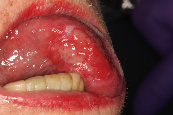
Erythroplakia: a mostly red, ulcerated plaque with a small white area on the superior aspect
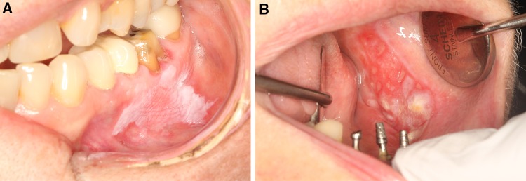
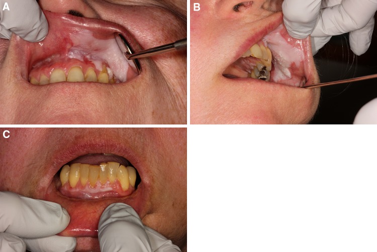
Leukoplakia which has a prevalence of 1–4% of the population, is defined as a “white plaque of questionable risk having excluded other (known) diseases or disorders that carry no increased risk for cancer” [1, 10]. As such, the clinical diagnosis of leukoplakia is made after exclusion of other white lesions which have clinico-histopathologic findings that are predictable and consistent (Table 1). Homogenous leukoplakia is generally demarcated and fissured, while non-homogenous leukoplakia are sub-typed as verrucous/nodular or erythro-leukoplakia, and these latter lesions have a higher risk for malignant transformation (Fig. 2a, b). Proliferative verrucous leukoplakia is, at this time, considered a subset of non-homogenous leukoplakia and is characterized by relentlessly progressive, multifocal leukoplakias, or a large leukoplakia at a single site or at contiguous sites (Fig. 3a–c) [11, 12]. A recent study suggests that proliferative leukoplakia, similar to solitary leukoplakia, may not only present with the classic verrucous/nodular pattern but may also have homogenous/fissured and erythroplakic components and suggested the term “proliferative leukoplakia (PL)” for accuracy and inclusiveness [13]. Furthermore, many differences, (including genomic alterations) exist between solitary leukoplakia and PL and it may be more appropriate for PL to stand alone as its own entity (Table 2) [13–15]. In studies performed after 1990, malignant transformation for localized leukoplakia ranged from 7.7 to 22.0% [16–18] while that for PL is 70–100% [11, 13].
Table 1.
Oral white lesions
| Developmental |
| Cannon white sponge nevus |
| Hereditary benign intraepithelial dyskeratosis |
| Other dyskeratotic genodermatoses (e.g. dyskeratosis congenita, pachyonychia congenita) |
| Reactive |
| Retention keratosis (e.g. hairy tongue) |
| Traumatic lesions/keratoses |
| Leukoedema |
| Morsicatio mucosae oris |
| Benign alveolar ridge keratosis |
| Others |
| Chemical-induced contact lesions |
| Contact desquamation |
| Smokeless tobacco keratosis |
| Nicotinic stomatitis |
| Others |
| Infectious |
| Candidiasis |
| Oral hairy leukoplakia |
| Oral secondary syphilis |
| Immune-mediated and autoimmune |
| Idiopathic oral lichen planus |
| Lichenoid hypersensitivity reaction (contact or drug-induced) |
| Chronic graft-vs.-host disease |
| Lupus erythematosus |
| Migratory glossitis |
| Others |
| Preneoplastic and neoplastic |
| Leukoplakia |
| Oral squamous cell carcinoma |
| Metabolic |
| Palifermin-induced keratosis |
| Uremic stomatitis |
Fig. 2.
a Homogenous and fissured leukoplakia, well-demarcated, involving the buccal mucosa and gingival margin—this represented mild epithelial dysplasia b Erythroleukoplakia of buccal mucosa—this represented squamous cell carcinoma
Fig. 3.
Proliferative mostly homogenous leukoplakia with involvement of contiguous sites namely a Upper lip mucosa, vestibule and gingiva b Buccal mucosa c Mandibular facial gingiva
Table 2.
Localized (conventional) leukoplakia versus proliferative leukoplakia
| Parameter | Localized leukoplakia | Proliferative leukoplakia |
|---|---|---|
| Gender | Mostly in men 2–3.5:1 | Mostly in women (2.5–5:1) |
| Smoking history | Strong > 60% | Weak < 30% |
| Number of sites | Single site | Usually multiple sites |
| Most common sites | Lateral/ventral tongue and floor of mouth | Gingiva and buccal mucosa |
| Prevalence of dysplasia at first biopsy | 40–45%; corollary is that 55–60% represent keratosis of unknown significance | < 10%; corollary is that > 90% represent keratosis of unknown significance |
| Malignant transformation | 8–22% | 70–100% |
| Deletion in p14arf exon 1B (within CDKN2A locus) | 3.8% | 40.0% |
| Management | Usually amenable to surgical excision, ablation because of its localized nature | Usually managed with follow-up and periodic biopsies with excision when cancer develops |
In the more recent literature, OED, CIS or invasive SCC is present in 40–46% of leukoplakia (Fig. 4) [19–23]. This begs the question: what then is the diagnosis of the non-dysplastic and non-carcinoma cases? In one study, 43% of leukoplakias were OED, CIS or SCC and the authors propose the concept of “keratosis of unknown significance (KUS)” for the other 57% of cases that were keratotic yet did not show OED (see below) [23]. Many cases submitted for histopathologic examination do not have adequate history or photographs and this hampers the ability of the pathologist to evaluate whether a keratotic lesion is reactive or not reactive. Historically, cases of so-called “benign hyperkeratosis” showed malignant transformation in 11–30% of cases (Fig. 4) [20, 21, 24].
Fig. 4.
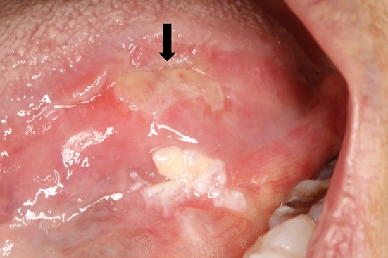
Verrucous-nodular leukoplakia diagnosed 4 years prior as “hyperkeratosis, no dysplasia”, now representing carcinoma-in situ, with invasive carcinoma on the superior aspect where the ulcer is (arrow)
Traumatic/frictional and reactive keratoses take two forms in general but both appear as white, poorly-demarcated, macerated, ragged-appearing keratotic papules and plaques often mistaken for leukoplakia. They occur most frequently on the nonkeratinized mucosa of the buccal mucosa, tongue and lip mucosa (morsicatio mucosae oris) or on the retromolar pad and edentulous alveolar ridge mucosa (Fig. 5a, b) [25]. A diagnosis of “hyperkeratosis, parakeratosis and acanthosis” without specifically stating that the changes are reactive, traumatic, frictional or factitial, relegates such lesions to the clinical entity leukoplakia and may be the reason that older studies showed a lower incidence of dysplasia in “leukoplakias”, a portion of were likely reactive/traumatic. In one recent study, 75% of all lesions submitted as leukoplakia or hyperkeratosis were traumatic/frictional or otherwise reactive lesions [23].
Fig. 5.
Morsicatio mucosae oris of a Lower lip mucosa with white, rough, macerated appearance b Benign alveolar ridge keratosis, poorly-demarcated, of the retromolar pad
The concept of classic oral lichen planus (OLP) as a premalignant condition is controversial and malignant transformation occurs in up to 1% of cases, a much lower rate when compared with leukoplakia, erythroplakia or submucous fibrosis [26, 27]. Classic OLP exhibits white reticulations present symmetrically and bilaterally on the buccal mucosa, gingiva (this could be purely erythematous) and tongue (Fig. 6a, b) [28]. Clinically, it is indistinguishable from medication-induced oral lichenoid hypersensitivity reaction, chronic graft-vs.-host disease and other immune-mediated and autoimmune conditions.
Fig. 6.
Lichen planus on the a Right and b Left buccal mucosa with reticulations and symmetry
Histopathology of Oral Lichen Planus, Reactive Keratoses and OED
Oral Lichen Planus
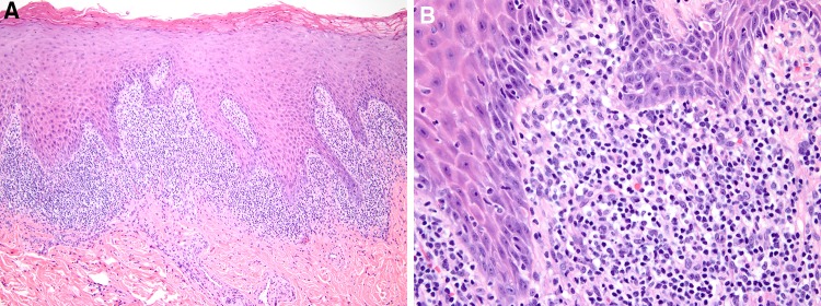
It is rare that all the features of OLP are noted on a biopsy. The most important features of this interface disorder are destruction of the basal cell layer often accompanied by colloid body formation, and a band of lymphocytes at the interface [28]. It may be keratotic or not (erythematous/erosive lesions of the gingiva are not), acanthotic or atrophic, show intra-epithelial inflammation or not, and exhibit saw-tooth rete ridges or not (Fig. 7a, b). All these features may be muted if the lesion is evolving or devolving, or if the patient has received therapy.
Fig. 7.
Lichen planus a Parakeratosis, acanthosis, saw-tooth rete ridges and lymphohistiocytic band at the interface b Spongiosis, leukocyte exocytosis, colloid bodies, loss of basal cell layer and lymphohistiocytic infiltrate at the interface
However, what it should not show, is substantial papillomatosis or verruciform architecture and OED, in particular, basal cell hyperplasia because OLP is by definition, an interface inflammatory process characterized by basal cell destruction. Whether there is reactive epithelial atypia or OED is subject to the pathologists’ interpretation with attendant inter-examiner variability. In such cases, it is the clinical appearance of the lesion which is the most helpful in arriving at an accurate diagnosis. When it is unclear from the requisition form whether this is a biopsy from a bilaterally symmetric and reticulated lesion, some pathologists have adopted the following diagnostic phrase: “lichenoid mucositis consistent with lichen planus in the appropriate clinical context” with appropriate clinical context to mean bilaterality, symmetry and reticulations. OED with a lymphocytic band at the interface will be discussed later.
Traumatic/Frictional and Reactive Keratoses
Reactive keratoses are by far, more common than leukoplakias because of the trauma-intense environment that is the oral cavity. Their histopathology is fairly distinctive and correlates with clinical findings.
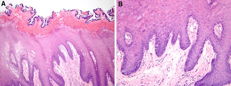
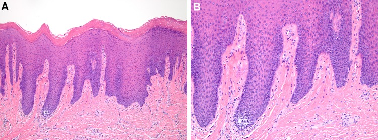
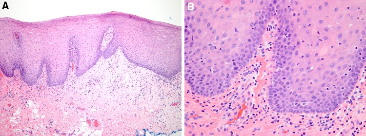
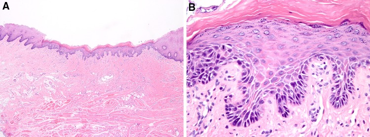
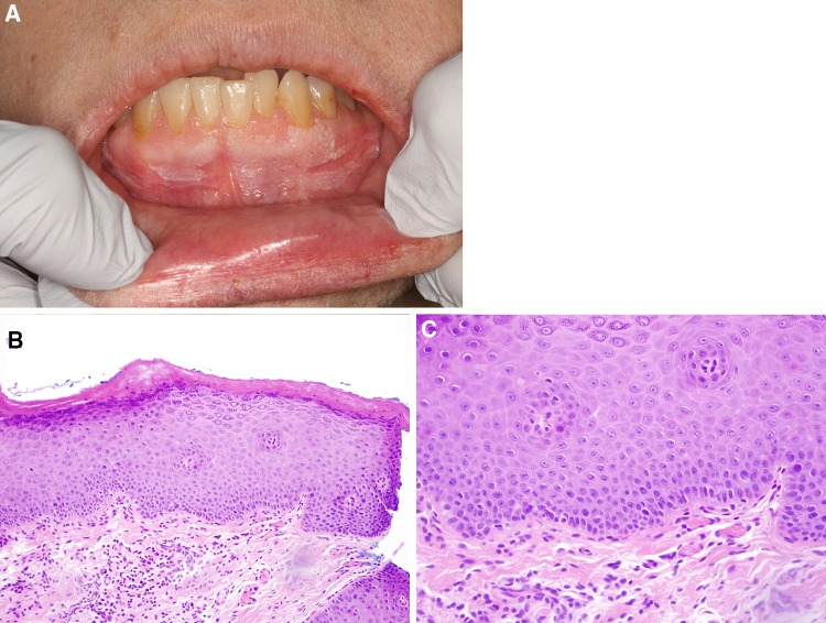
Chronic frictional/factitial/bite keratoses (corresponding to the clinical lesions of morsicatio mucosae oris). There is parakeratosis, sometimes with fissures and clefts rimmed by bacteria, acanthosis often with keratinocyte edema, tapered rete ridges and usually minimal to no inflammation unless the lesion has been ulcerated or recently traumatized in which case mild reactive epithelial atypia may be present (Fig. 8a, b) [25]. Fibrosis/scarring may be evident in the lamina propria with extension into the underlying skeletal muscle.
Benign alveolar ridge keratosis [(BARK), oral lichen simplex chronicus]. There is hyperkeratosis often with wedge-shaped hypergranulosis, and acanthosis with tapered rete ridges (Fig. 9a, b) [25, 29]. There may be parakeratosis if there has been recent trauma or ulceration.
Non-specific reactive lesions. These show hyperkeratosis or parakeratosis, acanthosis and chronic inflammation, and may represent evolving or devolving lesions of the conditions discussed above or other inflammatory conditions (Fig. 10a, b).
Fig. 8.
Chronic frictional/factitial keratosis a Parakeratosis with fissures and clefts rimmed by bacteria, acanthosis with keratinocyte edema, tapered rete ridges and scattered lymphocytes b No evidence of dysplasia
Fig. 9.
Benign alveolar ridge keratosis a Hyperkeratosis, acanthosis with tapered rete ridges and scattered lymphocytes b No evidence of dysplasia
Fig. 10.
Parakeratosis, acanthosis, chronic inflammation, reactive a Slight parakeratosis, acanthosis, tapered rete ridges and mild chronic inflammation b No evidence of dysplasia
Keratotic lesions with evidence of mild cytologic atypia that wax and wane are likely reactive keratoses and may explain the cases of so-called dysplasias that regress.
Histopathology of OED
There is no difference between the histopathologic features of OED of erythroplakia, leukoplakia and submucous fibrosis; the last always shows marked fibrosis of the lamina propria with associated history of use of areca nut. The criteria for diagnosis of OED are similar to those used at other mucosal sites and currently, they are divided into architectural and cytologic features (Table 3). OED is currently still graded mild, moderate and severe based on involvement of < 1/3, between 1/3 and 2/3, and > 2/3 (but not full thickness) of the epithelium [1]. There has been a proposal to use a binary low-grade and high-grade OED as is the convention at other mucosal sites [30]. The issue of inter-examiner variability remains a vexing issue [31, 32].
Table 3.
Histopathologic criteria for oral epithelial dysplasia
| Current criteria | Proposed criteria |
|---|---|
| Architectural changes | Architectural changes (low power) |
| Irregular epithelial stratification | Verrucous or papillary architecture |
| Loss of polarity of basal cells | Bulky exophytic or endophytic epithelial hyperplasia |
| Drop-shaped rete ridges | Hyperkeratosis with epithelial atrophy |
| Increased number of mitotic figures | Hyperkeratosis with “skip” segments of normal epithelium |
| Abnormally superficial mitoses | Drop- or bud-shaped rete ridges |
| Premature keratinization in single cells | Organizational changes (medium power) |
| Keratin pearls within rete ridges | Dyscohesion |
| Loss of epithelial cell cohesion | Loss of polarity and stratification |
| Cytologic changes | Basal cell hyperplasia |
| Abnormal variation in nuclear size | |
| Abnormal variation in nuclear shape | Superficial mitotic figures |
| Abnormal variation in cell size | Keratin pearls within rete ridges |
| Abnormal variation in cell shape | Cytologic changes (high power, on exfoliative cytology) |
| Increased nuclear: cytoplasmic ratio | Basal cell hyperplasia |
| Atypical mitotic figures | Increased nuclear:cytoplasmic ratio |
| Increased number and size of nucleoli | Cellular and nuclear pleomorphism |
| Hyperchromasia | Dyskeratosis/premature keratinization |
| Atypical mitotic figures and increased number of mitotic figures | |
| Hyperchromasia | |
| Cells with “glassy” cytoplasm |
However, beyond the conventional criteria shown in Table 3, there are other criteria that are just as important and these relate to the low-power architecture of the lesion. As such, I propose here a modified version of the existing criteria as noted in Table 3. These features often occur in combination rather than alone, and may be associated with cytologic features of OED or not.
Architectural Features of OED
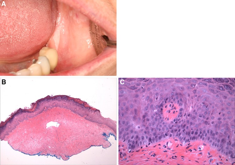
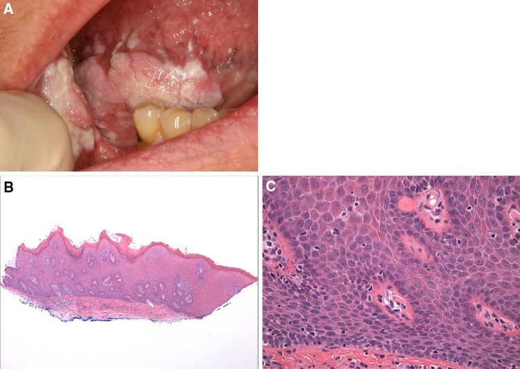
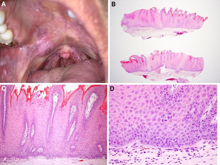
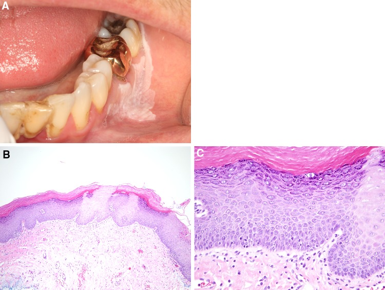
Verrucous and papillary architecture are well-known features of OED, and these may occur in the absence of significant cytologic features of OED and is in particular, associated with the verrucous variant of PL [33]. This feature may be mild presenting as papillomatosis (Figs. 11a–c, 12a–c). The term atypical verrucous hyperplasia with/out dysplasia is often applied to more florid epithelial hyperplasia (Fig. 13a–d) [34–36]. In general, the main architectural difference between atypical verrucous hyperplasia and verrucous carcinoma (both usually with minimal epithelial atypia) is the presence of an endophytic, bluntly-invasive growth pattern in verrucous carcinoma with bulbous frond-like rete ridges. However, in the uncommon scenario when the epithelial proliferation is bulky equating with a clinical mass lesion (see below), even if the lesion is primarily exophytic, the diagnosis of verrucous carcinoma should be considered.
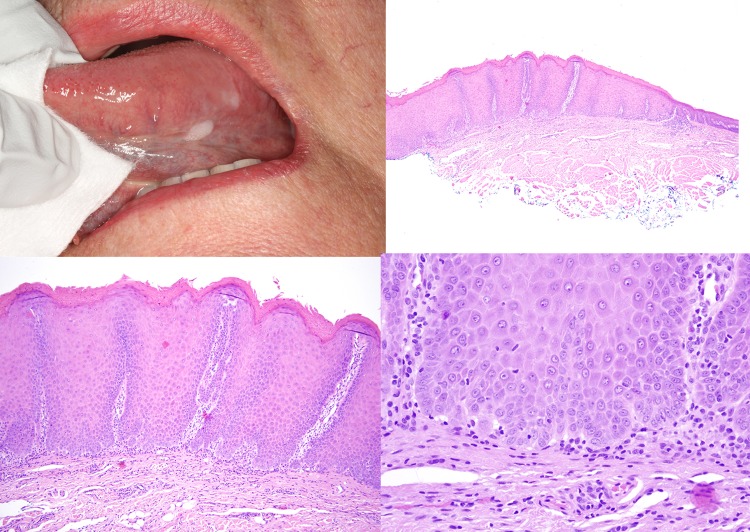
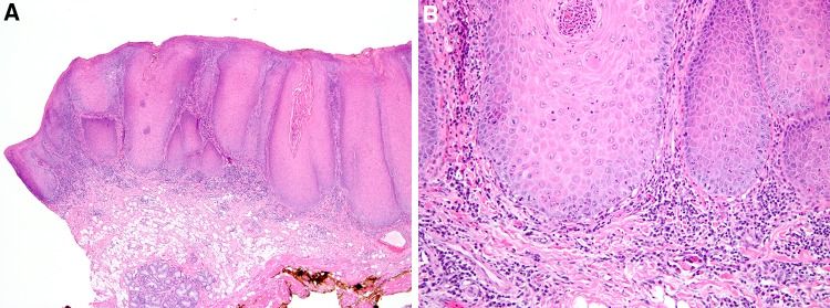
Bulky epithelial hyperplasia/proliferation is an important feature that is often over-looked, and this is closely associated with verrucous and papillary architecture. This is not mere acanthosis because the epithelium is multiple times the thickness of epithelium normal for that particular site. OED may or may not be present (Figs. 13a–d, 14a–d, 15a, b).
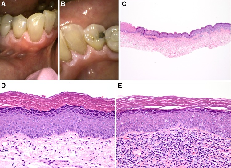
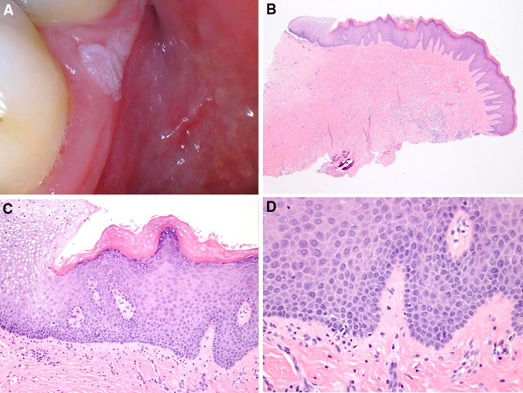
The literature on OED focuses on epithelial hyperplasia as an important precursor to dysplasia. However, epithelial atrophy with hyperkeratosis or parakeratosis, is a common feature of OED and also of KUS (Figs. 16a–e, 17a, b); this is also often associated with “skip” segments (see below). These often do not exhibit evidence of OED or show mild epithelial atypia at most. In general, reactive keratoses tend to exhibit acanthosis.
Sharp demarcation and skip segments are often seen in biopsies of leukoplakia (Figs. 16c, 18a–c, 19a–d). Sharp demarcation of thick keratin correlates with the clinical appearance of a sharply-demarcated clinical lesion. “Skip” segments, defined as alternating areas of thick keratosis with areas of normal-appearing nonkeratinized epithelium, are not infrequently encountered in biopsies of leukoplakia with or without OED.
Bud or drop-shaped rete ridges
Fig. 11.
Hyperkeratosis and papillomatosis, not reactive a White, sharply-demarcated fissured homogenous leukoplakia (not benign alveolar ridge keratosis) of the edentulous ridge mucosa b Hyperkeratosis, papillomatosis and slight epithelial atrophy for gingiva c Minimal evidence of dysplasia
Fig. 12.
Atypical verrucous hyperplasia with hyperkeratosis a Proliferative leukoplakia with verrucous plaques of the ventral tongue, floor of mouth and right buccal mucosa (with carcinoma) b Biopsy from ventral tongue shows atypical verrucous hyperplasia with hyperkeratosis c Minimal evidence of dysplasia
Fig. 13.
Atypical verrucous hyperplasia a Demarcated mucosal-colored verrucous leukoplakia of the soft palate b Atypical verrucous hyperplasia with primarily exophytic growth pattern c Atypical verrucous hyperplasia d Minimal evidence of dysplasia
Fig. 14.
Parakeratosis and bulky epithelial proliferation with mild epithelial dysplasia a Leukoplakia, slightly rough, of the ventral tongue b Slightly papillary bulky epithelial hyperplasia 3–4 times the thickness of the normal epithelium as noted on the right c Parakeratosis with epithelial hyperplasia and tapered rete ridges d Mild epithelial dysplasia
Fig. 15.
Parakeratosis and bulky endophytic squamous proliferation with mild epithelial dysplasia a Parakeratosis and bulky epithelial proliferation with bulbous rete ridges and lymphocytic band at the interface b Mild epithelial dysplasia
Fig. 16.
Hyperkeratosis, epithelial atrophy and chronic inflammation, not reactive a Linear demarcated leukoplakia of buccal and b Lingual gingiva c Hyperkeratosis (sharply-demarcated), epithelial atrophy and lymphocytic band at the interface d No evidence of dysplasia e A different area exhibiting mild epithelial atypia with lymphocytic band at the interface, unlikely reactive
Fig. 17.
Hyperkeratosis and moderate epithelial dysplasia a Hyperkeratosis and epithelial atrophy b Bud-shaped rete ridges and moderate epithelial dysplasia
Fig. 18.
Hyperkeratosis, epithelial atrophy and chronic inflammation, not reactive a Fissured demarcated leukoplakia of the buccal mucosa, vestibule and gingiva b Hyperkeratosis, epithelial atrophy with “skip” segments c No evidence of dysplasia
Fig. 19.
Hyperkeratosis, papillomatosis and mild chronic inflammation, not reactive a Demarcated leukoplakia of the lingual gingiva. Courtesy of Dr. John Duhaime, MA b Sharply demarcated hyperkeratosis corresponding with the clinically demarcated lesion, hyperkeratosis with papillomatosis c Papillomatosis d No evidence of dysplasia
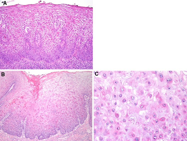
Organizational features of OED as defined here are changes that are readily seen on medium power and show how the keratinocytes are organized within the epithelium, and how they relate to each other within the spinous layer. Architectural features as defined here by the criteria above, and organizational features as defined here (historically referred to as architectural features) are not seen on exfoliative cytology, the last being best discerned on high power, and constitute the cytologic features of OED. For example, dyscohesion, an organizational feature where cell-to-cell contact is lost is an important feature of OED easily seen at medium power but obviously cannot be detected on exfoliative cytology (Fig. 20a). This must be distinguished from spongiosis caused by inflammation, associated with leukocyte exocytosis. Irregular epithelial stratification and loss of polarity of basal cells is combined as a single criterion of loss of polarity and stratification. Premature keratinization is better seen on high power and readily discerned on exfoliative cytology and has been moved to the section on cytologic features under “dyskeratosis”. Basal cell hyperplasia is both an organizational feature, because in normal epithelium basal cells should not extend beyond the basal two layers, as well as a cytologic feature because it is readily identified on exfoliative cytology. The presence of cells with “glassy” cytoplasm, similar to that seen in keratoacanthomatous pattern of SCC, is another feature that can be helpful (Fig. 20b, c).
Fig. 20.
a Severe epithelial dysplasia with dyscohesion and dyskeratosis b Acanthosis with severe epithelial dysplasia c Dyskeratotic cells and cells with “glassy” cytoplasm
The grading of OED currently relies on organizational (as defined here, see above) and cytologic features of dysplasia. However, the architectural features as defined here are just as important and should be factored into OED grading. Furthermore, it is difficult to use the division of epithelium into thirds for grading lesions that have bulky and verrucous architecture. The case shown in Fig. 15a, b would be graded as mild dysplasia but this does not represent the nature of the lesion which is a squamous proliferation.
Human Papillomavirus-Associated OED
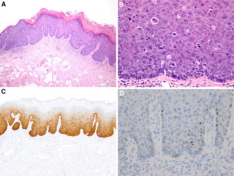
Human papillomavirus (HPV)-associated OED is a distinctive subset of OED that is characterized by parakeratosis containing hyperchromatic crenated nuclei, hyperkeratosis, karyorrhectic cells (“mitosoid” bodies) and apoptotic cells (Fig. 21a, b) [37]. The karyorrhectic cells have fragmented chromatin and a peri-cellular halo from loss of attachment to adjacent cells; ultimately, they develop pyknotic nuclei and brightly eosinophilic cytoplasm as they undergo apoptosis. The study for p16 is positive in a continuous band in almost all keratinocytes and lesions are positive for high-risk human papillomavirus (Fig. 21c, d). In one study of 54 cases, most were associated with HPV-16 as expected and 15% developed invasive SCC [37]. Because this particular form of OED usually presents clinically as a conventional leukoplakia, it is amenable to early diagnosis and excision.
Fig. 21.
Human papillomavirus-associated severe dysplasia a Hyperkeratosis, parakeratosis, acanthosis and severe epithelial dysplasia. b Many karyorrhectic cells with peri-cellular halo, and apoptotic cells c block staining for p16 d mRNA in situ hybridization positive for high risk human papillomavirus
Epithelial Atypia and Candidiasis
Primary candidiasis is frequently associated with secondary epithelial atypia, primary OED is often associated with secondary candidiasis, and it is sometimes difficult to differentiate between the two. Although chronic candidiasis is now listed as one of the oral potentially malignant lesions, this is not yet fully accepted [4]. If chronic candidiasis is a precancerous condition, there would be a high prevalence of SCC on the commissures, denture-bearing mucosae and dorsum of tongue and this is not the case. Many features of OED are mimicked by reactive epithelial atypia seen in candidiasis and adjacent to ulcers. When it is unclear whether a lesion is candidiasis with reactive epithelial atypia or OED with candidiasis, treating the patient with anti-fungal therapy is extremely useful. If the lesion resolves completely, the lesion represents candidiasis with reactive epithelial atypia. If the lesion improves somewhat but is still present, a rebiopsy is indicated. It may be that the epithelium in OED is locally immunocompromised, allowing for ready candidal colonization.
Keratosis of uncertain significance (KUS)
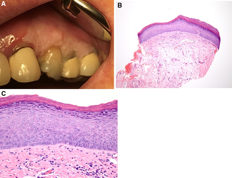
This is a category of lesions where biopsy of a leukoplakia shows hyperkeratosis and/or parakeratosis, acanthosis or more frequently atrophy, with minimal cytologic atypia, and variable to no inflammation (Figs. 16d, 18a–c, 22a–c, 23a–c). Often there is slight papillomatosis and “skip” segments. They do not have features that characterize traumatic/frictional keratoses. Adding the phrase “not likely reactive” to the diagnosis informs the clinician that the lesion is not likely caused by trauma or reaction to injury and is therefore suspicious for a very early dysplastic lesion for the following reasons:
Fig. 22.
Hyperkeratosis, not reactive a Demarcated, fissured leukoplakia of the mandibular facial gingiva b Hyperkeratosis c No evidence of dysplasia
Fig. 23.
Hyperkeratosis and epithelial atrophy, not reactive a Demarcated, slightly fissured leukoplakia of the left maxillary attached gingiva b Hyperkeratosis, epithelial atrophy, and mild chronic inflammation. c No evidence of dysplasia
Clinically, leukoplakias with a KUS histopathologic phenotype are indistinguishable from leukoplakias with OED
When followed over time, such “benign hyperkeratosis” developed OED and OSCC in 11–30% of cases [20, 21, 24].
Excisions of leukoplakias and oral SCC show areas of KUS in continuity with areas of OED and SCC
Cases of PL show KUS in early biopsies (12–50% of cases) that over time progress to dysplasia and exhibit aneuploidy [38, 39]. Twenty-nine (> 90%) of the original 30 cases of proliferative (verrucous) leukoplakia reported by Hansen et al. [33] exhibited KUS.
Whole genome sequencing showed the presence of mutations in KMT2C, TP53 and other mutations often found in OED at the same frequency (unpublished data).
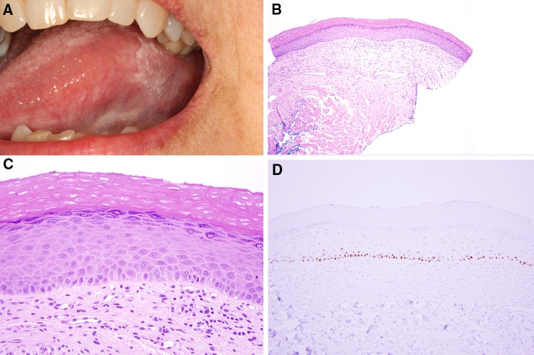
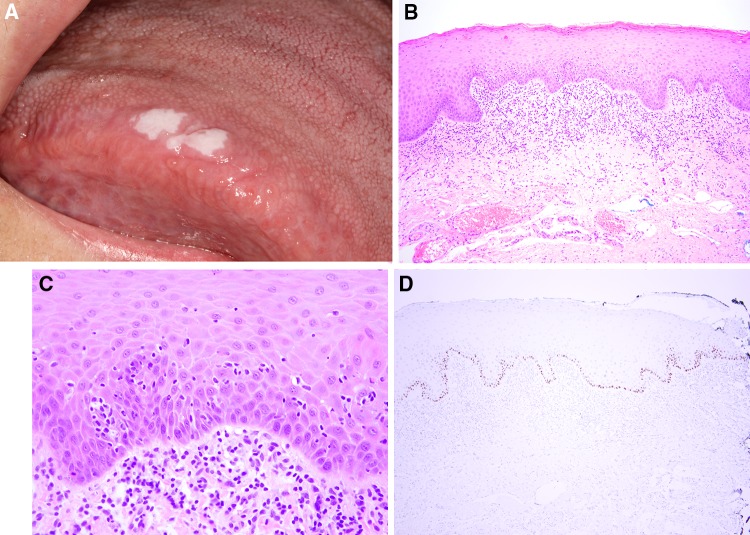
Wild-type p53 protein is expressed in < 5% of basal cells in normal epithelium [40]. Its expression is increased in OED although the degree of expression varied in studies by different studies [40–42]. However, if there is continuous positivity within > 90% of basal cell nuclei and in suprabasal nuclei, this is very strongly associated with the dysplastic phenotype; it is also strongly expressed in SCC. Positivity for p53 is not uncommon in cases of KUS (Fig. 24d). While routine use of p53 is unnecessary when OED is obvious, it may be helpful in differentiating reactive epithelial atypia in inflamed mucosa, from true OED (Fig. 24a–d).
Fig. 24.
Hyperkeratosis and mild chronic inflammation, not reactive a Partially demarcated leukoplakia of the left ventral tongue (she had proliferative leukoplakia with lesions on the right tongue and floor of mouth) b Hyperkeratosis with mild chronic inflammation. c. No evidence of dysplasia. d p53 is positive within basal cells in a continuous band
The concept of KUS, that is a lesion with likely genomic alterations, but that has not developed yet the dysplastic phenotype, is consistent with current theories on carcinogenesis, likely corresponding to the “first strike” of the “three strikes” of carcinogenesis [43]. Whether lesions with KUS at the surgical margins are more likely to develop a “recurrence” would be of interest.
Lichenoid Lesions With and Without Dysplasia
“Lichenoid mucositis” continues to be a frequent histopathologic diagnosis of many mucosal biopsies. A lymphocytic band at the interface is often referred to as “lichenoid mucositis” by many pathologists, but such a band often is seen beneath conventional OED within leukoplakia. One report noted this in 11–39% of cases of OED [3]. The question is: is this OLP with reactive atypia or is this OED with a lymphocytic (“lichenoid”) host response? Having a clinical image of the lesion and judicious use of p53 are often helpful in helping to distinguish between the two (Fig. 25a–d). The presence of papillomatosis, and substantial cytologic atypia are clues that the lesion may not represent OLP (Fig. 26a, b).
Fig. 25.
Parakeratosis, epithelial atrophy with mild dysplasia and chronic inflammation, not reactive a Demarcated leukoplakia of the right tongue dorsum (Courtesy of Dr. Alessandro Villa, MA). b Parakeratosis, epithelial atrophy and lymphocytic band at the interface c Reactive epithelial atypia vs mild epithelial dysplasia d p53 is positive within basal cells in a continuous band consistent with dysplasia
Fig. 26.
Parakeratosis and severe epithelial dysplasia a Parakeratosis, bud-shaped rete ridges and lymphocytic band at the interface b Dyscohesion and severe epithelial dysplasia
In one study, oral lichenoid lesions defined as either the clinical or histopathologic features being “compatible with” instead of “typical for” OLP was associated with 3.2% versus 0% malignant transformation in oral lichenoid lesion versus the OLP group [44]. If a biopsy taken from a solitary white/red plaque on the ventral tongue is diagnosed as lichenoid mucositis, the unfortunate result is that this lesion may then be diagnosed clinically as “plaque-type” OLP. Subsequent development of oral SCC results in OLP being categorized as a potentially malignant disorder, driving up the rate of malignant transformation for OLP. However, if instead, one views the solitary white and/or red plaque on the tongue as a leukoplakia or erythroleukoplakia, and the lymphocytic band as a host response to OED, then subsequent malignant transformation is expected.
Such a lymphocytic band occurs frequently at the interface of oral SCCs (Fig. 27) and current understanding suggests that a strong lymphocytic host response may be associated with better prognosis, and is the basis for checkpoint immunotherapy [45, 46]. The term “lichenoid dysplasia” should be avoided because clinicians may mistakenly interpret this as dysplasia arising within lichen planus.
Fig. 27.
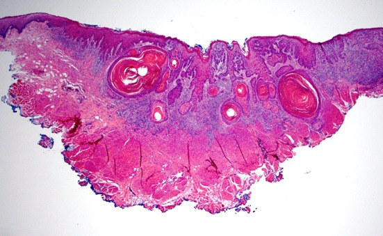
Infiltrative, keratinizing squamous cell carcinoma, well-differentiated. Note the lymphocytic band around tumor, as well as at the edges where epithelial dysplasia is present
Pathology Sign-Out
A pathology report that reads “hyperkeratosis and/or parakeratosis, acanthosis” with or without inflammation, applies to chronic traumatic/factitial/frictional keratoses, benign alveolar ridge (frictional) keratosis, other nonspecific reactive keratoses, cases of treated OLP or OLP not at its peak of presentation as well as KUS. The clinician is often at a loss as to how to proceed. The worst case scenario is when a patient with leukoplakia exhibiting KUS is told that she/he has nothing to worry about because there is no precancer or cancer and the patient is not followed (Fig. 4). An accurate diagnosis informs decision-making for the clinician and guides management which includes
Reassurance only for frictional and reactive lesions, with no further treatment necessary
Rebiopsy after a short treatment with anti-fungal and/or anti-inflammatory agents to reduce inflammation (cases of reactive atypia vs OED)
Excision and follow-up if clinically appropriate with acceptable morbidity (KUS and OED)
follow-up and periodic rebiopsy (lesions of PL)
As such, I suggest the following:
Chronic factitial/frictional keratoses or BARKs be diagnosed as such outright, or that the words “reactive”, “traumatic”, “frictional” or “factitial” appear at the end of the diagnostic phrase
That the words “not reactive” or “likely not reactive” be added to the diagnostic phrase if the lesions represent a KUS with a suitable note. Examples include “hyperkeratosis, epithelial atrophy and chronic inflammation, not reactive” or “atypical verrucous hyperplasia, not reactive”.
That clinicians be encouraged to include a photograph with all mucosal biopsies similar to enclosing radiographs for osseous lesions.
Conclusions
Leukoplakia, erythroplakia and submucous fibrosis are highly associated with the development of OED. However, other changes such as papillary/verrucous architecture, bulky epithelial hyperplasia, epithelial atrophy, and “skip” segments are criteria that must also be taken into account when evaluating for OED, especially when the conventional cytologic features of OED are minimal or absent. The concept of KUS is an important one, and more research on this group of lesions may shed further light on them. Finally, a lymphocytic band is frequently seen beneath OED and represents a lymphocytic host response to OED. It cannot over-emphasized that the clinical appearance of the lesion is almost as important as the histopathologic features especially in cases of “lichenoid mucositis” and KUS.
Compliance with Ethical Standards
Conflict of interest
Sook-Bin Woo received research funding by Bristol-Myers Squibb, New York
Ethical Approval
All procedures performed in studies involving human participants were in accordance with the ethical standards of the institutional and/or national research committee and with the 1964 Helsinki declaration and its later amendments or comparable ethical standards.
Footnotes
Publisher’s Note
Springer Nature remains neutral with regard to jurisdictional claims in published maps and institutional affiliations.
References
- 1.Reibel J, Gale N, Hille J, Hunt JL, Lingen M, Muller S, et al. Oral potentially malignant disorders and oral epithelial dysplasia. In: El-Naggar AK, Chan JKC, Grandis JR, Takata T, Slootweg P, et al., editors. WHO classification of head and neck tumours. 4. Lyon: iaRC; 2017. pp. 112–115. [Google Scholar]
- 2.Muller S. Oral lichenoid lesions: distinguishing the benign from the deadly. Mod Pathol. 2017;30(s1):54–67. doi: 10.1038/modpathol.2016.121. [DOI] [PubMed] [Google Scholar]
- 3.Fitzpatrick SG, Honda KS, Sattar A, Hirsch SA. Histologic lichenoid features in oral dysplasia and squamous cell carcinoma. Oral Surg Oral Med Oral Pathol Oral Radiol. 2014;117(4):511–520. doi: 10.1016/j.oooo.2013.12.413. [DOI] [PubMed] [Google Scholar]
- 4.Speight PM, Khurram SA, Kujan O. Oral potentially malignant disorders: risk of progression to malignancy. Oral Surg Oral Med Oral Pathol oral Radiol. 2018;125(6):612–627. doi: 10.1016/j.oooo.2017.12.011. [DOI] [PubMed] [Google Scholar]
- 5.Shafer WG, Waldron CA. Erythroplakia of the oral cavity. Cancer. 1975;36:1021–1028. doi: 10.1002/1097-0142(197509)36:3<1021::AID-CNCR2820360327>3.0.CO;2-W. [DOI] [PubMed] [Google Scholar]
- 6.Mashberg A, Feldman LJ. Clinical criteria for identifying early oral and oropharyngeal carcinoma: erythroplasia revisited. Am J Surg. 1988;156(4):273–275. doi: 10.1016/S0002-9610(88)80290-3. [DOI] [PubMed] [Google Scholar]
- 7.Reichart PA, Philipsen HP. Oral erythroplakia—a review. Oral Oncol. 2005;41(6):551–561. doi: 10.1016/j.oraloncology.2004.12.003. [DOI] [PubMed] [Google Scholar]
- 8.Guha N, Warnakulasuriya S, Vlaanderen J, Straif K. Betel quid chewing and the risk of oral and oropharyngeal cancers: a meta-analysis with implications for cancer control. Int J Cancer. 2014;135(6):1433–1443. doi: 10.1002/ijc.28643. [DOI] [PubMed] [Google Scholar]
- 9.Tilakaratne WM, Ekanayaka RP, Warnakulasuriya S. Oral submucous fibrosis: a historical perspective and a review on etiology and pathogenesis. Oral surgery, oral medicine. Oral Pathol Oral Radiol. 2016;122(2):178–191. doi: 10.1016/j.oooo.2016.04.003. [DOI] [PubMed] [Google Scholar]
- 10.Warnakulasuriya S, Johnson NW, van der Waal I. Nomenclature and classification of potentially malignant disorders of the oral mucosa. J Oral Pathol Med. 2007;36(10):575–580. doi: 10.1111/j.1600-0714.2007.00582.x. [DOI] [PubMed] [Google Scholar]
- 11.Pentenero M, Meleti M, Vescovi P, Gandolfo S. Oral proliferative verrucous leukoplakia: are there particular features for such an ambiguous entity? Systematic review. Br J Dermatol. 2014 doi: 10.1111/bjd.12853. [DOI] [PubMed] [Google Scholar]
- 12.Abadie WM, Partington EJ, Fowler CB, Schmalbach CE. Optimal management of proliferative verrucous leukoplakia: a systematic review of the literature. Otolaryngol Head Neck Surg. 2015;153(4):504–511. doi: 10.1177/0194599815586779. [DOI] [PubMed] [Google Scholar]
- 13.Villa A, Menon RS, Kerr AR, De Abreu Alves F, Guollo A, Ojeda D, et al. Proliferative leukoplakia: proposed new clinical diagnostic criteria. Oral Dis. 2018;24(5):749–760. doi: 10.1111/odi.12830. [DOI] [PubMed] [Google Scholar]
- 14.Kresty LA, Mallery SR, Knobloch TJ, Song H, Lloyd M, Casto BC, et al. Alterations of p16(INK4a) and p14(ARF) in patients with severe oral epithelial dysplasia. Cancer Res. 2002;62(18):5295–5300. [PubMed] [Google Scholar]
- 15.Okoturo EM, Risk JM, Schache AG, Shaw RJ, Boyd MT. Molecular pathogenesis of proliferative verrucous leukoplakia: a systematic review. Br J Oral Maxillofac Surg. 2018;56(9):780–785. doi: 10.1016/j.bjoms.2018.08.010. [DOI] [PubMed] [Google Scholar]
- 16.Lumerman HS, Freedman PD, Kerpel SM, Cale AE. Screening for oral hairy leukoplakia by cytologic examination. Diagn Cytopathol. 1990;6(3):225. doi: 10.1002/dc.2840060319. [DOI] [PubMed] [Google Scholar]
- 17.Arduino PG, Bagan J, El-Naggar AK, Carrozzo M. Urban legends series: oral leukoplakia. Oral Dis. 2013;19(7):642–659. doi: 10.1111/odi.12065. [DOI] [PubMed] [Google Scholar]
- 18.Ho MW, Risk JM, Woolgar JA, Field EA, Field JK, Steele JC, et al. The clinical determinants of malignant transformation in oral epithelial dysplasia. Oral Oncol. 2012;48(10):969–976. doi: 10.1016/j.oraloncology.2012.04.002. [DOI] [PubMed] [Google Scholar]
- 19.Feller L, Altini M, Slabbert H. Pre-malignant lesions of the oral mucosa in a South African sample—a clinicopathological study. J Dent Assoc S Afr. 1991;46(5):261–265. [PubMed] [Google Scholar]
- 20.Schepman KP, van der Meij EH, Smeele LE, van der Waal I. Malignant transformation of oral leukoplakia: a follow-up study of a hospital-based population of 166 patients with oral leukoplakia from The Netherlands. Oral Oncol. 1998;34(4):270–275. doi: 10.1016/S1368-8375(98)80007-9. [DOI] [PubMed] [Google Scholar]
- 21.Brouns E, Baart J, Karagozoglu K, Aartman I, Bloemena E, van der Waal I. Malignant transformation of oral leukoplakia in a well-defined cohort of 144 patients. Oral Dis. 2014;20(3):e19–e24. doi: 10.1111/odi.12095. [DOI] [PubMed] [Google Scholar]
- 22.Lee JJ, Hung HC, Cheng SJ, Chen YJ, Chiang CP, Liu BY, et al. Carcinoma and dysplasia in oral leukoplakias in Taiwan: prevalence and risk factors. Oral Surg Oral Med Oral Pathol Oral Radiol Endod. 2006;101(4):472–480. doi: 10.1016/j.tripleo.2005.07.024. [DOI] [PubMed] [Google Scholar]
- 23.Woo SB, Grammer RL, Lerman MA. Keratosis of unknown significance and leukoplakia: a preliminary study. Oral Surg Oral Med Oral Pathol oral Radiol. 2014;118(6):713–724. doi: 10.1016/j.oooo.2014.09.016. [DOI] [PubMed] [Google Scholar]
- 24.Silverman S, Gorsky M, Lozada F. Oral leukoplakia and malignant transformation. Cancer. 1984;53:563–568. doi: 10.1002/1097-0142(19840201)53:3<563::AID-CNCR2820530332>3.0.CO;2-F. [DOI] [PubMed] [Google Scholar]
- 25.Woo SB, Lin D. Morsicatio mucosae oris–a chronic oral frictional keratosis, not a leukoplakia. J Oral Maxillofac Surg. 2009;67(1):140–146. doi: 10.1016/j.joms.2008.08.040. [DOI] [PubMed] [Google Scholar]
- 26.Fitzpatrick SG, Hirsch SA, Gordon SC. The malignant transformation of oral lichen planus and oral lichenoid lesions: a systematic review. J Am Dent Assoc. 2014;145(1):45–56. doi: 10.14219/jada.2013.10. [DOI] [PubMed] [Google Scholar]
- 27.Aghbari SMH, Abushouk AI, Attia A, Elmaraezy A, Menshawy A, Ahmed MS, et al. Malignant transformation of oral lichen planus and oral lichenoid lesions: a meta-analysis of 20095 patient data. Oral Oncol. 2017;68:92–102. doi: 10.1016/j.oraloncology.2017.03.012. [DOI] [PubMed] [Google Scholar]
- 28.Cheng YS, Gould A, Kurago Z, Fantasia J, Muller S. Diagnosis of oral lichen planus: a position paper of the American Academy of Oral and Maxillofacial Pathology. Oral Surg Oral Med Oral Pathol oral Radiol. 2016;122(3):332–354. doi: 10.1016/j.oooo.2016.05.004. [DOI] [PubMed] [Google Scholar]
- 29.Natarajan E, Woo SB. Benign alveolar ridge keratosis (oral lichen simplex chronicus): a distinct clinicopathologic entity. J Am Acad Dermatol. 2008;58(1):151–157. doi: 10.1016/j.jaad.2007.07.011. [DOI] [PubMed] [Google Scholar]
- 30.Kujan O, Oliver RJ, Khattab A, Roberts SA, Thakker N, Sloan P. Evaluation of a new binary system of grading oral epithelial dysplasia for prediction of malignant transformation. Oral Oncol. 2006;42(10):987–993. doi: 10.1016/j.oraloncology.2005.12.014. [DOI] [PubMed] [Google Scholar]
- 31.Kujan O, Khattab A, Oliver RJ, Roberts SA, Thakker N, Sloan P. Why oral histopathology suffers inter-observer variability on grading oral epithelial dysplasia: an attempt to understand the sources of variation. Oral Oncol. 2007;43(3):224–231. doi: 10.1016/j.oraloncology.2006.03.009. [DOI] [PubMed] [Google Scholar]
- 32.Warnakulasuriya S, Reibel J, Bouquot J, Dabelsteen E. Oral epithelial dysplasia classification systems: predictive value, utility, weaknesses and scope for improvement. J Oral Pathol Med. 2008;37(3):127–133. doi: 10.1111/j.1600-0714.2007.00584.x. [DOI] [PubMed] [Google Scholar]
- 33.Hansen LS, Olson JA., Jr Proliferative verrucous leukoplakia. Oral Surg Oral Med Oral Pathol. 1985;60:285–298. doi: 10.1016/0030-4220(85)90313-5. [DOI] [PubMed] [Google Scholar]
- 34.Wang YP, Chen HM, Kuo RC, Yu CH, Sun A, Liu BY, et al. Oral verrucous hyperplasia: histologic classification, prognosis, and clinical implications. J Oral Pathol Med. 2009;38(8):651–656. doi: 10.1111/j.1600-0714.2009.00790.x. [DOI] [PubMed] [Google Scholar]
- 35.Zain RB, K TG, Ramanathan A, Kim J, Tilakaratne WM, Takata T, et al. Exophytic verrucous hyperplasia of the oral cavity—application of standardized criteria for diagnosis from a consensus report. Asian Pac J Cancer Prevent: APJCP. 2016;17(9):4491–4501. doi: 10.7314/APJCP.2016.17.(9).4491. [DOI] [PubMed] [Google Scholar]
- 36.Muller S. Oral epithelial dysplasia, atypical verrucous lesions and oral potentially malignant disorders: focus on histopathology. Oral Surg Oral Med Oral Pathol Oral Radiol. 2018;125(6):591–602. doi: 10.1016/j.oooo.2018.02.012. [DOI] [PubMed] [Google Scholar]
- 37.Lerman MA, Almazrooa S, Lindeman N, Hall D, Villa A, Woo SB. HPV-16 in a distinct subset of oral epithelial dysplasia. Mod Pathol. 2017;30(12):1646–1654. doi: 10.1038/modpathol.2017.71. [DOI] [PubMed] [Google Scholar]
- 38.Klanrit P, Sperandio M, Brown AL, Shirlaw PJ, Challacombe SJ, Morgan PR, et al. DNA ploidy in proliferative verrucous leukoplakia. Oral Oncol. 2007;43(3):310–316. doi: 10.1016/j.oraloncology.2006.03.016. [DOI] [PubMed] [Google Scholar]
- 39.Gouvea AF, Santos Silva AR, Speight PM, Hunter K, Carlos R, Vargas PA, et al. High incidence of DNA ploidy abnormalities and increased Mcm2 expression may predict malignant change in oral proliferative verrucous leukoplakia. Histopathology. 2013;62(4):551–562. doi: 10.1111/his.12036. [DOI] [PubMed] [Google Scholar]
- 40.Abbas NF, Labib El-Sharkawy S, Abbas EA, Abdel Monem El-Shaer M. Immunohistochemical study of p53 and angiogenesis in benign and preneoplastic oral lesions and oral squamous cell carcinoma. Oral Surg Oral Med Oral Pathol Oral Radiol Endod. 2007;103(3):385–390. doi: 10.1016/j.tripleo.2005.11.008. [DOI] [PubMed] [Google Scholar]
- 41.Shahnavaz SA, Regezi JA, Bradley G, Dube ID, Jordan RC. p53 gene mutations in sequential oral epithelial dysplasias and squamous cell carcinomas. J Pathol. 2000;190(4):417–422. doi: 10.1002/(SICI)1096-9896(200003)190:4<417::AID-PATH544>3.0.CO;2-G. [DOI] [PubMed] [Google Scholar]
- 42.Nikitakis NG, Rassidakis GZ, Tasoulas J, Gkouveris I, Kamperos G, Daskalopoulos A, et al. Alterations in the expression of DNA damage response-related molecules in potentially preneoplastic oral epithelial lesions. Oral Surg Oral Med Oral Pathol Oral Radiol. 2018;125(6):637–649. doi: 10.1016/j.oooo.2018.03.006. [DOI] [PubMed] [Google Scholar]
- 43.Vogelstein B, Kinzler KW. The path to cancer—three strikes and you’re out. N Engl J Med. 2015;373(20):1895–1898. doi: 10.1056/NEJMp1508811. [DOI] [PubMed] [Google Scholar]
- 44.van der Meij EH, Mast H, van der Waal I. The possible premalignant character of oral lichen planus and oral lichenoid lesions: a prospective five-year follow-up study of 192 patients. Oral Oncol. 2007;43(8):742–748. doi: 10.1016/j.oraloncology.2006.09.006. [DOI] [PubMed] [Google Scholar]
- 45.Brandwein-Gensler M, Smith RV, Wang B, Penner C, Theilken A, Broughel D, et al. Validation of the histologic risk model in a new cohort of patients with head and neck squamous cell carcinoma. Am J Surg Pathol. 2010;34(5):676–688. doi: 10.1097/PAS.0b013e3181d95c37. [DOI] [PubMed] [Google Scholar]
- 46.Hanna GJ, Kofman ER, Shazib MA, Woo SB, Reardon B, Treister NS, et al. Integrated genomic characterization of oral carcinomas in post-hematopoietic stem cell transplantation survivors. Oral Oncol. 2018;81:1–9. doi: 10.1016/j.oraloncology.2018.04.007. [DOI] [PubMed] [Google Scholar]


