Abstract
The BRAF gene and the TERT promoter are among the most frequently altered genomic loci in low-grade (LGG) and high-grade-glioma (HGG), respectively. The coexistence of BRAF and TERT promoter aberrations characterizes a subset of aggressive glioma. Therefore, we investigated interactions between those alterations in malignant glioma. We analyzed co-occurrence of BRAFV600E and TERT promoter mutations in our clinical data (n = 8) in addition to published datasets (n = 103) and established a BRAFV600E-positive glioma cell panel (n = 9) for in vitro analyses. We investigated altered gene expression, signaling events and TERT promoter activity upon BRAF- and E-twenty-six (ETS)-factor inhibition by qRT-PCR, chromatin immunoprecipitation (ChIP), Western blots and luciferase reporter assays. TERT promoter mutations were significantly enriched in BRAFV600E-mutated HGG as compared to BRAFV600E-mutated LGG. In vitro, BRAFV600E/TERT promoter double-mutant glioma cells showed exceptional sensitivity towards BRAF-targeting agents. Remarkably, BRAF-inhibition attenuated TERT expression and TERT promoter activity exclusively in double-mutant models, while TERT expression was undetectable in BRAFV600E-only cells. Various ETS-factors were broadly expressed, however, only ETS1 expression and phosphorylation were consistently downregulated following BRAF-inhibition. Knock-down experiments and ChIP corroborated the notion of a functional role for ETS1 and, accordingly, all double-mutant tumor cells were highly sensitive towards the ETS-factor inhibitor YK-4-279. In conclusion, our data suggest that concomitant BRAFV600E and TERT promoter mutations synergistically support cancer cell proliferation and immortalization. ETS1 links these two driver alterations functionally and may represent a promising therapeutic target in this aggressive glioma subgroup.
Electronic supplementary material
The online version of this article (10.1186/s40478-019-0775-6) contains supplementary material, which is available to authorized users.
Keywords: BRAF, TERT promoter, Glioma, Brain tumor, ETS-factors, ETS1
Introduction
Glioma represents the most common tumor type in the central nervous system (CNS) across all age groups [37]. The biology and clinical behavior of glioma are highly heterogeneous as reflected by WHO grades ranging from I to IV [27]. Generally, they are divided into low-grade glioma (LGG), comprised of WHO grades I/II, and WHO grade III/IV tumors which are referred to as high-grade glioma (HGG). Moreover, glioma encompasses a variety of histologic subtypes some of which can present either as LGG or HGG [27].
BRAF is a serine/threonine kinase and central mediator in the well-described oncogenic mitogen-activated protein kinase (MAPK) signaling pathway [14]. Various alterations such as activating mutations of BRAF are commonly found in cancerous tissues [14]. In the pediatric patient population, more than half of LGG are characterized by genetic alterations of the BRAF gene resulting in increased cellular proliferation due to hyperactivation of downstream signaling [16, 39]. Moreover, the missense mutation BRAFV600E is present in a considerable amount of LGG namely pleomorphic xanthoastrocyma (PXA) and ganglioglioma (GG), but also other subtypes of astrocytoma [43]. With respect to HGG, BRAFV600E has been described in anaplastic PXA or anaplastic GG [43], as well as pediatric (6–12%) [8, 43] and adult (7.7%) glioblastoma (GBM), often accompanied by an epithelioid phenotype [8, 20]. The biological differences between BRAF-mutant LGG and HGG remain poorly understood. To date, only concomitant deletion of the CDKN2A locus has been described to synergistically promote glioma development [15] and to define inferior outcome in BRAFV600E-positive glioma [21, 34]. Small-molecule inhibitors of BRAF and its downstream-target MEK have already been approved for other BRAF-driven cancer types, such as melanoma [14] and have been shown to effectively inhibit glioma growth both in preclinical models [5, 9, 22, 36] and small patient cohorts [7, 17, 21]. Consequently, phase I/II trials with BRAF- or MEK-inhibitors either as single agent (NCT01677741, NCT01748149, NCT03363217, NCT01089101, NCT02285439, NCT03213691) or in combination (NCT02684058, NCT03340506, NCT02034110) have already been initiated. First analyses show promising results in both the pediatric [3, 7, 21, 42] and the adult patient population [17].
The telomerase reverse transcriptase (TERT) gene codes for the core catalytic subunit of telomerase, an enzyme which is responsible for elongating the telomeric ends of chromosomes, thereby enabling cancer cells to bypass senescence. Hence, telomerase re-activation is a frequent mechanism, used in malignant tissues to render replicative immortality and is associated with worse prognosis in various types of brain tumors [11, 26]. Specific mutations within the TERT promoter, C250T (−146C > T), C228T (−124C > T) and A161C (−57A > C), have been identified to play an important role in telomerase re-activation in multiple tumor types including HGG [13, 46]. C228T represents the most frequent of either mutation in both LGG as well as HGG [18]. Functionally, all three non-coding mutations open new binding-sites for e-twenty-six (ETS/TCF) family transcription factors involved in TERT promoter hyperactivation [4, 13]. In addition to a major role of GABPA [4], contribution of MAPK-activated ETS-factors have been reported in BRAF-mutant melanoma and thyroid cancer [45, 50].
Pathologic activation of the MAPK signaling pathway in cancer cells is well-known to cause oncogene induced senescence (OIS), a tumor suppressing mechanism [38] which has also been described in BRAF-altered glioma [2]. Interestingly, re-expression of TERT has been shown to promote escape from OIS in BRAF-mutant cancer cells [38]. Moreover, TERT promoter and BRAF double-mutant papillary thyroid cancer exhibits a particularly aggressive course of disease, suggesting an important interaction of these two prominent oncogenic genomic aberrations [35, 52]. In brain tumors, cases with concurrent mutations of BRAF and the TERT promoter have been identified and appear to be associated with an aggressive tumor biology [29, 33, 34, 40, 54].
Hence, in this study we sought to elucidate the role of concomitant BRAFV600E and TERT promoter mutations in the malignant phenotype of glioma, to dissect the involvement of different ETS-factors and investigate potential therapeutic implications.
Materials and methods
Clinical samples and patient data
Tumor tissues for analyses and establishment of patient-derived cell models were derived from patients treated at the General Hospital of Vienna or the Department of Neurosurgery at the Neuromed Campus, Kepler University Hospital in Linz. The histopathological diagnoses were assessed by experienced neuropathologist teams according to the 2016 WHO classification. Clinical histories and characteristics were obtained from patient charts available at the respective hospitals.
Cell culture
All cell models were kept under humidified conditions containing 5% CO2 at 37 °C (normal cell culture conditions) and were regularly checked for mycoplasma contamination. Cell authentication was performed by short tandem repeat (STR) analysis. All primary glioma cell lines originating from the Department of Neurosurgery, Neuromed Campus, Kepler University Hospital, Linz (BTL53, BTL1333, BTL1304, BTL2231, BTL2176) and from the Medical University of Vienna (VBT4, VBT92, VBT125, VBT150, VBT172) were cultured in RPMI-1640 medium (Sigma-Aldrich, Missouri, USA) supplemented with 10% fetal calf serum (FCS, Gibco, Thermo Fisher Scientific, MA, USA).
NMC-G1, and AM38 cells were purchased from the Japanese Collection of Research Bioresources Cell Bank (Japan) and were cultured according to the distributor’s recommendations. DBTRG-05MG was purchased from the “Deutsche Sammlung von Mikroorganismen und Zellkulturen GmbH” (Braunschweig, Germany) and cultured in RPMI-1640 medium supplemented with 10% FCS. Neither antibiotics nor any other anti-microbial substances were used during this study. All experiments with both primary and stable cell models were performed between passages 5 and 15.
Molecular characterization
DNA of tumor tissues and cell cultures was extracted and characterized for the respective BRAFV600E and TERT promoter mutation status by direct sequencing as previously published [47]. For international cell models, available genetic information was extracted from the COSMIC database [48]. CDKN2A status was assessed by Ion Torrent sequencing and qRT-PCR, whilst activation of CDK4/6-signaling was estimated by detecting the phosphorylation of the Retinoblastoma-associated protein (Rb) on immunoblots. Copy number variants of the CDNK2A and TERT locus were confirmed using array comparative genome hybridization data either derived from COSMIC database (NMC-G1, DBTRG-05MG, AM38) [48] or analyzed in house as previously published (BTL1333, BTL53, BTL2176, VBT92, VBT125) [32]. The p53-pathway was evaluated through expression analysis of total p53 and the downstream target p21 by Western blot as well as sequencing data from COSMIC database (NMC-G1, DBTRG-05MG, AM38) [48] or established by Ion Torrent sequencing (BTL53, BTL1333, BTL2176, BTL2231, VBT92, VBT125, BTL1304). The Ion Torrent PGM System, the “Ion AmpliSeq Cancer Hotspot Panel v2 with 207 Amplicons” library and “Ion Torrent Suite Software (Version 5.10.1)” software were used to sequence tumor hotspot mutations. Sequencing data from NMC-G1, DBTRG-05MG and AM38 were derived from COSMIC, however, only previously reported pathogenic mutations were included in the manuscript.
In silico analyses
RNA sequencing data derived from the cancer genome atlas (TCGA) from GBM (n = 166), skin cutaneous melanoma (n = 469) and bladder urothelial carcinoma (n = 408) were stratified according to their BRAF mutation status. Average logarithmic relative expression values of TERT and different ETS-factors in the respective subgroups were calculated by RNA sequencing algorithms (DESeq2). A dataset of glioma cases including information on BRAF and TERT promoter status, WHO grade and histologic subtype was compiled from COSMIC [48] database and a previously published dataset [18].
Colony formation assay
Dabrafenib, vemurafenib, and YK-4-279 were purchased from Selleck Chemicals (Houston, TX, USA). Low densities of cells ranging from 0.5 × 10³-3 × 10³ cells/well depending on the respective cell proliferation time were seeded in 500 μl growth media in duplicates in 24-well plates and settled for 24 h under normal cell culture conditions. Upon the recovery time, the indicated drug concentration was added in 100 μl growth medium. Following drug exposure time of 7 days, cells were washed with phosphate-buffered saline (PBS) and fixed with ice-cold methanol for 30 min at 4 °C before cells were stained using crystal violet. Digital photographs were taken using a Nikon D3200 camera and processed with ImageJ software. For quantification, crystal violet was eluted using 2% sodium dodecyl sulfate and color absorbance was measured at 560 nm at the Tecan infinite 200Pro (Zurich, Switzerland). Values were analyzed using GraphPad Prism software 5.0 and are given in arbitrary units (AU) as mean +/− standard deviation (SD) normalized to untreated control.
Cell viability assay – ATP assay
Dabrafenib, vemurafenib, and YK-4-279 were purchased from Selleck Chemicals (Houston, TX, USA). Cells were seeded in 100 μl of the respective growth medium in triplicates of 96-well plates at cell densities ranging from 2 × 10³-4 × 10³/well cells depending on the respective cell proliferation time. Following a 24 h recovery time under normal cell culture conditions, cells were treated with different drug concentrations in 100 μl growth medium. Upon 72 h, cell viability was analyzed based on the cellular ATP content following manufacturer’s instructions (“CellTiter-Glo® Luminescent Cell Viability Assay”, Promega, Madison, WI, USA). Luminescence was measured at 1000 nm at the Tecan infinite 200Pro. Raw data were analyzed using GraphPad Prism software 5.0. Results are given as mean +/− SD and were normalized to untreated control cells.
Protein isolation and Western blotting
4 × 10⁵-6 × 10⁵ cells/well were seeded in 2 ml of growth medium in 6-well plates and left under normal cell culture conditions for recovery. On the next day when 80–90% confluence was reached, cells were treated with 1 μM dabrafenib for 6 h. Upon scraping and washing the cells in PBS, cells were mechanically (ultrasound) and chemically lysed (lysis buffer: 50 mM Tris/HCl (pH 7.6), 300 mM NaCl, 0.5% Triton X-100, supplemented with protease inhibitors PMSF and complete and phosphatase inhibitor PhosSTOP; all supplements from Roche, Rotkreuz, Switzerland).
Total protein concentrations were determined following manufacturer’s instructions (“Pierce™ BCA Protein Assay Kit”, Rockford, IL, USA). 15 μg of proteins were loaded onto 10% polyacrylamide-gels and polyacrylamide gel electrophoresis was run at 90 V. Proteins were blotted onto polyvinylidenfluorid membranes via semidry Western blotting. Blotting efficiency was checked with Ponceau protein staining. Antibodies detecting target proteins (Additional file 1: Table S1) were diluted 1:1000 in 3% BSA in Tris-buffered saline with 0.1% Tween20.
RNA-extraction, reverse transcription and qRT-PCR
4 × 10⁵-6 × 10⁵ cells/well were seeded in 6-well plates in 2 ml of the respective growth medium. Upon 1 day, cells were exposed to 1 μM dabrafenib for 16 h. Total RNA was isolated using TRIzol reagent (Thermo Fisher Scientific, Waltham, MS, USA) and chloroform isolation according to standard protocols and checked for purity (260/280 ratio > 1.8) and concentration (100-500 ng/μl) using Nanodrop 1000 (Thermo Fisher Scientific). 1 μg of RNA was reverse transcribed into cDNA using Revert aid reverse transcriptase (Thermo Fisher Scientific). cDNA was diluted 1:25 and mixed to the same parts with 2x GoTaq Green Master Mix (Promega) and 10 nM of both forward and reverse primers (Eurofins Scientific, Luxembourg, Luxembourg; primer table in Additional file 1: Table S2). CFX Connect Real-Time PCR Detection System and analysis software (BioRad, Hercules, CA, USA) was used for running quantitative PCR. Raw data were normalized to internal control RPL-41 (dCT) and converted to a linear form using 2-dCT (mean from triplicates). In treatment experiments, expression values were normalized to the housekeeping gene RPL-41 as well as to the respective untreated control (ΔΔCT), set as 1, and were converted to a linear form using 2-ΔΔCT (mean, +/− SEM from triplicates).
siRNA-mediated knock-down of ETS1
2 × 10⁵-3 × 10⁵ cells/ml were seeded in 500 μl or 2 ml growth medium into 24-well or 6-well plates, respectively, and incubated for 24 h under standard cell culture conditions in order to recover. On the following day, knock-down was performed using 50 nM ETS1-targeting SMARTpool siRNA (UCAUUAGCUAUGGUAUUGA, GUCUCAAGCAUUAAAAGCU, CCCCAAGGUUUAAAUACAA, GGUUGGACUCUGAAUUUUG) or 50 nM Accell Green non-targeting siRNA (GE Healthcare Little Chalfont, UK). Transfection was performed using Xfect RNA transfection reagent (Takara Bio, Kyoto, Japan) according to company’s recommendations. Upon 48 h of incubation under normal cell culture conditions, total RNA was isolated and qRT-PCR was performed as described above.
Luciferase reporter assay
4 × 10⁵-6 × 10⁵ cells were seeded in 2 ml of growth medium into 6-well plates and incubated under standard cell culture conditions for 24 h. Upon recovery, cells were transfected with the indicated plasmids as described previously [47] using Lipofectamine 3000 according to manufacturer’s recommendations. After incubation for 24 h, cells were treated with 1 μM dabrafenib. Following 16 h drug exposure, proteins were isolated and luciferase signals were analyzed using the Dual-Glo Luciferase Assay System (Promega) according to manufacturer’s instructions.
ChIP
Protein crosslinking was performed using 1% methanol-free paraformaldehyde (Thermo Fisher Scientific) and reaction was stopped with glycine. Dynabeads Protein A (Thermo Fisher Scientific) were precleared, subsequently blocked using bovine serum albumin and finally loaded with antibodies targeting ETS1, GABPA, IgG and AcH3K27 described in Additional file 1: Table S1. Chromatin was sonicated and validated for suitable fragment size via agarose gels. Crosslinked protein-chromatin suspension was added to the antibody pre-loaded beads and incubated over night at 4 °C on an overhead rotator. The next day, beads were washed to remove unbound fragments and DNA was eluted from the beads upon heat-induced reverse-crosslinking. DNA was isolated by phenol-chloroform (Sigma Aldrich) purification and genomic fragments were quantified with qRT-PCR as described above using primers adjacent to the prominent TERT promoter mutations C228T and C250T. The used primer sequences are listed in Additional file 1: Table S2.
Ectopic TERT expression using adenoviral constructs
The HA-tagged TERT adenoviral construct (HA-TERT, human, #349917A) was purchased from ABM (Richmond, BC, CAN) and multiplied by several rounds of HEK-293 cell amplification. GFP adenovirus was constructed using AdEasy Adenoviral Vector System (Agilent, La Jolla, CA, USA) and served as infection control. Cells were cracked by three freeze and thaw cycles in Tris/HCl (pH 8). DBTRG-05MG cells were infected using 30 moi of the respective viruses. RNA for qRT-PCR (primer sequences are listed in Additional file 1: Table S2) were isolated 48 h upon infection. For viability test by ATP-assay, cells were counted and seeded 48 h after virus infection. 24 h later, cells were treated with YK-4-279 and cell viability was measured after 72 h (see cell viability assay section above).
Results
TERT promoter mutations are associated with TERT expression and enhanced aggressiveness in BRAFV600E-mutated glioma
We analyzed the TERT promoter mutation status in a small cohort of pediatric cases with BRAFV600E-mutated glioma (n = 8, Additional file 1: Table S3) treated at the General Hospital of Vienna. TERT promoter mutation status of these BRAFV600E-positive glioma patients was correlated to clinical parameters including gender, age, WHO grade and overall survival. Interestingly, the single patient harboring a tumor with additional TERT promoter mutation showed the most aggressive course of disease (Additional file 1: Table S3). To further analyze this clinical finding on a broader basis, we curated a dataset of 103 BRAFV600E-mutated glioma with information on tumor grade and TERT promoter mutation from publicly available datasets (Additional file 2: Table S4). Corroboratively, double-mutant tumors were significantly enriched (Fisher’s exact test, p = 0.003) in HGG (WHO grade III/IV; 19/69, 28%) as compared to LGG (WHO grade I/II, 1/34, 3%).
To investigate the underlying oncogenic mechanisms of concomitant TERT promoter and BRAFV600E mutations in glioma, we analyzed the expression of different ETS-factors (ETS1, GABPA, GABPB-1S, GABPB-1 L, GABPB-2) and their downstream targets cyclin D1 and TERT in tissues of BRAFV600E-positive glioma (n = 8) (Additional file 3: Figure S1). Strikingly, TERT mRNA was only expressed in the tumor with concomitant TERT promoter and BRAFV600E mutations, whereas neither differences in expression of ETS-factors nor the downstream target cyclin D1 were observed (Additional file 3: Figure S1). This was confirmed by analysis of in silico RNA sequencing data, additionally including the ETS-factors ETV1, ETV4 and ETV5 (Additional file 3: Figure S2A). Interestingly, investigation of in silico data rather showed a trend towards lower TERT mRNA expression in BRAF-mutant GBM, but not other BRAF-mutant tumor types (Additional file 3: Figure S2B).
Based on these data, we sought to elucidate the interplay of BRAFV600E signaling and downstream effects on the TERT promoter in more detail. Therefore, we established a panel of twelve glioma-derived cell lines with different BRAF and TERT promoter status, containing nine BRAFV600E-mutant and three BRAF wild-type cell models (Table 1). The latter were considered as references to dissect the effect of oncogenic BRAF activation in the background of both a mutated (BTL2176) and a wild-type (BTL1333, BTL53) TERT promoter. Consistent with our previous tissue analyses, only BRAFV600E/TERT promoter-mutant cell lines, but not the models with an isolated BRAFV600E mutation, expressed TERT mRNA (Table 1). Notably, all available BRAFV600E-positive stable cell lines derived from our neurosurgical departments or from commercial sources turned out to be TERT promoter-mutated. In contrast, primo-cell cultures from three BRAFV600E-positive glioma specimens not expressing TERT mRNA and not developing into stable cell models were TERT promoter-wild-type (Table 1). Consequently, only one of these BRAFV600E/TERT promoter wild-type primo-cell models could be propagated sufficiently for further in vitro experiments. Moreover, none of the double-mutant cell lines showed a gain of the TERT gene locus (Additional file 3: Figure S3), supporting our hypothesis that promoter mutation is the primary driver of telomerase re-activation in these tumors.
Table 1.
Histopathological and molecular characteristics of the cell models
| Histology | BRAF V600E | TERT prom. mutation | TERT mRNA expression | CDKN2A expression | Additional genetic aberrations | Stable cell line | |
|---|---|---|---|---|---|---|---|
| BTL1333 | GBM* | wt | wt | neg | pos | TP53(D228V*) | yes |
| BTL53 | GBM | wt | wt | pos | pos |
TP53(V173M) RB1 deletion |
yes |
| BTL2176 | GBM | wt | C228T | pos | neg | PIK3CA(N1044K) | yes |
| NMC-G1 | GBM | pos | C228T (homozygous) | pos | neg | – | yes |
| DBTRG-05MG | GBM | pos | C228T | pos | neg | POT1(G40*) | yes |
| AM38 | GBM | pos | C250T | pos | neg | ALK(S737 L) | yes |
| VBT92 | aPXA+ | pos | C228T | pos | neg | – | yes |
| VBT125 | GSo | pos | C228T | pos | neg | – | yes |
| BTL1304 | GS | pos | C228T | pos | neg | PTEN(K266E) | yes |
| BTL2231 | PXA# | pos | wt | neg | neg | – | no |
| VBT150 | PXA | pos | wt | neg | pos | n.a. | no |
| VBT172 | aPXA | pos | wt | neg | pos | n.a | no |
* GBM = glioblastoma multiforme
+aPXA = anaplastic pleomorphic xanthoastrocytoma
oGS = gliosarcoma
#PXA = pleomorphic xanthoastrocytoma
pos positive, neg negative, wt wild-type, mut mutated, n.a. not analyzed
We further characterized the panel for the respective CDKN2A and TP53 status. All analyzed BRAFV600E cell models lacked TP53 mutations and, in contrast to BRAF wild-type cells, homogenously expressed the p53 downstream target p21 (Additional file 3: Figure S3). Notably, only tumor cell explants with a BRAFV600E-mutated background harboring loss of CDKN2A expression in combination with TERT promoter mutation developed into stable, immortalized cell lines (Table 1, Additional file 3: Figure S3). In line with CDKN2A loss-of-function and consecutive activation of the cyclin D1/cyclin dependent kinase (CDK) 4/6 complex [15], Rb was hyperphosphorylated in the majority of double-mutant glioma cells, indicating functional inhibition (Additional file 3: Figure S3).
ETS-factors are hyperactivated in BRAFV600E-mutated glioma cells
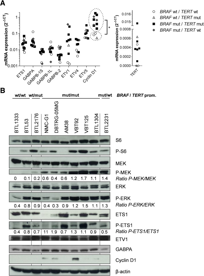
Based on the well-described activation of ETS-factors via MAPK-signaling [51] and having confirmed ETS-factor expression in the respective tumor tissues, we hypothesized that BRAFV600E mutant glioma cells were characterized by hyperactivated ETS-signaling. Analyses of ETS-factor mRNA expression confirmed that the transcription factors ETS1, GABPA, GABPB, ETV1, ETV4 and ETV5 were widely expressed throughout the entire cell panel, however, no differences between the genotypes were observed. Similarly, the distinct splice-variants of GABPB (GABPB-1S, GABPB-1 L, GABPB-2) showed no differences in expression between the genotypes. In contrast, cyclin D1 displayed significantly higher expression in BRAFV600E positive cell models as compared to wild-type cells. No differences between the investigated genotypes were observed for TERT mRNA expression (Fig. 1a).
Fig. 1.
Expression patterns of TERT, ETS-factors and activation of associated signaling cascades. a mRNA expression of ETS-factors and the ETS-downstream targets cyclin D1 and TERT were analyzed in the indicated genotypes. Means of three independent experiments are shown. Cyclin D1 mRNA expression of BRAF wild-type versus BRAFV600E mutated cell lines was quantified by unpaired student’s t-test (*p < 0.05). b Western blot analyses of cell lines with different BRAF and TERT promoter status as indicated are depicted. Proteins of S6 and MAPK pathway as well as selected ETS-factors and downstream targets are shown. Ratios between phosphorylated and total proteins as indicated were calculated after normalization to β-actin. wt = wild-type, mut = mutated
Next, we investigated expression and signaling activation of MAPK pathway members on protein level. Consistent with oncogenic BRAF signaling, phosphorylation levels of MEK were markedly enhanced in BRAFV600E-mutant as compared to BRAF wild-type cell models. In contrast, no distinct differences were observed for activation of S6 and ERK indicating activation of the respective pathways by alternative mechanisms in the BRAF wild-type models, an effect which has previously been described in both glioma [36] and melanoma [55]. With respect to ETS-factors, ETS1 was variably expressed throughout the cell panel and showed phosphorylation in all genotypes (Fig. 1b, Additional file 3: Figure S4). GABPA was widely and ETV1 constitutively expressed in the investigated cell line panel. In line with our qRT-PCR results, cyclin D1 was predominantly detectable in cell models harboring BRAF alterations (Fig. 1b).
BRAFV600E/TERT promoter double-mutant glioma cells are highly sensitive towards BRAF-inhibitors
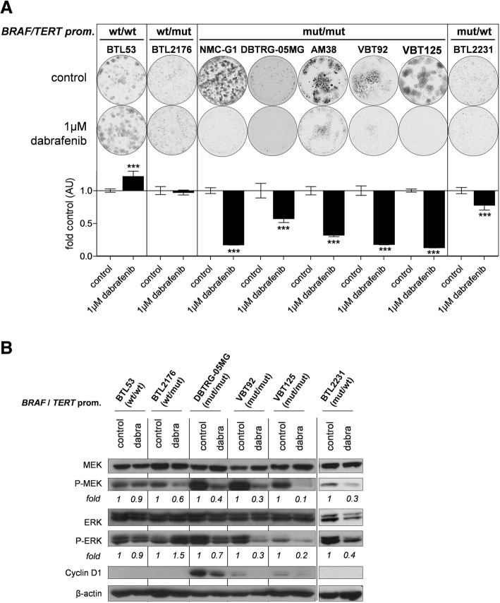
In order to validate the expected dependency of BRAFV600E-mutant glioma on hyperactivated MAPK-signaling, we tested the anti-proliferative effects of the BRAF-inhibitor dabrafenib. As predicted, dabrafenib was only effective in BRAF-mutated cell models (Additional file 3: Figure S5a, Fig. 2a). Interestingly, sensitivity was highest in those cell lines harboring additional TERT promoter mutations (Fig. 2a). In contrast, cell proliferation of cell models lacking BRAFV600E mutation was not inhibited but rather increased (Fig. 2a). In addition, these results were confirmed with vemurafenib, another BRAF-inhibitor (Additional file 3: Figure S5b). In addition to basal expression levels, we further investigated downstream effects of BRAF-inhibition in the respective genotypes. Dabrafenib effectively decreased both MEK- as well as ERK-phosphorylation levels in all BRAFV600E-mutated cell lines. In contrast, despite inhibition of MEK-phosphorylation, ERK-phosphorylation was stable or even increased in BRAF wild-type cell models (Fig. 2b). This finding is well in agreement with the so-called RAF-paradox by BRAF-inhibition under wild-type conditions [14]. Moreover, dabrafenib treatment resulted in downregulation of cyclin D1 in double-mutant cell models (Fig. 2b). No effect on cell proliferation was observed upon short term BRAF-inhibitor treatment (data not shown).
Fig. 2.
Anti-proliferative effects and altered downstream-signaling upon BRAF-inhibition. a Clone formation assays of with different BRAF and TERT promoter status as indicated are shown. Cells were seeded at low density and treated with 1 μM dabrafenib for 7 days. The upper panel depicts one representative well per condition. The lower panel shows the quantitative results represented as mean +/− SD of the respective untreated control. ***p < 0.001 (unpaired student’s t-tests) (b) Western blot analyses of cell models with different BRAF and TERT promoter status are depicted. Cell models were treated with 1 μM dabrafenib for 6 h. Expression and phosphorylation of the indicated MAPK pathway mediators as well as cyclin D1 are shown. Fold values are given as normalized expression to β-actin followed by activated kinase/total kinase and are normalized to the respective control. wt = wild-type, mut = mutated, dabra = dabrafenib
Oncogenic MAPK-signaling mediates TERT expression in BRAFV600E/TERT promoter double-mutant glioma
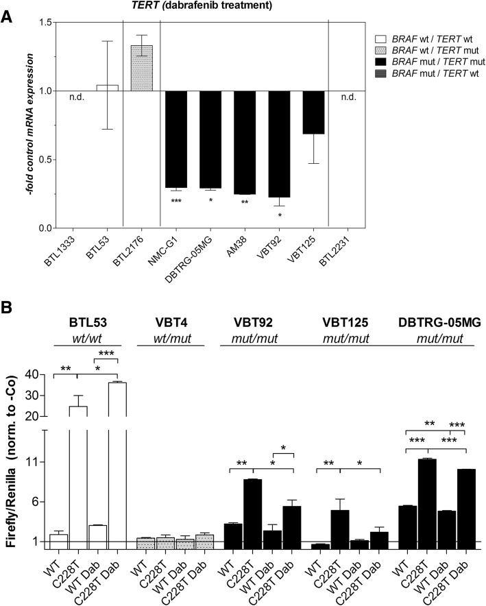
After confirming the respective impact of BRAF-inhibition on downstream signaling in the different genotypes, we tested the effect of dabrafenib on TERT expression. TERT mRNA was constitutively downregulated by dabrafenib (Fig. 3a) or vemurafenib (Additional file 3: Figure S6) in double-mutant glioma models only. Conversely, dabrafenib rather stimulated TERT expression in BRAF wild-type/TERT promoter-mutant cells and had no effect in the double wild-type background. No effect on cell proliferation was observed upon short term BRAF-inhibitor treatment (data not shown). In order to clarify the role of TERT promoter mutation in telomerase re-activation in the respective genotypes, we analyzed activation of the wild-type and mutant promoter sequences in each genotype via luciferase reporter assays. As expected, the C228T-mutated TERT promoter construct was significantly more active as compared to the wild-type promoter in most of the cell models. This high promoter activity could be suppressed by treatment with dabrafenib solely in the BRAFV600E-mutated cell models. In contrast, BRAF-inhibition increased the activity of the mutated TERT promoter in a double wild-type background (Fig. 3b).
Fig. 3.
Regulation of TERT expression and TERT promoter activity upon BRAF-inhibition. a TERT mRNA expression following dabrafenib treatment (1 μM, 16 h) of the indicated cell models is shown. Mean +/− SD; unpaired student’s t-tests (b) Luciferase reporter assays were performed in cell lines with different BRAF and TERT promoter status as indicated using wild-type or mutated (C228T) TERT promoter sequences. Cells were treated with 1 μM dabrafenib for 16 h. Results are given as ratio of firefly to renilla luciferase (internal control) and were normalized to a promoter-less construct (−Co, set to 1). Values are given as mean +/− SD from duplicates. One representative experiment out of three, delivering comparable results, is shown. Tukey’s multi-comparison one-way ANOVA was applied for statistical analysis. *p < 0.05, **p < 0.01, ***p < 0.001; wt = wild-type, mut = mutated, n.d. = not detected, dab = dabrafenib
ETS1 activation is impaired upon BRAF-inhibition in BRAFV600E-mutant glioma
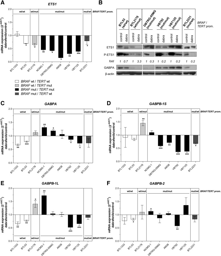
In order to investigate the impact of BRAF-mediated downstream signaling on ETS-factors in more detail, we selected ETS1, which is well described for being transcriptionally and post-translationally activated by MAPK signaling, for further analyses [30, 44, 50]. In correspondence to the expression patterns observed for TERT, ETS1 was more effectively downregulated by dabrafenib in all BRAFV600E/TERT promoter-mutant cell models (Fig. 4a). Moreover, ETS1 activation via phosphorylation was blocked in response to the BRAF-inhibitors dabrafenib (Fig. 4b) and vemurafenib (Additional file 3: Figure S7) in all BRAFV600E-mutant cells hence paralleling phosphorylation of ERK. In contrast, the levels of ETS1 expression and phosphorylation in BRAF wild-type cell lines were not efficiently blocked, obviously based on the above described paradoxical ERK-activation by the BRAF-inhibitors under wild-type conditions. GABPA protein expression, however, was not affected by BRAF-inhibition in double-mutant glioma cells (Fig. 4b). To exclude whether this was a side-effect of cell growth inhibition we tested short-term BRAF-inhibitor treatment and observed no effect on cell proliferation (data not shown).
Fig. 4.
Regulation of ETS-factors by oncogenic BRAF signaling. a mRNA and (b) protein expression/phosphorylation levels of ETS1/GABPA upon 16 h at 1 μM (qRT-PCR) and 6 h at 1 μM (Western blot) dabrafenib treatment. Fold values are given as normalized expression to β-actin and subsequent calculation of the ratio phospho/total ETS1 and are normalized to the respective untreated controls. mRNA expression levels of (c) GABPA, (d) GABPB-1S, (e) GABPB-1 L, and (f) GABPB-2 are depicted for the indicated cell models upon dabrafenib treatment (1 μM, 16 h). *p < 0.05, **p < 0.01, ***p < 0.001 (unpaired students’ t-tests); All values are given as mean +/− SD; wt = wild-type, mut = mutated, dabra = dabrafenib
With respect to other ETS-factors, only GABPB-1S expression was also predominately inhibited by dabrafenib in a BRAFV600E-mutated background whereas GABPA, GABPB-1 L and GABPB-2 showed variable response patterns (Fig. 4c-f). In line with the data from our cell models, a residual tumor of an anaplastic PXA case operated during combination treatment of dabrafenib and the MEK-inhibitor trametinib had lost expression of ETS1, cyclin D1 and TERT (Additional file 3: Figure S8).
ETS1 mediates TERT expression in BRAFV600E and TERT promoter-mutated glioma cells
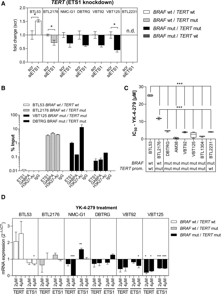
To more specifically dissect the link between MAPK-signaling and telomerase re-activation, we applied siRNA mediated knock-down of ETS1. Knock-down consistently decreased TERT mRNA expression across BRAFV600E/TERT promoter-mutated glioma cells as well as in a TERT promoter-mutated cell model (Fig. 5a). Notably, no effect on cell proliferation was observed upon siRNA-mediated knock-down (data not shown). Moreover, by ChIP-qRT-PCR we confirmed strong binding of ETS1 and GABPA selectively to the mutant TERT promoter locus, paralleled by activating H3K27-acetylation. In one double mutant cell model ETS1 even was the dominant of the investigated ETS-factors bound to the mutant promoter site (Fig. 5b). To clarify, whether ETS-factors play a central role in the malignant phenotype of BRAFV600E-mutant glioma, we assessed sensitivity to the ETS-inhibitor YK-4-279 [10] across our cell panel. Indeed, BRAF-mutant cell models were hypersensitive (IC50 < 5 μM) towards this compound suggesting a wide dependency of cell viability/proliferation on ETS-mediated signals (Fig. 5c). Accordingly, in contrast to TERT promoter wild-type glioma, ETS-factor inhibition by YK-4-279 reduced TERT mRNA expression in double-mutant cell models whereas ETS1 expression was more variable (Fig. 5d). Moreover, TERT re-expression partly rescued double-mutant glioma cells from the YK-4-279-induced growth inhibitory effect (Additional file 3: Figure S9). Combination of the BRAF inhibitor dabrafenib and the ETS-factor inhibitor YK-4-279 revealed additive to rather antagonistic effects, especially in the double-mutant glioma cell models (Additional file 3: Figure S10).
Fig. 5.
Activation of the mutant TERT promoter by ETS1. a An siRNA approach was applied to ETS1 knock-down in cell models with different BRAF and TERT promoter status as indicated. TERT mRNA expression was detected using qRT-PCR. Significance levels were evaluated by unpaired students’ t-tests (mean +/− SEM). b Binding of ETS1 and GABPA to the different TERT promoter variants and presence of the activating histone mark H3K27-Ac was analyzed by ChIP-qRT-PCR. IgG served as negative control. Values are given as % Input, depicted mean +/− SD from duplicates. c Cytotoxicity assay were performed in cell models of different BRAF and TERT promoter background as depicted. Half-maximal inhibitory concentration (IC50) after YK-4-279 treatment for 72 h was calculated. Tukey’s multi-comparison one-way ANOVA was applied (mean +/− SD). d TERT and ETS1 mRNA expression levels were analyzed by qRT-PCR after 16 h treatment with the indicated concentrations of YK-4-279. Ordinary one-way ANOVAs for every cell line were calculated (Dunnet correction, 0.05 (95% confidence interval) as controls vs. treatments (mean +/− SEM). *p < 0.05, **p < 0.01, ***p < 0.001, wt = wild-type, mut = mutated, n.d. = not detected
Discussion
The discovery of genomic aberrations in BRAF as drivers of certain glioma subtypes has resulted in a magnificent extension of the therapeutic repertoire for these patients [17, 39, 43]. Recent studies, however, indicate that additional factors such as loss of CDKN2A or telomerase re-activation may significantly influence the clinical outcome of glioma patients with oncogenic BRAF, suggesting a biological heterogeneity within this subgroup [34, 40]. In this context, OIS and replicative senescence have been described as central obstacles for proliferation in BRAF-altered glioma cells [2]. Furthermore, TERT derepression has been demonstrated to promote escape of cancer cells from OIS [38]. Previous clinical observations and case-report studies delivered preliminary evidence that TERT promoter mutations indicate tumors with higher aggressiveness within BRAFV600E-mutated glioma [33, 34, 40, 54]. Consequently, we systematically investigated the cellular factors contributing to the interplay of BRAFV600E and TERT promoter mutations in glioma.
To begin with, we analyzed the clinical impact of telomerase re-activation in a small cohort of BRAFV600E-mutated glioma. In accordance with previous reports, we observed a highly aggressive course in one anaplastic PXA with both BRAFV600E and TERT promoter mutations in our patient collective [18, 28]. Moreover, only this double-mutant tumor expressed detectable levels of TERT mRNA. Consistent with this observation, an in silico analysis of 103 BRAFV600E-mutated glioma showed that TERT promoter mutations are significantly enriched in WHO grade III/IV tumors. Within this dataset, 28% of BRAFV600E-mutated HGG harbored additional TERT promoter mutations, which is consistent with earlier reports of secondary HGG and anaplastic PXA [18, 20, 28, 33, 34, 40]. Particular aggressiveness of double-mutant tumors has already been described for other tumor-types such as melanoma and thyroid cancer [23, 35, 52].
In order to elucidate the potential role of TERT promoter mutations in the malignant phenotype of certain BRAFV600E-mutated glioma, we curated a unique set of cell models containing nine BRAFV600E-mutant gliomas, the largest panel reported to date. Notably, all stable BRAFV600E-positive cell lines harbored TERT promoter mutations corroborating the aggressive biology of double-mutant tumors. Moreover, double-mutant models were further characterized by loss of the CDKN2A locus and BRAF-induced cyclin D1 expression. In a previous reported study, depletion of CDKN2A, CDKN1A, and TP53 has been demonstrated as essential factor to overcome OIS in a patient-derived model of pilocytic astrocytoma [2]. These findings support our data as all stable BRAFV600E-mutant cell models harbor a loss at the CDKN2A locus. Moreover, the same study showed that TERT re-expression in this BRAF-driven cell model enabled escape from replicative senescence [2]. This supports the notion that the aggressive behavior of these tumors is fueled by a synergistic activation of MAPK-signaling in addition to CDK4/6-activation and telomerase re-expression.
The pathogenic mechanism of TERT promoter mutations is widely mediated by transcriptional activation which involves the binding of ETS-factors [13]. Specifically, the ETS-factors ETS1, GABPA, GABPB, and the splice variant GABPB-1 L, have been linked to activation of the mutated TERT promoter [4, 13, 31, 53]. Interestingly, both overall expression (e.g. GABPB, ETS1) as well as posttranscriptional activation (e.g. ETS1) can be stimulated via the MAPK-pathway [6, 25, 41, 51]. Accordingly, with respect to BRAFV600E-mutated melanoma, both phosphorylation of ETS1 and upregulation of GABPB via Fos have been shown to link oncogenic BRAF signaling to activation of the mutant TERT promoter [25, 50]. We analyzed the expression of the major ETS-factors ETS1, GABPA, and GABPB with its splice variants, ETV1, ETV4 and ETV5 all of which were widely expressed across our glioma cell panel. In contrast, TERT mRNA was only detectable in BRAFV600E cells with additional TERT promoter mutations. Cell proliferation assays with BRAF-inhibitors confirmed the selective activity against BRAFV600E-mutated glioma cells, as already reported by previous preclinical studies and case series [7, 12, 19, 21, 22]. Strikingly, BRAF-inhibition also blocked activating phosphorylation of ETS1 selectively in BRAFV600E-mutant glioma cells. This effect has previously been described in other BRAF-mutated tumor types like melanoma [50], but not yet in glioma. Additionally, we could detect an inhibitory effect on cyclin D1 expression, confirming previous data, suggesting it as a downstream target of oncogenic MAPK-signaling in BRAF-mutated glioma [39].
In a following step, we demonstrated that activation of TERT transcription is dependent on BRAF signaling solely in the background of BRAFV600E and TERT promoter double-mutation. Moreover, the clinical relevance of this finding is supported by investigation of the tumor material derived from a single patient treated with a combination of BRAF- and MEK-inhibitors resulting in undetectable levels of both ETS1 and TERT during therapy. Oncogenic BRAF signaling has been shown to bridge telomerase re-activation via the mutated TERT promoter sequence. With respect to ETS-factor activation, GABPB expression has been described to be upregulated via Fos, a well described downstream effector of MAPK and ETS1 signals [25, 41], and ETS1 is directly phosphorylated via the MAPK-pathway [50]. Next, we tested the impact of BRAF-inhibition on the expression of the respective ETS-factors and found that only ETS1 expression, but not expression of GABPA or the different GABPB splice variants, were significantly and consistently downregulated throughout all BRAFV600E-mutated glioma. These findings are well in agreement with a feed-forward loop on ETS1 expression exerted by MAPK-signaling and ETS1 as predominant effector molecule [30]. Additionally, we show that knock-down of ETS1 reduces TERT expression confirming it as mediator of BRAFV600E-driven TERT promoter activation. Accordingly, ChIP-analysis revealed ETS1 and GABPA-binding specifically to the mutant TERT promoter site. Previous studies in BRAF wild-type glioblastoma models have already demonstrated a central role of GABPA in activation of the mutant TERT promoter [4]. Our data point towards cooperation of GABPA with ETS1, especially in a BRAFV600E-mutant glioma background. Accordingly, a recent study in melanoma has shown that the C228T variant, the predominant mutation detected in our panel, is more efficiently activated by ETS1 as compared to the other TERT promoter mutations [1]. The potential role of other MAPK- induced ETS-factors is of high interest to be investigated in further studies.
BRAF-inhibition has become the standard of care in BRAFV600E-mutated melanoma [14]. However, acquired insensitivity plays an important role in therapy failure [24]. Additionally, first results of clinical application in BRAFV600E-positive glioma indicate that also a proportion of these tumors exhibit intrinsic BRAF-inhibitor resistance [17]. Consequently, inhibition of ETS-factors as downstream MAPK-signal transmitters have been suggested to provide a novel therapeutic opportunity to overcome upstream resistance development [49]. Therefore, we tested the effect of YK-4-279, a well-described ETS-factor inhibitor [10], in our cell line panel. In line with previous reports from our group concerning meningioma [47], also glioma cell models harboring mutant TERT promoters were hypersensitive towards YK-4-279 treatment [47] and YK-4-279 distinctly reduced TERT mRNA expression. Strikingly, BRAFV600E-mutant models were even more sensitive towards YK-4-279, irrespective of the underlying TERT promoter status. TERT re-expression from a viral promoter partially rescued double-mutated glioma cells from the cytotoxic effect of YK-4-279. As we found that both dabrafenib as well as YK-4-279 were highly active against BRAFV600E mutated glioma, we aimed to investigate interactions between the two drugs. No synergistic, but rather antagonistic effects were identified particularly in the double-mutant glioma cell models. These results correspond to our findings that dabrafenib alone already reduced the expression levels of ETS1, the target of YK-4-279. Taken together, our data suggest that apart from the demonstrated TERT promoter activating properties, ETS-factors appear to play an important role in tumor biology of BRAFV600E-mutated glioma.
Conclusions
Summarizing, we prove that telomerase re-activation based on a mutant TERT promoter sequence in BRAFV600E-mutant glioma is driven by oncogenic BRAF signaling predominantly via downstream activation of ETS-factors. Accordingly, ETS-factor inhibition is a promising therapeutic option for therapy-resistant BRAF-mutant glioma.
Additional files
Table S1. Antibodies. Table S2. Primers. Table S3. Histopathological, molecular and clinical parameters of the Vienna patient cohort. (DOCX 16 kb)
Table S4. Dataset of 103 BRAFV600E-mutated glioma with information on tumor grade and TERT promoter mutation. (XLSX 10 kb)
Figure S1. mRNA expression of ETS transcription factors and downstream targets in tumor tissue. Figure S2. Expression of Ets-factors and TERT in TCGA RNA sequencing data sets. Figure S3. Evaluation of TP53 and CDKN2A/Rb signaling pathways. Figure S4. Activation levels of MEK, ERK and ETS1. Figure S5. Anti-proliferative effects of BRAF inhibitors. Figure S6. Inhibition of TERT expression upon vemurafenib treatment. Figure S7. ETS1 inhibition upon vemurafenib treatment. Figure S8. Changes in expression patterns in patient tissue upon targeted treatment. Figure S9. Ectopic TERT re-expression partly rescues double-mutant glioma cells from YK-4-279- mediated cytotoxicity. Figure S10. Combined BRAF and Ets-factor inhibition. (PDF 786 kb)
Acknowledgements
The authors thank Thomas Mohr, Mirjana Stojanovic and Petra Lapsanska for excellent technical assistance.
Abbreviations
- CDK
cyclin dependent kinase
- ChIP
chromatin immunoprecipitation
- CNS
central nervous system
- ETS/TCF
E-twenty-six transcription factor
- GBM
glioblastoma multiforme
- GG
ganglioglioma
- HGG
high-grade glioma
- LGG
low-grade glioma
- MAPK
mitogen-activated protein kinase
- OIS
oncogene induced senescence
- PXA
pleomorphic xanthoastrocyma
- Rb
retinoblastoma protein
- STR
short tandem repeat
- TCGA
The Cancer Genome Atlas
- TERT
telomerase reverse transcriptase
Authors’ contributions
JG, LG and WB designed the study, interpreted the data and wrote the manuscript. LG and JG planned and LG, JG, DK, KN and HMS executed experiments. DL, SSK, LK, CDinhof and CP contributed to data analyses and experiments. LM, AA, CDorfer, TC, AP and IS managed and delivered clinical material. CH performed the histological assessments. RK provided technical support and reviewed the manuscript. All authors read and approved the final manuscript.
Funding
The work was supported by the “Fellinger Krebsforschung” (to J.G.), the “Jubiläumsfonds der Österreichischen Nationalbank” (project # 15173 to I.S.), Innovative Medicines Initiative (ITCC-P4 to W.B.), and the Austrian Science Fund FWF (project number: P30105 to W.B., project number: T906 to D.L.). Carina Dinhof is a recipient of a DOC fellowship of the Austrian Academy of Sciences at the Institute of Cancer Research (Medical University of Vienna).
Availability of data and materials
All data generated or analyzed during this study are included in this published article.
Ethics approval and consent to participate
The study was approved by the ethics committee of the Medical University of Vienna and by the local ethics committee of Upper Austria in Linz. Informed consent was obtained from all participating patients and/or legal representatives.
Consent for publication
Not applicable.
Competing interests
The authors declare that they have no competing interests.
Footnotes
Publisher’s Note
Springer Nature remains neutral with regard to jurisdictional claims in published maps and institutional affiliations.
Contributor Information
Lisa Gabler, Email: lisa.gabler@meduniwien.ac.at.
Daniela Lötsch, Email: daniela.loetsch@meduniwien.ac.at.
Dominik Kirchhofer, Email: dominik.kirchhofer@meduniwien.ac.at.
Sushilla van Schoonhoven, Email: sushilla.vanschoonhoven@meduniwien.ac.at.
Hannah M. Schmidt, Email: hanni_schmidt2@gmx.de
Lisa Mayr, Email: lisa.mayr@meduniwien.ac.at.
Christine Pirker, Email: christine.pirker@meduniwien.ac.at.
Katharina Neumayer, Email: katharina.neumayer@kepleruniklinikum.at.
Carina Dinhof, Email: carina.dinhof@meduniwien.ac.at.
Lucia Kastler, Email: lucia.kastler@meduniwien.ac.at.
Amedeo A. Azizi, Email: amedeo.azizi@meduniwien.ac.at
Christian Dorfer, Email: christian.dorfer@meduniwien.ac.at.
Thomas Czech, Email: thomas.czech@meduniwien.ac.at.
Christine Haberler, Email: christine.haberler@meduniwien.ac.at.
Andreas Peyrl, Email: andreas.peyrl@meduniwien.ac.at.
Rajiv Kumar, Email: r.kumar@dkfz.de.
Irene Slavc, Email: irene.slavc@meduniwien.ac.at.
Sabine Spiegl-Kreinecker, Email: sabine.spiegl-kreinecker@kepleruniklinikum.at.
Johannes Gojo, Email: johannes.gojo@meduniwien.ac.at.
Walter Berger, Email: walter.berger@meduniwien.ac.at.
References
- 1.Andrés-Lencina JJ, Rachakonda S, García-Casado Z, Srinivas N, Skorokhod A, Requena C, Soriano V, Kumar R, Nagore E. TERT promoter mutation subtypes and survival in stage I and II melanoma patients. Int J Cancer. 2018;04:1–10. doi: 10.1002/ijc.31780. [DOI] [PubMed] [Google Scholar]
- 2.Bächli H, Milde T, Schuhmann MU, Brabetz S, Gronych J, Jones DTW, Capper D, von Deimling A, Marquardt V, Pauck D, Oehme I, Hielscher T, Ridinger J, Stiles CD, Hohloch J, Sahm F, Pfister SM, Ecker J, Witt O, Korshunov A, Remke M, Usta D, Brummer T, Selt F. Establishment and application of a novel patient-derived KIAA1549:BRAF-driven pediatric pilocytic astrocytoma model for preclinical drug testing. Oncotarget. 2016;8:11460–11479. doi: 10.18632/oncotarget.14004. [DOI] [PMC free article] [PubMed] [Google Scholar]
- 3.Bautista F, Paci A, Minard-Colin V, Dufour C, Grill J, Lacroix L, Varlet P, Valteau-Couanet D, Geoerger B. Vemurafenib in pediatric patients with BRAFV600E mutated high-grade gliomas. Pediatr Blood Cancer. 2014;61:1101–1103. doi: 10.1002/pbc.24891. [DOI] [PubMed] [Google Scholar]
- 4.Bell R. J. A., Rube H. T., Kreig A., Mancini A., Fouse S. D., Nagarajan R. P., Choi S., Hong C., He D., Pekmezci M., Wiencke J. K., Wrensch M. R., Chang S. M., Walsh K. M., Myong S., Song J. S., Costello J. F. The transcription factor GABP selectively binds and activates the mutant TERT promoter in cancer. Science. 2015;348(6238):1036–1039. doi: 10.1126/science.aab0015. [DOI] [PMC free article] [PubMed] [Google Scholar]
- 5.Burger MC, Ronellenfitsch MW, Lorenz NI, Wagner M, Voss M, Capper D, Tzaridis T, Herrlinger U, Steinbach JP, Stoffels G, Langen KJ, Brandts C, Senft C, Harter PN, Bähr O. Dabrafenib in patients with recurrent, BRAF V600E mutated malignant glioma and leptomeningeal disease. Oncol Rep. 2017;38:3291–3296. doi: 10.3892/or.2017.6013. [DOI] [PMC free article] [PubMed] [Google Scholar]
- 6.Callaway K, Waas WF, Rainey MA, Ren P, Dalby KN. Phosphorylation of the transcription factor ets-1 by ERK2: rapid dissociation of ADP and phospho-ets-1. Biochemistry. 2010;49:3619–3630. doi: 10.1021/bi100199q. [DOI] [PMC free article] [PubMed] [Google Scholar]
- 7.Chamberlain MC. Salvage therapy with BRAF inhibitors for recurrent pleomorphic xanthoastrocytoma: a retrospective case series. J Neuro-Oncol. 2013;114:237–240. doi: 10.1007/s11060-013-1176-5. [DOI] [PubMed] [Google Scholar]
- 8.Dahiya S, Emnett RJ, Haydon DH, Leonard JR, Phillips JJ, Perry A, Gutmann DH. BRAF-V600E mutation in pediatric and adult glioblastoma. Neuro-Oncology. 2014;16:318–319. doi: 10.1093/neuonc/not146. [DOI] [PMC free article] [PubMed] [Google Scholar]
- 9.Dasgupta T, Olow AK, Yang X, Hashizume R, Nicolaides TP, Tom M, Aoki Y, Berger MS, Weiss WA, Stalpers LJA, Prados M, David James C, Mueller S, Haas-Kogan DA. Survival advantage combining a BRAF inhibitor and radiation in BRAF V600E-mutant glioma. J Neuro-Oncol. 2016;126:385–393. doi: 10.1007/s11060-015-1939-2. [DOI] [PMC free article] [PubMed] [Google Scholar]
- 10.Erkizan HV, Kong Y, Merchant M, Schlottmann S, Barber-Rotenberg JS, Yuan L, Abaan OD, Chou TH, Dakshanamurthy S, Brown ML, Ren A, Toretsky JA. A small molecule blocking oncogenic protein EWS-FLI1 interaction with RNA helicase a inhibits growth of Ewing’s sarcoma. Nat Med. 2009;15:750–756. doi: 10.1038/nm.1983. [DOI] [PMC free article] [PubMed] [Google Scholar]
- 11.Gojo J, Lötsch D, Spiegl-Kreinecker S, Pajtler KW, Neumayer K, Korbel P, Araki A, Brandstetter A, Mohr T, Hovestadt V, Chavez L, Kirchhofer D, Ricken G, Stefanits H, Korshunov A, Pfister SM, Dieckmann K, Azizi AA, Czech T, Filipits M, Kool M, Peyrl A, Slavc I, Berger W, Haberler C. Telomerase activation in posterior fossa group a ependymomas is associated with dismal prognosis and chromosome 1q gain. Neuro-Oncology. 2017;19:1183–1194. doi: 10.1093/neuonc/nox027. [DOI] [PMC free article] [PubMed] [Google Scholar]
- 12.Grossauer S, Koeck K, Murphy NE, Meyers ID, Daynac M, Truffaux N, Truong AY, Nicolaides TP, McMahon M, Berger MS, Phillips JJ, James CD, Petritsch CK. Concurrent MEK targeted therapy prevents MAPK pathway reactivation during BRAFV600E targeted inhibition in a novel syngeneic murine glioma model. Oncotarget. 2016;7:75839–75853. doi: 10.18632/oncotarget.12419. [DOI] [PMC free article] [PubMed] [Google Scholar]
- 13.Heidenreich B, Kumar R. TERT promoter mutations in telomere biology. Mutat Res. 2017;771:15–31. doi: 10.1016/j.mrrev.2016.11.002. [DOI] [PubMed] [Google Scholar]
- 14.Holderfield M, Deuker MM, McCormick F, McMahon M. Targeting RAF kinases for cancer therapy: BRAF-mutated melanoma and beyond. Nat Rev Cancer. 2014;14:455–467. doi: 10.1038/nrc3760. [DOI] [PMC free article] [PubMed] [Google Scholar]
- 15.Huillard E, Hashizume R, Phillips JJ, Griveau A, Ihrie RA, Aoki Y, Nicolaides T, Perry A, Waldman T, McMahon M, Weiss WA, Petritsch C, James CD, Rowitch DH. Cooperative interactions of BRAFV600E kinase and CDKN2A locus deficiency in pediatric malignant astrocytoma as a basis for rational therapy. Proc Natl Acad Sci. 2012;109:8710–8715. doi: 10.1073/pnas.1117255109. [DOI] [PMC free article] [PubMed] [Google Scholar]
- 16.Jones DTW, Kocialkowski S, Liu L, Pearson DM, Bäcklund LM, Ichimura K, Collins VP. Tandem duplication producing a novel oncogenic BRAF fusion gene defines the majority of pilocytic astrocytomas. Cancer Res. 2008;68:8673–8677. doi: 10.1158/0008-5472.CAN-08-2097. [DOI] [PMC free article] [PubMed] [Google Scholar]
- 17.Kaley T, Touat M, Subbiah V, Hollebecque A, Rodon J, Lockhart AC, Keedy V, Bielle F, Hofheinz R-D, Joly F, Blay J-Y, Chau I, Puzanov I, Raje NS, DeAngelis LM, Makrutzki M, Riehl T, Pitcher B, Baselga J, Hyman DM, Wolf J, DeAngelis LM, Makrutzki M, Riehl T, Pitcher B, Baselga J, Hyman DM. BRAF inhibition in BRAFV600-mutant gliomas: results from the VE-BASKET study. J Clin Oncol. 2018;36:3477–3484. doi: 10.1200/JCO.2018.78.9990. [DOI] [PMC free article] [PubMed] [Google Scholar]
- 18.Koelsche C, Sahm F, Capper D, Reuss D, Sturm D, Jones DTW, Kool M, Northcott PA, Wiestler B, Bohmer K, Meyer J, Mawrin C, Hartmann C, Mittelbronn M, Platten M, Brokinkel B, Seiz M, Herold-Mende C, Unterberg A, Schittenhelm J, Weller M, Pfister S, Wick W, Korshunov A, von Deimling A, Böhmer K, Meyer J, Mawrin C, Hartmann C, Mittelbronn M, Platten M, Brokinkel B, Seiz M, Herold-Mende C, Unterberg A, Schittenhelm J, Weller M, Pfister S, Wick W, Korshunov A, von Deimling A, Bohmer K, Meyer J, Mawrin C, Hartmann C, Mittelbronn M, Platten M, Brokinkel B, Seiz M, Herold-Mende C, Unterberg A, Schittenhelm J, Weller M, Pfister S, Wick W, Korshunov A, von Deimling A. Distribution of TERT promoter mutations in pediatric and adult tumors of the nervous system. Acta Neuropathol. 2013;126:907–915. doi: 10.1007/s00401-013-1195-5. [DOI] [PubMed] [Google Scholar]
- 19.Kogiso M, Qi L, Lindsay H, Huang Y, Zhao X, Liu Z, Braun FK, Du Y, Zhang H, Bae G, Zhao S, Injac SG, Sobieski M, Brunell D, Mehta V, Tran D, Murray J, Baxter PA, Yuan X-J, Su JM, Adesina A, Perlaky L, Chintagumpala M, Parsons DW, Lau CC, Stephan CC, Lu X, Li X-N, Kogiso M, Qi L, Lindsay H, Huang Y, Zhao X, Liu Z, Braun FK, Du Y, Zhang H, Bae G, Zhao S, Injac SG, Sobieski M, Brunell D, Mehta V, Tran D, Murray J, Baxter PA, Yuan X-J, Su JM, Adesina A, Perlaky L, Chintagumpala M, Williams Parsons D, Lau CC, Stephan CC, Lu X, Li X-N. Xenotransplantation of pediatric low grade gliomas confirms the enrichment of BRAF V600E mutation and preservation of CDKN2A deletion in a novel orthotopic xenograft mouse model of progressive pleomorphic xanthoastrocytoma. Oncotarget. 2017;8:87455–87471. doi: 10.18632/oncotarget.20713. [DOI] [PMC free article] [PubMed] [Google Scholar]
- 20.Korshunov A, Chavez L, Sharma T, Ryzhova M, Schrimpf D, Stichel D, Capper D, Sturm D, Kool M, Habel A, Kleinschmidt-DeMasters BK, Rosenblum M, Absalyamova O, Golanov A, Lichter P, Pfister SM, Jones DTW, Perry A, von Deimling A. Epithelioid glioblastomas stratify into established diagnostic subsets upon integrated molecular analysis. Brain Pathol. 2018;28:656–662. doi: 10.1111/bpa.12566. [DOI] [PMC free article] [PubMed] [Google Scholar]
- 21.Lassaletta A, Zapotocky M, Mistry M, Ramaswamy V, Honnorat M, Krishnatry R, Stucklin AG, Zhukova N, Arnoldo A, Ryall S, Ling C, McKeown T, Loukides J, Cruz O, De Torres C, Ho CY, Packer RJ, Tatevossian R, Qaddoumi I, Harreld JH, Dalton JD, Mulcahy-Levy J, Foreman N, Karajannis MA, Wang S, Snuderl M, Rao AN, Giannini C, Kieran M, Ligon KL, Garre ML, Nozza P, Mascelli S, Raso A, Mueller S, Nicolaides T, Silva K, Perbet R, Vasiljevic A, Conter CF, Frappaz D, Leary S, Crane C, Chan A, Ng HK, Shi ZF, Mao Y, Finch E, Eisenstat D, Wilson B, Carret AS, Hauser P, Sumerauer D, Krskova L, Larouche V, Fleming A, Zelcer S, Jabado N, Rutka JT, Dirks P, Taylor MD, Chen S, Bartels U, Huang A, Ellison DW, Bouffet E, Hawkins C, Tabori U. Therapeutic and prognostic implications of BRAF V600E in pediatric low-grade gliomas. J Clin Oncol. 2017;35:2934–2941. doi: 10.1200/JCO.2016.71.8726. [DOI] [PMC free article] [PubMed] [Google Scholar]
- 22.Levy JMM, Thompson JC, Griesinger AM, Amani V, Donson AM, Birks DK, Morgan MJ, Mirsky DM, Handler MH, Foreman NK, Thorburn A. Autophagy inhibition improves chemosensitivity in BRAFV600E brain tumors. Cancer Discov. 2014;4:773–780. doi: 10.1158/2159-8290.CD-14-0049. [DOI] [PMC free article] [PubMed] [Google Scholar]
- 23.Li Y, Cheng HS, Chng WJ, Tergaonkar V. Activation of mutant TERT promoter by RAS-ERK signaling is a key step in malignant progression of BRAF-mutant human melanomas. Proc Natl Acad Sci. 2016;113:14402–14407. doi: 10.1073/pnas.1611106113. [DOI] [PMC free article] [PubMed] [Google Scholar]
- 24.Lito P, Rosen N, Solit DB. Tumor adaptation and resistance to RAF inhibitors. Nat Med. 2013;19:1401–1409. doi: 10.1038/nm.3392. [DOI] [PubMed] [Google Scholar]
- 25.Liu R, Zhang T, Zhu G, Xing M (2018) Regulation of mutant TERT by BRAF V600E/MAP kinase pathway through FOS/GABP in human cancer. Nat Commun 9. 10.1038/s41467-018-03033-1 [DOI] [PMC free article] [PubMed]
- 26.Lötsch D, Ghanim B, Laaber M, Wurm G, Weis S, Lenz S, Webersinke G, Pichler J, Berger W, Spiegl-Kreinecker S. Prognostic significance of telomerase-associated parameters in glioblastoma: effect of patient age. Neuro-Oncology. 2013;15:423–432. doi: 10.1093/neuonc/nos329. [DOI] [PMC free article] [PubMed] [Google Scholar]
- 27.Louis David N., Perry Arie, Reifenberger Guido, von Deimling Andreas, Figarella-Branger Dominique, Cavenee Webster K., Ohgaki Hiroko, Wiestler Otmar D., Kleihues Paul, Ellison David W. The 2016 World Health Organization Classification of Tumors of the Central Nervous System: a summary. Acta Neuropathologica. 2016;131(6):803–820. doi: 10.1007/s00401-016-1545-1. [DOI] [PubMed] [Google Scholar]
- 28.Ma C, Feng R, Chen H, Hameed NUFUF, Aibaidula A, Song Y, Wu J. BRAF V600E, TERT, and IDH2 mutations in pleomorphic Xanthoastrocytoma: observations from a large case-series study. World Neurosurg. 2018;120:e1225–e1233. doi: 10.1016/j.wneu.2018.09.050. [DOI] [PubMed] [Google Scholar]
- 29.Mackay A, Burford A, Molinari V, Jones DTW, Izquierdo E, Brouwer-Visser J, Giangaspero F, Haberler C, Pietsch T, Jacques TS, Figarella-Branger D, Rodriguez D, Morgan PS, Raman P, Waanders AJ, Resnick AC, Massimino M, Garrè ML, Smith H, Capper D, Pfister SM, Würdinger T, Tam R, Garcia J, Das Thakur M, Vassal G, Grill J, Jaspan T, Varlet P, Jones C. Molecular, pathological, radiological, and immune profiling of non-brainstem pediatric high-grade glioma from the HERBY phase II randomized trial. Cancer cell. 2018;33:829–842.e5. doi: 10.1016/j.ccell.2018.04.004. [DOI] [PMC free article] [PubMed] [Google Scholar]
- 30.Majérus M-A, Bibollet-Ruche F, Telliez J-B, Wasylyk B, Bailleul B. Serum, AP-1 and Ets-1 stimulate the human Ets-1 promoter. Nucleic Acids Res. 1992;20:2699–2703. doi: 10.1093/nar/20.11.2699. [DOI] [PMC free article] [PubMed] [Google Scholar]
- 31.Mancini A, Xavier-Magalhães A, Woods WS, Nguyen KT, Amen AM, Hayes JL, Fellmann C, Gapinske M, McKinney AM, Hong C, Jones LE, Walsh KM, Bell RJA, Doudna JA, Costa BM, Song JS, Perez-Pinera P, Costello JF. Disruption of the β1L isoform of GABP reverses glioblastoma replicative immortality in a TERT promoter mutation-dependent manner. Cancer Cell. 2018;34:513–528. doi: 10.1016/j.ccell.2018.08.003. [DOI] [PMC free article] [PubMed] [Google Scholar]
- 32.Mathieu V, Pirker C, Schmidt WM, Spiegl-Kreinecker S, Lotsch D, Heffeter P, Hegedus B, Grusch M, Kiss R, Berger W. Aggressiveness of human melanoma xenograft models is promoted by aneuploidy-driven gene expression deregulation. Oncotarget. 2012;3:399–413. doi: 10.18632/oncotarget.473. [DOI] [PMC free article] [PubMed] [Google Scholar]
- 33.Matsumura N, Nakajima N, Yamazaki T, Nagano T, Kagoshima K, Nobusawa S, Ikota H, Yokoo H. Concurrent TERT promoter and BRAF V600E mutation in epithelioid glioblastoma and concomitant low-grade astrocytoma. Neuropathology. 2017;37:58–63. doi: 10.1111/neup.12318. [DOI] [PubMed] [Google Scholar]
- 34.Mistry M, Zhukova N, Merico D, Rakopoulos P, Krishnatry R, Shago M, Stavropoulos J, Alon N, Pole JD, Ray PN, Navickiene V, Mangerel J, Remke M, Buczkowicz P, Ramaswamy V, Stucklin AG, Li M, Young EJ, Zhang C, Castelo-Branco P, Bakry D, Laughlin S, Shlien A, Chan J, Ligon KL, Rutka JT, Dirks PB, Taylor MD, Greenberg M, Malkin D, Huang A, Bouffet E, Hawkins CE, Tabori U. BRAF mutation and CDKN2A deletion define a clinically distinct subgroup of childhood secondary high-grade glioma. J Clin Oncol. 2015;33:1015–1022. doi: 10.1200/JCO.2014.58.3922. [DOI] [PMC free article] [PubMed] [Google Scholar]
- 35.Moon S, Song YS, Kim YA, Lim JA, Cho SW, Moon JH, Hahn S, Park DJ, Park YJ. Effects of coexistent BRAFV600E and TERT promoter mutations on poor clinical outcomes in papillary thyroid Cancer: a meta-analysis. Thyroid. 2017;27:651–660. doi: 10.1089/thy.2016.0350. [DOI] [PubMed] [Google Scholar]
- 36.Nicolaides TP, Li H, Solomon D, Hariono S, Hashizume R, Barkovich K, Baker SJ, Paugh BS, Jones C, Forshew T, Hindley GF, Hodgson JG, Kim J-S, Rowitch DH, Weiss W, Waldman T, James CD. Targeted therapy for BRAFV600E malignant astrocytoma. Clin Cancer Res. 2011;17:7595–7604. doi: 10.1158/1078-0432.CCR-11-1456. [DOI] [PMC free article] [PubMed] [Google Scholar]
- 37.Ostrom Quinn T., Gittleman Haley, Fulop Jordonna, Liu Max, Blanda Rachel, Kromer Courtney, Wolinsky Yingli, Kruchko Carol, Barnholtz-Sloan Jill S. CBTRUS Statistical Report: Primary Brain and Central Nervous System Tumors Diagnosed in the United States in 2008-2012. Neuro-Oncology. 2015;17(suppl 4):iv1–iv62. doi: 10.1093/neuonc/nov189. [DOI] [PMC free article] [PubMed] [Google Scholar]
- 38.Patel PL, Suram A, Mirani N, Bischof O, Herbig U. Derepression of hTERT gene expression promotes escape from oncogene-induced cellular senescence. Proc Natl Acad Sci. 2016;113:E5024–E5033. doi: 10.1073/pnas.1602379113. [DOI] [PMC free article] [PubMed] [Google Scholar]
- 39.Pfister S, Janzarik WG, Remke M, Ernst A, Werft W, Becker N, Toedt G, Wittmann A, Kratz C, Olbrich H, Ahmadi R, Thieme B, Joos S, Radlwimmer B, Kulozik A, Pietsch T, Herold-mende C, Gnekow A, Reifenberger G. BRAF gene duplication constitutes a mechanism of MAPK pathway activation in low-grade astrocytomas. J Clin Invest. 2008;118:1739–1749. doi: 10.1172/JCI33656DS1. [DOI] [PMC free article] [PubMed] [Google Scholar]
- 40.Phillips JJ, Gong H, Chen K, Joseph NM, van Ziffle J, Bastian BC, Grenert JP, Kline CN, Mueller S, Banerjee A, Nicolaides T, Gupta N, Berger MS, Lee HS, Pekmezci M, Tihan T, Bollen AW, Perry A, Shieh JTC, Solomon DA. The genetic landscape of anaplastic pleomorphic xanthoastrocytoma. Brain Pathol. 2018;29:85–96. doi: 10.1111/bpa.12639. [DOI] [PMC free article] [PubMed] [Google Scholar]
- 41.Plotnik JP, Budka JA, Ferris MW, Hollenhorst PC. ETS1 is a genome-wide effector of RAS/ERK signaling in epithelial cells. Nucleic Acids Res. 2014;42:11928–11940. doi: 10.1093/nar/gku929. [DOI] [PMC free article] [PubMed] [Google Scholar]
- 42.Robinson GW, Orr BA, Gajjar A. Complete clinical regression of a BRAF V600E-mutant pediatric glioblastoma multiforme after BRAF inhibitor therapy. BMC Cancer. 2014;14:1–5. doi: 10.1186/1471-2407-14-258. [DOI] [PMC free article] [PubMed] [Google Scholar]
- 43.Schindler G, Capper D, Meyer J, Janzarik W, Omran H, Herold-Mende C, Schmieder K, Wesseling P, Mawrin C, Hasselblatt M, Louis DN, Korshunov A, Pfister S, Hartmann C, Paulus W, Reifenberger G, Von Deimling A. Analysis of BRAF V600E mutation in 1,320 nervous system tumors reveals high mutation frequencies in pleomorphic xanthoastrocytoma, ganglioglioma and extra-cerebellar pilocytic astrocytoma. Acta Neuropathol. 2011;121:397–405. doi: 10.1007/s00401-011-0802-6. [DOI] [PubMed] [Google Scholar]
- 44.Selvaraj N, Kedage V, Hollenhorst PC. Comparison of MAPK specificity across the ETS transcription factor family identifies a high-affinity ERK interaction required for ERG function in prostate cells. Cell Commun Signal. 2015;13:12. doi: 10.1186/s12964-015-0089-7. [DOI] [PMC free article] [PubMed] [Google Scholar]
- 45.Song Young Shin, Yoo Seong-Keun, Kim Hwan Hee, Jung Gyeongseo, Oh Ah-Reum, Cha Ji-Young, Kim Su-jin, Cho Sun Wook, Lee Kyu Eun, Seo Jeong-Sun, Park Young Joo. Interaction of BRAF-induced ETS factors with mutant TERT promoter in papillary thyroid cancer. Endocrine-Related Cancer. 2019;26(6):629–641. doi: 10.1530/ERC-17-0562. [DOI] [PubMed] [Google Scholar]
- 46.Spiegl-Kreinecker S, Lötsch D, Ghanim B, Pirker C, Mohr T, Laaber M, Weis S, Olschowski A, Webersinke G, Pichler J, Berger W, Lötsch D, Ghanim B, Pirker C, Mohr T, Laaber M, Weis S, Olschowski A, Webersinke G, Pichler J, Berger W. Prognostic quality of activating TERT promoter mutations in glioblastoma: interaction with the rs2853669 polymorphism and patient age at diagnosis. Neuro-Oncology. 2015;17:1231–1240. doi: 10.1093/neuonc/nov010. [DOI] [PMC free article] [PubMed] [Google Scholar]
- 47.Spiegl-Kreinecker S, Lötsch D, Neumayer K, Kastler L, Gojo J, Pirker C, Pichler J, Weis S, Kumar R, Webersinke G, Gruber A, Berger W. TERT promoter mutations are associated with poor prognosis and cell immortalization in meningioma. Neuro-Oncology. 2018;20:1584–1593. doi: 10.1093/neuonc/noy104. [DOI] [PMC free article] [PubMed] [Google Scholar]
- 48.Tate John G, Bamford Sally, Jubb Harry C, Sondka Zbyslaw, Beare David M, Bindal Nidhi, Boutselakis Harry, Cole Charlotte G, Creatore Celestino, Dawson Elisabeth, Fish Peter, Harsha Bhavana, Hathaway Charlie, Jupe Steve C, Kok Chai Yin, Noble Kate, Ponting Laura, Ramshaw Christopher C, Rye Claire E, Speedy Helen E, Stefancsik Ray, Thompson Sam L, Wang Shicai, Ward Sari, Campbell Peter J, Forbes Simon A. COSMIC: the Catalogue Of Somatic Mutations In Cancer. Nucleic Acids Research. 2018;47(D1):D941–D947. doi: 10.1093/nar/gky1015. [DOI] [PMC free article] [PubMed] [Google Scholar]
- 49.Tetsu O, McCormick F. ETS-targeted therapy: can it substitute for MEK inhibitors? Clin Transl Med. 2017;6:16. doi: 10.1186/s40169-017-0147-4. [DOI] [PMC free article] [PubMed] [Google Scholar]
- 50.Vallarelli AF, Rachakonda PS, André J, Heidenreich B, Riffaud L, Bensussan A, Kumar R, Dumaz N, Sivaramakrishna Rachakonda P, André J, Heidenreich B, Riffaud L, Bensussan A, Kumar R, Dumaz N. TERT promoter mutations in melanoma render TERT expression dependent on MAPK pathway activation. Oncotarget. 2016;7:53127–53136. doi: 10.18632/oncotarget.10634. [DOI] [PMC free article] [PubMed] [Google Scholar]
- 51.Wasylyk B, Hagman J, Gutierrez-Hartmann A. Ets transcription factors: nuclear effectors of the Ras-MAP-kinase signaling pathway. Trends Biochem Sci. 1998;23:213–216. doi: 10.1016/S0968-0004(98)01211-0. [DOI] [PubMed] [Google Scholar]
- 52.Xing M, Liu R, Liu X, Murugan AK, Zhu G, Zeiger MA, Pai S, Bishop J. BRAF V600E and TERT promoter mutations cooperatively identify the most aggressive papillary thyroid cancer with highest recurrence. J Clin Oncol. 2014;32:2718–2726. doi: 10.1200/JCO.2014.55.5094. [DOI] [PMC free article] [PubMed] [Google Scholar]
- 53.Xu X, Li Y, Bharath SR, Ozturk MB, Bowler MW, Loo BZL, Tergaonkar V, Song H. Structural basis for reactivating the mutant TERT promoter by cooperative binding of p52 and ETS1. Nat Commun. 2018;9:3183. doi: 10.1038/s41467-018-05644-0. [DOI] [PMC free article] [PubMed] [Google Scholar]
- 54.Yang RR, Aibaidula A, Wang W, Chan AKY, Z feng S, Z yu Z, DTM C, Poon WS, X zhi L, W cai L, R qi Z, Li YX, NYF C, Chen H, Wu J, Zhou L, KKW L, Ng HK. Pediatric low-grade gliomas can be molecularly stratified for risk. Acta Neuropathol. 2018;136:641–655. doi: 10.1007/s00401-018-1874-3. [DOI] [PubMed] [Google Scholar]
- 55.Zuidervaart W, Van Nieuwpoort F, Stark M, Dijkman R, Packer L, Borgstein AM, Pavey S, Van Der Velden P, Out C, Jager MJ, Hayward NK, Gruis NA. Activation of the MAPK pathway is a common event in uveal melanomas although it rarely occurs through mutation of BRAF or RAS. Br J Cancer. 2005;92:2032–2038. doi: 10.1038/sj.bjc.6602598. [DOI] [PMC free article] [PubMed] [Google Scholar]
Associated Data
This section collects any data citations, data availability statements, or supplementary materials included in this article.
Supplementary Materials
Table S1. Antibodies. Table S2. Primers. Table S3. Histopathological, molecular and clinical parameters of the Vienna patient cohort. (DOCX 16 kb)
Table S4. Dataset of 103 BRAFV600E-mutated glioma with information on tumor grade and TERT promoter mutation. (XLSX 10 kb)
Figure S1. mRNA expression of ETS transcription factors and downstream targets in tumor tissue. Figure S2. Expression of Ets-factors and TERT in TCGA RNA sequencing data sets. Figure S3. Evaluation of TP53 and CDKN2A/Rb signaling pathways. Figure S4. Activation levels of MEK, ERK and ETS1. Figure S5. Anti-proliferative effects of BRAF inhibitors. Figure S6. Inhibition of TERT expression upon vemurafenib treatment. Figure S7. ETS1 inhibition upon vemurafenib treatment. Figure S8. Changes in expression patterns in patient tissue upon targeted treatment. Figure S9. Ectopic TERT re-expression partly rescues double-mutant glioma cells from YK-4-279- mediated cytotoxicity. Figure S10. Combined BRAF and Ets-factor inhibition. (PDF 786 kb)
Data Availability Statement
All data generated or analyzed during this study are included in this published article.


