Abstract
Ceramic bearing surfaces are being increasingly used in young patients undergoing total hip arthroplasty. However, failures have been reported including fractures even with the newer third generation ceramics. The recommended treatment for fracture of ceramic bearing surfaces is complete synovectomy and revision total hip arthroplasty. However, disappointing results have also been reported with this approach. The residual ceramic particles may lead to complications. We report a fatal case of cobalt toxicity leading to cardiomyopathy secondary to the catastrophic failure of a Cobalt-Chrome femoral head, which followed the revision of a fractured ceramic-on-ceramic total hip arthroplasty.
Key Words: Ceramic head fracture, Cobalt Toxicity, Fatal cardiomyopathy, Metal on Polyethylene, Revision hip arthroplasty
Introduction
Ceramic bearing surfaces are being increasingly used in total hip arthroplasties. Fractures of ceramics have been frequently reported. Despite the recommended treatment for fracture of ceramic bearing surfaces i.e., complete synovectomy and revision total hip arthroplasty, disappointing results have also been reported with this approach (1, 2). We report a fatal case of cobalt toxicity induced cardiomyopathy secondary to catastrophic failure of a Cobalt-Chrome femoral head, which followed the revision of a fractured ceramic-on-ceramic total hip arthroplasty.
Case presentation
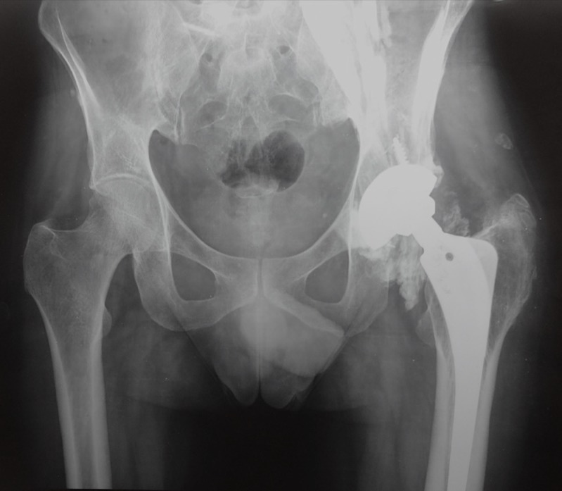
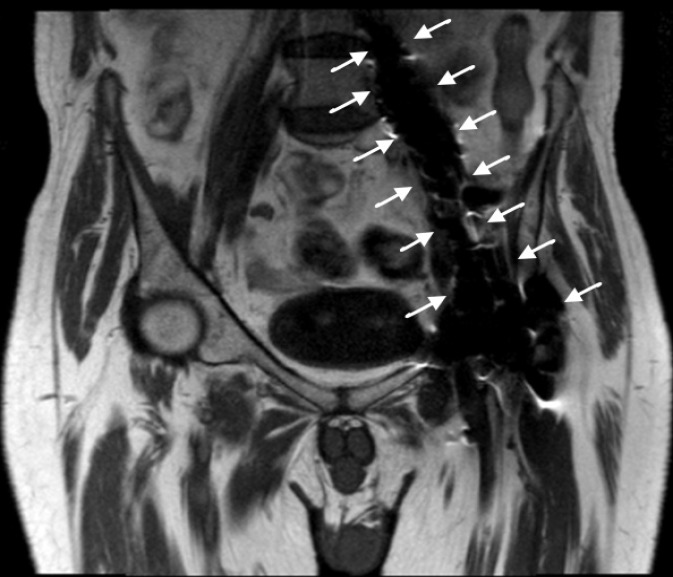
37 years old male sustained a fracture neck femur of left side at the age of 21 years, for which he underwent closed reduction and internal fixation with cannulated cancellous screws. The fracture united but he developed avascular necrosis of femoral head for which he underwent primary total hip arthroplasty five years after the first surgery in an Orthopaedic centre outside our institute. The clinical records showed the implants used to be Plasma cup (B Braun Aesculap®) and Bicontact stem (B Braun Aesculap®) with ceramic on ceramic articulation. He remained asymptomatic for 5 years down the line after the primary total hip arthroplasty when he developed sudden onset difficulty in walking. Radiographs revealed fracture of the ceramic head. He underwent a revision surgery with removal of acetabular liner and ceramic head and change of articulation to a Cobalt-Chrome metallic head on Ultra High Molecular Weight Polyethylene (UHMWPE) insert at a different tertiary care Orthopaedic centre outside our institute. The acetabular shell and the stem were not changed. After the revision surgery, the patient continued with his routine activities without any discomfort till 3 years later when he started having dull aching pain in his left hip, shortness of breath and easy fatigability. He was then referred to our institute. The fresh X-rays showed severe osteolysis and ill-defined increased radio opacities around the neck and periacetabular region extending upto the iliac region [Figure 1]. Ultrasonography (USG) revealed an ill-defined hypoechoic collection with few hyperechoic foci (metal debris) showing distal acoustic shadowing around the neck and the supraacetabular region extending along the iliopsoas tendon upto the muscle bulk. Magnetic Resonance Imaging (MRI) with Metal Artifact Reduction Sequence (MARS) showed periprosthetic collections around the neck of the femoral component and the surrounding soft tissues extending upto the iliopsoas muscle [Figure 2]. The collections were typically isointense to muscle on T1W images and hypointense On T2W images. The capsule and the lining synovium was thickened and appeared isointense on T1W and very low signal intensity on T2W. In addition, there was left-sided fatty atrophy of the gluteus medius and minimus muscles with oedema of the iliopsoas muscle. He was evaluated by a cardiologist and diagnosed to have dilated cardiomyopathy with severe Left Ventricular Dysfunction with an ejection fraction of 20% only. Positron Emission Tomography Computed Tomography (PET CT) showed abnormal metabolic activity in left ventricular myocardium. On investigation, he was found to have highly raised serum Cobalt level of 373 µg/L (Normal range: 0.03- 0.29 µg/L). Based on the above evaluation, he was diagnosed to have Cobalt induced Cardiomyopathy. Considering the deleterious effect of cobalt toxicity and poor cardiac function, he was taken up for revision surgery. Aspiration of the hip before capsulotomy yielded blackish non-purulent fluid with granular material. During revision surgery, the capsule was thickened. The synovium was hypertrophied and deposits of blackish metal debris could be macroscopically visible. Exposure of the articulating components revealed grossly distorted femoral head and the polyethylene liner with black granular deposition in the neck as well as the polyethylene liner and the surrounding soft tissue [Figure 3]. Retrieved components showed femoral stem with damaged trunnion with black deposition over the coated surface in the proximal part. The femoral head was grossly deformed with scratches all over the articulating surface. The polyethylene liner was also deformed with flattening of the rim and wear in the superior aspect of the inner surface along with deposition of black granular material. A thorough debridement was done. Removal of all the black tinged soft tissue led us proximally where the iliopsoas muscle was tattooed (correlating the radioopacities seen in the radiographs). All the accessible black soft tissues were removed and samples were sent for Histopathological examination. The hip was revised with Trabecular MetalTM cup (Zimmer®, Warsaw, IN), Wagner Stem (Zimmer®, Warsaw, IN), Cemented Longevity Liner (Zimmer®, Warsaw, IN) and a ceramic head. Histopathology of the tissue specimen showed extensively hyalinsed fibrocollagenous tissue with blackish amorphous fine granular extracellular material deposition in the interstitial soft tissue. No inflammatory cell infiltrate of any chronic inflammatory cell infiltrate was seen [Figure 4]. Four weeks following the surgery, the serum cobalt level declined to 173 µg/L. However, his cardiac function continued to deteriorate and he had to be admitted in Intensive Care Unit. He succumbed to death three months following the revision surgery while waiting for cardiac transplantation.
Figure 1.
X-ray both hips with pelvis showing severe osteolysis and ill-defined increased radio opacities around the neck and periacetabular region of left prosthetic side extending upto the iliac region
Figure 2.
Magnetic Resonance Imaging (MRI) with Metal Artifact Reduction Sequence (MARS) showing periprosthetic collections around the neck of the femoral component and the surrounding soft tissues extending upto the iliopsoas muscle (white arrows).
Figure 3.
Intraoperative picture showing grossly distorted femoral head and the polyethylene liner. Note the black granular deposition in the surrounding soft tissue
Figure 4.
Histopathology of the tissue specimen showed extensively hyalinsed fibrocollagenous tissue with blackish amorphous fine granular extracellular material deposition in the interstitial soft tissue
Discussion
Ceramic bearing surfaces are being increasingly used in young patients undergoing total hip arthroplasty because of enhanced scratch resistance and wettability which produce excellent fluid-film lubrication and negligible. The risk of periprosthetic osteolysis is also less as the ceramic particles are said to be biologically inert and hence decreased requirement of revision (1). However, the main issue related to ceramic materials is their intrinsic brittleness. Fractures of ceramic heads have been reported from 0% to 13% for first and second-generation ceramics (2). The recommended treatment for fracture of ceramic bearing surfaces is complete synovectomy and revision total hip arthroplasty. Disappointing results have been reported following revision total hip arthroplasty with various bearing surfaces done for ceramic head fracture (3). Most failures are due to incomplete synovectomy and the residual ceramic particles, which in turn causes third body wear and osteolysis. This can lead to local metallosis, and, in more severe cases, systemic manifestations like neuro-endocrine disorder, hearing and visual impairment and cardiomyopathy (4). We report a fatal case of cobalt toxicity secondary to the catastrophic failure of a Cobalt-Chrome femoral head, which followed the revision of a fractured ceramic-on-ceramic total hip arthroplasty.
Case reports of Cobalt cardiomyopathy attributed occupational exposure have been described in the literature. However, fatal cardiomyopathy is limited to an isolated case report (5). Similarly, reports of cobalt toxicity have been described secondary to failed Metal on Polyethylene and Metal on Metal, articulations (6). S M Bradberry et al, in their review article have mentioned that 11 patients had cardiotoxicity out of 18 patients reported in the literature (7). Ten of those 18 patients had undergone revision with one containing a metal component. We could find only two such case reports where cardiomyopathy occurred following revision to a metal on polyethylene articulation for fractured ceramic component (8, 9). Both were revised for fractured acetabular ceramic component. Our case is first to report the revision for the fractured ceramic head. Table 1 describes the various points common in the reports and compared to our report.
Table 1.
Cases of Cobalt induced cardiomyopathy reported in the literature
| SN | Authors | Patient particulars * | Cause for revision | Clinical Features | Serum Cobalt level | Treatment given | Final result |
|---|---|---|---|---|---|---|---|
| 1 | Andrew Harris et al (14) | 57/F | Fractured ceramic liner | Cardiomyopathy, Hypothyroidism, Weakness, Fatigue, Neuropathy |
788.1 μg/L | Implant removal, Synovectomy, Thorough debridement and revision to a Ceramic on Polyethylene articulation |
Survived and doing well |
| 2 | Zywiel et al (15) | 46/M | Fractured ceramic liner | Fatigue, Hypothyroidism, Cardiomyopathy, Neuropathy |
6521 μg/L | - | Death |
| 3 | Current case report | 37/M | Fractured ceramic head | Fatigue Weakness Dyspnoea Cardiomyopathy |
373 μg/L | Implant removal, Synovectomy, Thorough debridement and revision to a Ceramic on Polyethylene articulation |
Death |
Abbreviations: M= Male, F= Female,
Age in years
The risk of Cobalt toxicity is not limited to a single cause, but in fact to all total hip arthroplasties that include at least one Cobalt-Chrome component. Revision of ceramic fracture by a metal is one of the causes. The treatment of failed ceramic-on-ceramic articulations in patients with well-fixed components is challenging. Surgeons may be reluctant to revise the components to another ceramic-on-ceramic articulation because of the concern of possible re-fracture in the future. There have been reports of revision of a fracture of a ceramic component, to metal-on-polyethylene articulations with an extensive anterior and posterior synovectomy with good results and rates of radiological wear similar to those who underwent primary metal on- polyethylene total hip arthroplasty without revision. However, even after synovectomy and thorough debridement ceramic particles in significant number are left behind which may cause substantial scratches and hence excessive wear or osteolysis (10). This could be one of the reason in our patient that led to this devastating complication. Secondly, we assume that the trunnion might have been damaged during the ceramic head fracture. As the revision surgery was done with a metal on polythylene articulation, the damaged trunnion in turn eroded the metallic head leading to trunnionosis and hence the failure again. The deformed polyethylene liner and the metallic head in the second revision surgery also indicates eccentric loading over the articular surfaces leading to accelerated wear and hence failure.
Hence, we recommend the following points to be considered while revising a fractured ceramic component:
1. The fractured ceramic component should be revised with a ceramic component only (new generation) ensuring that the other risk factors for fracture such as component malpositioning, damaged trunnion etc. are not present.
2. The trunnion should be carefully examined and when in doubt about its damage, it should rather be revised during a revision for the fractured ceramic components.
3. Patients who have undergone Metal on Polyethylene revision for a ceramic fracture should be routinely followed up clinically and blood tests for serum Cobalt levels, especially if they have any symptoms.
Disclaimer: The views expressed in this article are our own and not an official position of the institution.
Source of support:
This work was funded by the Wellcome Trust/DBT India Alliance Fellowship Grant (No. IA/RTF/15/1/1003). Deepak Gautam is the recipient of the grant
Conflict of Interest:
Rest of the authors do not take any benefits in any form have been received or will be received from a commercial party related directly or indirectly to the subject of this article.
References
- 1.Fisher J, Jin Z, Tipper J, Stone M, Ingham E. Tribology of alternative bearings. Clin Orthop Relat Res. 2006;453(1):25–34. doi: 10.1097/01.blo.0000238871.07604.49. [DOI] [PubMed] [Google Scholar]
- 2.McLean CR, Dabis H, Mok D. Delayed fracture of the ceramic femoral head after trauma. J Arthroplasty. 2002;17(4):503–4. doi: 10.1054/arth.2002.32185. [DOI] [PubMed] [Google Scholar]
- 3.Allain J, Roudot-Thoraval F, Delecrin J, Anract P, Migaud H, Goutallier D. Revision total hip arthroplasty performed after fracture of a ceramic femoral head A multicenter survivorship study. J Bone Joint Surg Am. 2003;85-A(5):825–30. doi: 10.2106/00004623-200305000-00009. [DOI] [PubMed] [Google Scholar]
- 4.Kwon YM, Jacobs JJ, MacDonald SJ, Potter HG, Fehring TK, Lombardi AV. Evidence-based understanding of management perils for metal-on-metal hip arthroplasty patients. J Arthroplasty. 2010;27(8 Suppl):20–5. doi: 10.1016/j.arth.2012.03.029. [DOI] [PubMed] [Google Scholar]
- 5.Kennedy A, Dornan JD, King R. Fatal myocardial disease associated with industrial exposure to cobalt. Lancet. 1981;1(8217):412–4. doi: 10.1016/s0140-6736(81)91793-1. [DOI] [PubMed] [Google Scholar]
- 6.Mao X, Wong AA, Crawford RW. Cobalt toxicity: an emerging clinical problem in patients with metal-on-metal hip prostheses? Med J Aust. 2011;194(12):649–51. doi: 10.5694/j.1326-5377.2011.tb03151.x. [DOI] [PubMed] [Google Scholar]
- 7.Bradberry SM, Wilkinson JM, Ferner RE. Systemic toxicity related to metal hip prostheses. Clin Toxicol. 2014;52(8):837. doi: 10.3109/15563650.2014.944977. [DOI] [PubMed] [Google Scholar]
- 8.Harris A, Johnson J, Mansuripur PK, Limbird R. Cobalt toxicity after revision to a metal-on-polyethylene total hip arthroplasty for fracture of ceramic acetabular component. Arthroplasty Today. 2015;1(4):89–91. doi: 10.1016/j.artd.2015.09.002. [DOI] [PMC free article] [PubMed] [Google Scholar]
- 9.Zywiel MG, Brandt JM, Overgaard CB, Cheung AC, Turgeon TR, Syed KA. Fatal cardiomyopathy after revision total hip replacement for fracture of a ceramic liner. Bone Joint J. 2013;95-B(1):31–7. doi: 10.1302/0301-620X.95B1.30060. [DOI] [PubMed] [Google Scholar]
- 10.Sharma V, Ranawat AS, Rasquinha VJ, Weiskopf J, Howard H, Ranawat CS. Revision total hip arthroplasty for ceramic head fracture: a long-term follow-up. J Arthroplasty. 2010;25(3):342–7. doi: 10.1016/j.arth.2009.01.014. [DOI] [PubMed] [Google Scholar]


