Key Points
Question
Is prefrontal brain activation a novel biomarker for stress resilience in surgeons?
Findings
In this cohort study, 33 surgical residents were neuromonitored during 330 laparoscopic suturing drills performed within both self-paced and time-pressured conditions (order randomized). Analysis of 7920 channels of regional brain data demonstrated sustained prefrontal cortical activation in the most stress-resilient residents and deactivation responses among the least resilient residents.
Meaning
Technical performance stability under stressful operating conditions is associated with optimal functioning of executive centers in the brain, indicative of preserved attention and concentration.
Abstract
Importance
Intraoperative stressors may compound cognitive load, prompting performance decline and threatening patient safety. However, not all surgeons cope equally well with stress, and the disparity between performance stability and decline under high cognitive demand may be characterized by differences in activation within brain areas associated with attention and concentration such as the prefrontal cortex (PFC).
Objective
To compare PFC activation between surgeons demonstrating stable performance under temporal stress with those exhibiting stress-related performance decline.
Design, Setting, and Participants
Cohort study conducted from July 2015 to September 2016 at the Imperial College Healthcare National Health Service Trust, England. One hundred two surgical residents (postgraduate year 1 and greater) were invited to participate, of which 33 agreed to partake.
Exposures
Participants performed a laparoscopic suturing task under 2 conditions: self-paced (SP; without time-per-knot restrictions), and time pressure (TP; 2-minute per knot time restriction).
Main Outcomes and Measures
A composite deterioration score was computed based on between-condition differences in task performance metrics (task progression score [arbitrary units], error score [millimeters], leak volume [milliliters], and knot tensile strength [newtons]). Based on the composite score, quartiles were computed reflecting performance stability (quartile 1 [Q1]) and decline (quartile 4 [Q4]). Changes in PFC oxygenated hemoglobin concentration (HbO2) measured at 24 different locations using functional near-infrared spectroscopy were compared between Q1 and Q4. Secondary outcomes included subjective workload (Surgical Task Load Index) and heart rate.
Results
Of the 33 participants, the median age was 33 years, the range was 29 to 56 years, and 27 were men (82%). The Q1 residents demonstrated task-induced increases in HbO2 across the bilateral ventrolateral PFC (VLPFC) and right dorsolateral PFC in the SP condition and in the VLPFC in the TP condition. In contrast, Q4 residents demonstrated decreases in HbO2 in both conditions. The magnitude of PFC activation (change in HbO2) was significantly greater in Q1 than Q4 across the bilateral VLPFC during both SP (mean [SD] left VLPFC: Q1, 0.44 [1.30] μM; Q4, −0.21 [2.05] μM; P < .001; right VLPFC: Q1, 0.46 [1.12] μM; Q4, −0.15 [2.14] μM; P < .001) and TP (mean [SD] left VLPFC: Q1, 0.44 [1.36] μM; Q4, −0.03 [1.83] μM; P = .001; right VLPFC: Q1, 0.49 [1.70] μM; Q4, −0.32 [2.00] μM; P < .001) conditions. There were no significant between-group differences in Surgical Task Load Index or heart rate in either condition.
Conclusions and Relevance
Performance stability within TP is associated with sustained prefrontal activation indicative of preserved attention and concentration, whereas performance decline is associated with prefrontal deactivation that may represent task disengagement.
This cohort study compares prefrontal cortex activation between surgeons demonstrating stable performance under temporal stress with those exhibiting stress-related performance decline.
Introduction
Estimates from the United States place the mean death rate from medical error at 251 454 per annum.1 Numerous studies and high-profile cases (eg, Hadiza Bawa-Garba, MD)2 implicate stress and fatigue as contributing to medical errors.2,3,4 Surgery is inherently stressful, and patient safety may be jeopardized when demanding operating conditions precipitate deterioration in both technical (eg, errors and dexterity) and nontechnical performance (eg, judgment and decision-making).5,6 Conversely, stress resilience, the ability to maintain performance in the face of escalating mental demands, ought to be a fundamental attribute of a practice-ready surgeon, and yet is a characteristic still to be objectively defined.
Despite developments in objective assessment of technical7 and nontechnical skills,8 credentialing of general surgical residents relies in part on a framework of proficiency benchmarks such as the Fundamentals of Laparoscopic Surgery program,9 against which residents are assessed within arguably benign conditions that poorly emulate real-world scenarios. This notwithstanding, high-fidelity crisis simulations in virtual operating theaters enable technical and communication skills to be objectively assessed.10 Unfortunately, virtual operating theater crisis simulations fundamentally fail to define operator stress sensitivity or resilience because they lack baseline performance assessments.11,12 Defining stress sensitivity or its counterpart (resilience) requires that individual performance gradients be measured, and yet instead absolute performance data have been compared at the cohort level.6 Hence, while an appreciation of the effect of a given stressor on technical performance is gained, individual-level differences in the sensitivity to the stressor are lost. Reliably discriminating resilient residents (copers) from those with poor stress tolerance (chokers) could have an effect on graduate credentialing, surgical selection, and targeted stress-coping interventions.
Performance gradients per se offer little explanation as to why certain residents with similar training cope while others choke under mental demand. Mechanistic explanations for these between-resident differences may lie not in motor processing but rather in variation within the central nervous system. Observed differences in central nervous system processing may account for disparity in motor learning,13,14 cognitive capabilities,15 and responses to workload and stress.16,17,18 The brain’s prefrontal cortex (PFC), important for attention, concentration, and regulation of goal-directed behavior, initially scales linearly with cognitive load16 but then diminishes at excessive workloads.17,18 Despite several studies investigating surgeons’ brain function,19 few specifically address stress-induced changes in operator cognition, and hence, whether similar stress-related changes in PFC activation that are seen in other domains occur in surgeons remains unknown.
We sought to address this major research gap by constructing performance gradients in surgical residents performing laparoscopic suturing under varying temporal demands. The PFC responses of residents whose performance substantially deteriorated under time constraints (chokers) were contrasted with residents whose performance remained stable (copers). We hypothesize that an ability to stabilize laparoscopic performance under temporal stress is associated with sustained significant prefrontal activation, implying preserved attention and concentration, whereas significant performance decline would manifest as prefrontal attenuation, signifying task disengagement.
Methods
Participants
Following approval by the London Research Ethics Committee (LREC: 05/Q0403/142), 33 surgical residents (median age, 33 years; range, 29-56 years; 27 men [82%]) in general surgery residency training programs (postgraduate year [PGY] 1 or greater) gave written informed consent to participate. Participants were screened for handedness and neuropsychiatric illness and were asked to abstain from alcohol and caffeine consumption for 24 hours prior to participation.
Task Paradigm and Experimental Design
Participants were asked to perform intracorporeal laparoscopic suturing on a box trainer (iSim2; iSurgicals). Participants sutured a defect in a Penrose drain placing 2-0 polyglactin 910 sutures (Ethicon) as close as possible to premarked entry and exit points, producing a double throw followed by 2 single throws of a reef knot. Each participant executed the task under self-paced (SP; no time restriction on knot tying) and time pressure (TP; 2 minutes per knot time restriction) conditions. A 2-minute limit for task completion was imposed during the TP condition because it mirrors the Fundamentals of Laparoscopic Surgery proficiency time for intracorporeal knot tying (112 seconds).9 For each condition, participants tied 5 interrupted knots with a 30-second intertrial rest period.20 The condition that each participant encountered first (SP or TP) was randomly allocated using a coin-toss technique to minimize confounding time-on-task effects.
Functional Brain Imaging
Functional near-infrared spectroscopy is a noninvasive functional neuroimaging technique measuring cortical absorption of near-infrared light to estimate local concentration changes of oxygenated (HbO2) and deoxygenated hemoglobin. Hemodynamic responses depicting brain activation comprise task-evoked HbO2 increases and lower amplitude deoxygenated hemoglobin decreases. An ETG-4000 Optical Topography System (Hitachi Medical Co) captured activation across 24 prefrontal locations (channels), spatially defined according to the international 10-5 system of probe placement (eFigure 1 and eTable 1 in the Supplement). The probes were placed on each participant’s head by the same investigator (H.N.M), after which the participants were given 10 minutes warm-up time to become familiar with the box trainer and get used to wearing the device. The experiment was started immediately following the warm-up period.
Subjective Workload and Physiological Stress
Subjective workload was assessed using the Surgical Task Load Index (SURG-TLX).21 A wireless monitor (Bioharness; Zephyr Technology) continually recorded heart rate (HR). Change in HR from rest to task (ΔHR) was calculated per equation 1:
Technical Performance Assessment and Composite Deterioration Score
As previously described,22 technical performance was assessed using task progression scores (arbitrary units), error scores (millimeters), leak volume (milliliters), and knot tensile strength (newtons). For every participant, between-condition differences in performance metrics were calculated. These differences were subsequently normalized (0 to 1) per equation 2:
 |
A score of 1 represents the smallest deterioration in performance from SP to the TP condition, whereas 0 represents the greatest performance decline. To calculate a composite performance deterioration score for each participant, a weighted average of the normalized deterioration in all performance metrics was calculated per equation 3:
 |
Weights were assigned to each metric based on survey responses from 24 attending surgeons (task progression score = 30%; error score = 25%; leak volume = 20%; knot tensile strength = 25%). Participants were ranked based on the composite performance deterioration score, and analysis of brain function was undertaken to compare responses between the top (Q1) and bottom (Q4) quartiles.
Data Processing and Statistical Analysis
Statistical analysis was performed using SPSS version 23.0 (IBM Corp). A threshold P less than .05 was deemed statistically significant, and all P values were 2-sided.
Stress and Workload Data
For each group (Q1 or Q4), the paired-samples t test compared between-condition differences in SURG-TLX scores to determine whether the 2-minute time restriction recreated a sense of urgency, and the Wilcoxon signed rank test compared between-condition changes in ΔHR to establish whether cortical hemodynamic signals were influenced by changes in the systemic stress response. For each condition (SP or TP), the independent-samples t test compared SURG-TLX scores between Q1 and Q4, and the Mann-Whitney U test compared ΔHR.
Functional Neuroimaging Data
Functional neuroimaging data were preprocessed using a customized MATLAB-based toolbox (HOMER2) as previously described22 and a hybrid motion correction technique based on spline interpolation and Savitzky-Golay filtering.23 One participant was excluded from analysis owing to excessive noise in all channels. From the remaining 7680 channels, low signal-to-noise ratio prompted exclusion of 80 further channels (data rejection rate = 4%).
Identification of Channel Activation
For each quartile and condition, channel activation was determined by comparing the mean baseline HbO2 data sampled over 10 seconds before task onset (HbO2Rest) with mean task HbO2 data sampled over 60 seconds from task onset (HbO2Task) using the paired-samples t test. Channels in which there was a statistically significant (P < .05) increase in HbO2 were considered activated, and those in which there was a significant decrease in HbO2 were considered deactivated.
Comparisons of Activation Responses
For each channel, a variable ΔHbO2 was computed (defined as HbO2Task–HbO2Rest). For each quartile, ΔHbO2 in each channel was compared between the conditions using the paired-samples t test. Similarly, for a given experimental condition and each channel, ΔHbO2 was compared between Q1 and Q4 using the independent-samples t test.
General Linear Model (GLM)
To extract the evoked brain response (change in HbO2 concentration) to the laparoscopic suturing task, a boxcar function was designed on the basis of stimulus duration and condition. A linear model of an evoked response is given as Y = Xβ, where X is the design matrix and β is the estimate of the magnitude of brain activity. Consecutive gaussian functions were used to estimate the GLM and linear least squares were used to estimate the weights of these functions.24 A simple boxcar was used to estimate the expected response.25 The Cartesian coordinates of the near-infrared light emitters and detectors were obtained using a 3-dimensional digitizer (Polhemus; Hitachi Medical Corp) and coregistered with the Colin 27 magnetic resonance imaging brain atlas26 based on standard electroencephalogram 10-5 landmarks. Anatomical overlay of prefrontal oxygenation changes was performed using the AtlasViewer toolbox.27
Expertise and Performance Decline Relationship
Participants were categorized as junior (PGY 1 and 2), intermediate (PGY 3 and 4), or senior (PGY ≥5). The Pearson χ2 test was used to investigate the association between expertise and performance deterioration.
Results
Within-Group Comparisons
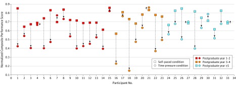
Within each group (eg, Q1 and Q4), subjective workload, performance, and optical data were compared between self-paced and time pressure conditions (eFigures 2 and 3 in the Supplement). There was no significant association between performance deterioration and expertise (χ2 = 9.11; P = .17) (Figure 1).
Figure 1. Participant-Level Performance Change From Self-paced to Time Pressure Conditions.
Changes in performance are depicted (arrowheads) in junior residents (postgraduate year 1 to 2; red), intermediate residents (postgraduate year 3 to 4; orange), and senior residents (postgraduate year 5 or higher; blue). Decrements in performance were apparent in all expertise groups and there was no statistically significant association between the degree of performance deterioration and level of training (χ2 = 9.11; P = .17).
Subjective Workload and Heart Rate
Subjective workload was significantly greater within TP compared with SP for both Q1 (mean [SD] SURG-TLX score: SP, 144.25 [51.22]; TP, 176.00 [59.32]; P = .01) and Q4 residents (SP, 142.25 [43.51]; TP, 194.63 [47.58]; P = .01) (Table). No significant between-condition differences in ΔHR were observed regardless of performance quartile (Table).
Table. SURG-TLX Score, Heart Rate, and ΔHbO2a.
| Condition | Mean (SD) | P Value | |
|---|---|---|---|
| Q1 | Q4 | ||
| SURG-TLX | |||
| SP | 144.25 (51.22) | 142.25 (43.51) | .93b |
| TP | 176.00 (59.32) | 194.63 (47.58) | .50b |
| P valuec | .01 | .01 | NA |
| HR (rest) | |||
| SP | 80.00 (20.00) | 84.00 (29.00) | <.001d,e |
| TP | 80.00 (13.00) | 83.00 (28.00) | <.001d,e |
| HR (task) | |||
| SP | 77.00 (18.00) | 76.00 (28.00) | <.001d,e |
| TP | 80.00 (17.00) | 81.00 (31.00) | <.001d,e |
| ΔHR | |||
| SP | −1.59 (3.97) | 0.09 (4.77) | .15e |
| TP | −2.51 (7.32) | −1.80 (5.27) | .69e |
| P valuef | .40 | .50 | NA |
| ΔHbO2 | |||
| Left VLPFC | |||
| SP | 0.44 (1.30) | −0.21 (2.05) | <.001b,d |
| TP | 0.44 (1.36) | −0.03 (1.83) | .001b,d |
| Right VLPFC | |||
| SP | 0.46 (1.12) | 0.15 (2.14) | <.001b,d |
| TP | 0.49 (1.70) | −0.32 (2.00) | <.001b,d |
| Right DLPFC | |||
| SP | 0.29 (0.80) | 0.06 (1.31) | .11b |
| TP | 0.22 (1.60) | 0.02 (1.46) | .31b |
| Left DMPFC | |||
| SP | 0.36 (1.13) | −0.18 (1.81) | .001b,d |
| TP | 0.35 (1.29) | −0.28 (2.17) | .001b,d |
Abbreviations: DLPFC, dorsolateral prefrontal cortex; DMPFC, dorsomedial prefrontal cortex; ΔHbO2; change in oxygenated hemoglobin concentration; HR, heart rate; ΔHR, change in heart rate from rest to task; IQR, interquartile range; NA, not applicable; SP, self-paced; SURG-TLX, Surgical Task Load Index; TP, time pressure; VLPFC, ventrolateral prefrontal cortex.
SURG-TLX and ΔHbO2 data are mean (SD) and measured in arbitrary units and μM × cm, respectively. HR and ΔHR data are median (IQR) and measured in beats per minute.
Paired-samples t test.
Independent-samples t test.
Significant P value (< .05).
Mann-Whitney U test.
Wilcoxon signed rank test.
Prefrontal Activation
Regarding Q1 residents, significant task-induced increases in HbO2 were observed in channels located in the bilateral ventrolateral PFC (VLPFC) and right dorsolateral PFC (DLPFC) during the SP condition and in channels situated in the bilateral VLPFC during the TP condition (eTables 2 and 3 in the Supplement). In Q4, task-evoked HbO2 decreases were observed in both conditions, particularly in channels located in the bilateral VLPFC and left dorsomedial PFC during the TP condition (eTables 4 and 5 in the Supplement). In Q1 residents, ΔHbO2 was greater in TP vs SP in 9 of 24 channels (channels 2-5, 8, 13-15, and 17), and greater in SP vs TP in 15 of 24 channels (channels 1, 6, 7, 9-12, 16, and 18-24). In Q4 residents, ΔHbO2 was greater in TP vs SP in 11 of 24 channels (channels 1-3, 5, 6, 8, 17-19, 21, and 22, and greater in SP vs TP in 13 of 24 channels (channels 4, 7, 9-16, 20, 23, and 24) (eTables 6 and 7 in the Supplement).
Between-Group Comparisons
Subjective Workload and Heart Rate
Within each experimental condition (eg, self-paced and time pressure), subjective workload, performance, and optical data were compared between Q1 and Q4 groups. Regardless of condition (SP or TP), there was no significant difference between Q1 and Q4 in mean (SD) SURG-TLX scores (SP: Q1 = 144.25 [51.22] vs Q4 = 142.25 [43.51]; P = .93; TP: Q1 = 176.00 [59.32] vs Q4 = 194.63 [47.58]; P = .50) (Table). There was no significant difference in ΔHR between Q1 and Q4 in either condition (SP: Q1 [IQR], −1.59 [3.97] beats/minute vs Q4 [IQR], 0.09 [4.77]; P = .15; TP: Q1, −2.51 [7.32] vs Q4, −1.80 [5.27], P = .69) (Table). Furthermore, there was no significant between-group difference in ΔHR change from SP to TP conditions.
Prefrontal Activation
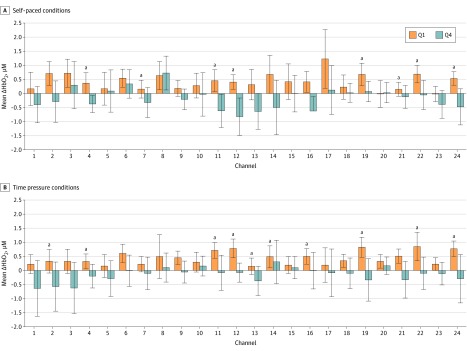
In the SP condition, Q1 residents exhibited significantly greater ΔHbO2 responses than Q4 in bilateral VLPFC channels (Figure 2A). Similarly, Q1 residents demonstrated significantly greater ΔHbO2 responses than Q4 in the bilateral VLPFC and left dorsomedial PFC in the TP condition (Figure 2B).
Figure 2. Channel Activation in Quartile 1 (Q1) and Quartile 4 (Q4) Residents.
Between-group channelwise differences in prefrontal change in oxygenated hemoglobin concentration (ΔHbO2) during self-paced (A) and time pressure (B) conditions. The Q1 residents demonstrate significantly greater activation compared with Q4 residents in 8 channels in the bilateral ventrolateral prefrontal cortex in the self-paced condition and in 10 channels in the bilateral ventrolateral prefrontal cortex and left dorsomedial prefrontal cortex in the time pressure condition. Error bars represent the 95% confidence interval.
aP < .05 (independent-samples t test).
General Linear Model
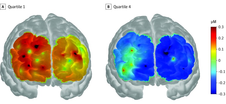
During both experimental conditions, the morphology of the task-induced HbO2 response in Q1 residents consisted of an initial increase in HbO2 concentration followed by a steady decline (eFigures 4 and 5 in the Supplement). In contrast, in Q4 residents, an initial decrease in HbO2 concentration was observed followed by a gradual increase to baseline (eFigures 4 and 5 in the Supplement). The general linear model mirrored these findings, particularly in the TP condition, during which there was widespread significant activation in the bilateral DLPFC and VLPFC in Q1 residents and deactivation in Q4 residents (Figure 3).
Figure 3. Activation and Deactivation Responses in Quartile 1 (Q1) and Quartile 4 (Q4) Residents.
Group-averaged oxygenated hemoglobin concentration (HbO2) concentration change in the prefrontal cortex among Q1 and Q4 residents during the time pressure condition. The displayed concentration change was averaged from 10 to 50 seconds after stimulus onset. The color bar indicates the scale of the concentration change in micromoles. The anatomical coregistration for probes was anchored using standard electroencephalogram 10-5 landmarks onto the Colin 27 head model.
Discussion
To our knowledge, this is the first study to expose disparate responses in surgeons’ brains based on sensitivity to temporal stress. The results highlight gross temporal and spatial disparities in the patterns of evoked prefrontal responses depending on stress-coping capabilities. Fascinatingly, stress sensitivity and performance degradation are not associated with postgraduate years of training and/or the grade of the operator. Moreover, traditional physiologic measures of stress (ΔHR) and subjective workload indices (SURG-TLX) do not discriminate between high performing, resilient residents (copers) and those with acute performance decline (chokers). Instead, performance stability within temporal stress is characterized by a pattern of prefrontal responses that are spatially more extensive, of greater amplitude intensity, and whose temporal characteristics map more closely to the typical hemodynamic response function (HRF). Conversely, neuroimaging features typifying stress sensitivity and heralding performance decline include low-amplitude responses, statistically significant decreases in cortical oxygenation change, and responses whose temporal characteristics follow an inverted HRF.
Performance Stability and Brain Activation
Resilient residents demonstrated typical activation responses that comprised an initial increase in HbO2 concentration followed by a decline. This pattern was most prominent in the bilateral VLPFC and right DLPFC in the SP condition, and the bilateral VLPFC in TP. Furthermore, compared with the least resilient residents, top quartile residents demonstrated on average 0.6μM greater ΔHbO2 in the bilateral VLPFC and right DLPFC in the SP condition and in the bilateral VLPFC and left dorsomedial PFC in the TP condition (Table). Sustained VLPFC activation in the TP condition would suggest an ability to maintain attentional control28,29 and vigilance30 during task performance, the corollary of which is improved task engagement and technical performance stabilization under stress.
Performance Decline and Brain Deactivations
Residents found to be highly sensitive to stress exhibit an inverted HRF, comprising an initial task-induced decrease in HbO2 concentration followed by a subsequent lower amplitude increase. While inverted HRF responses were observed during self-paced performance, they were detected more readily and across a wider distributed VLPFC and DLPFC network under temporal stress. Inverted HRF responses are coined “deactivation”; by virtue, they mirror the classic HRF.31 The VLPFC deactivations have been observed in other experiments involving TP and negative feedback32 and in the DLPFC in stressful working memory tasks,33 monetary incentive delay tasks,34 and video gaming.35
Cognitive Processes and Psychological Models
Psychological, cognitive, and physiological mechanisms may explain why resilient and stress-sensitive residents occupy 2 ends of an activation-deactivation continuum. Top-down control of motor behavior is dependent on optimal functioning of executive centers, such as the PFC.36 However, further functional granularity can be defined based on PFC regional anatomy. For example, the DLPFC is important for motor planning,37 decision-making,38 and attention,39 whereas the VLPFC is crucial for vigilance,30 resistance to environmental distraction,40 and attentional control.28,29 When a resident is faced with temporal pressure (eg, intraoperative bleeding), engagement of both VLPFC and DLPFC would work in consort to facilitate motor task success (ie, hemostasis). Conversely, deactivations observed among the least resilient residents may represent cognitive overload, in which executive control is disrupted30,38 to the detriment of technical performance.41,42 Task-irrelevant thoughts and concerns about failure may create distraction leading to loss of cognitive engagement, prefrontal deactivation, and performance decline.42 Indeed, the distraction-conflict model suggests that diversion of attentional resources to task-irrelevant cues precipitates stress-induced performance decline.42 Poor-performing residents may have been maximally distracted by temporal pressure, resulting in impaired concentration, PFC deactivation, and a propensity to choke.41
Systemic Stress and Hormone Responses
Under stressful conditions, noradrenergic neurons projecting from the brainstem inhibit prefrontal synaptic activity owing to noradrenaline preferentially binding to α-1 and β receptors.43,44 This is exacerbated by activation of the hypothalamus-pituitary-adrenal axis, which leads to greater glucocorticoid release.45 The complex interplay between stress, hormone release, and PFC activation is supported by other studies in which a salivary cortisol response to stress was associated with diminished PFC activation.46,47 While salivary cortisol was not measured, a plausible explanation for current deactivation(s) is hormone-mediated inhibition of PFC function, leading to poor attentional control during task performance under stress.48,49,50
Subjective Workloads
Regardless the degree of stress resilience or performance instability, subjective workload was significantly greater under temporal demand, suggesting the 2-minute time limit imposed a genuine sense of temporal urgency. Interestingly, cognitive workload escalation has been found to lead to stepwise increases in PFC hemodynamic change16 until a critical workload threshold, beyond which cortical oxygenation declines and performance decreases.17,18 We previously demonstrated that regardless of seniority, all residents have a degree of performance decline and PFC attenuation under acute temporal demand,22 implying that sudden workload increases cannot be easily be matched by PFC hemodynamic change. Critically, here we extend these findings by demonstrating that outwith of expertise, the magnitude of PFC attenuation reflects stress sensitivity.
Expertise Effects on Stress Sensitivity
Although all residents demonstrated a deterioration in absolute performance under TP, we observed no association between resident seniority and the degree of performance deterioration, which suggests that the ability to cope with intraoperative temporal stress is not simply a function of experience or time on task. However, it remains unknown whether cognitive training can ameliorate the effects of these individual differences. The surgical community has introduced training and assessments of teamwork and situational awareness to help residents manage stressful intraoperative events.51 Moreover, high-fidelity simulated operating environments have enabled crisis management programs to be successfully trialed,11,52 paving the way for a structured intraoperative stress management program for residents.
Clinical Translation and Future Work
This study suggests that systemic physiology and subjective workload measures cannot readily discern stress resilience. However, functional neuroimaging identified quantifiable differences in temporal and spatial prefrontal cortical hemodynamics, reflecting stress resilience and stress sensitivity. These differences in prefrontal function may indicate disparities in attention and concentration at times of temporal stress, highlighting the need to develop stress-coping training strategies. A number of techniques that foster stress resilience have been reported. For example, in a prospective study of resident physicians, mindfulness-based resilience training was shown to reduce anxiety and stress in some participants.53 Moreover, mindfulness has been shown to be both feasible and acceptable to surgical interns as a means of managing stress.54 Metacognition, ie, the awareness of one’s thoughts and cognitive processes, has been nurtured in athletes to overcome psychological stress during a competition55 and may be applied in the surgical domain to improve performance under stressful conditions.56 Similarly, mental practice, the cognitive rehearsal of a task without overt physical movement, is a widely recognized strategy used to enhance psychomotor performance in sports and music57 and has been shown to improve technical skills and reduce stress among surgeons.52,58,59 Coupling such training interventions with assessment of brain behavior will help determine whether they have the desired effect on operator cognition(s). Furthermore, the results highlight the importance of minimizing unnecessary time pressure in the operating room by, for example, avoiding overbooking cases to allow for training during the operating list and managing workflow processes.
Limitations
A sample size estimation was not feasible because data from similar previous neuroimaging studies were insufficient for a pre hoc power calculation.16,17,18 Indeed, studies investigating the association of stress with cognitive function have not incorporated sample size calculations.16,17,18 This notwithstanding, the sample size in our study compares favorably with the literature.16,17,18 Recruitment of participants may have been vulnerable to a selection bias. All general surgical residents in a single postgraduate training region were invited to participate, and it is possible that only residents who felt confident in their laparoscopic suturing ability agreed to participate. The subspecialty interest of participants was not recorded, and there may have been a disproportionate number of residents with a specialist interest in general surgical disciplines in which laparoscopic suturing is a fundamental part of training (eg, upper gastrointestinal tract surgery). Therefore, performance and brain behavior of this self-selected group may not be representative of the wider surgical community.
Data were collected in a controlled laboratory environment to precisely replicate the paradigm in this block-design experiment. While this may have influenced face validity, SURG-TLX scores under temporal demand suggests the experimental paradigm adequately taxed the residents by recreating a sense of temporal stress. Our study only investigated the effect of a single stressor (TP). While surgeons may experience multiple competing demands simultaneously, there are circumstances in which the primary stressor is temporal in nature (eg, major bleeding). In this regard, this study aimed to elucidate the neurophysiologic changes associated with performance decline during times of temporal stress. Nonetheless, future work will explore the effects of competing cognitive demands on prefrontal function and technical performance. Changes in systemic physiology may confound cortical hemodynamics60 with stress-induced HR increases misregistered as artificial HbO2 responses. However, this is juxtaposed with the observed decrease in HbO2 observed in the group displaying the greatest increase in absolute HR. Moreover, there was no differences between the most and least resilient residents in the magnitude task-evoked ΔHR (Table).
Conclusions
Residents whose technical performance is sensitive to intraoperative temporal demands exhibit prefrontal deactivations, whereas those with stable performance demonstrate sustained activation, particularly in brain regions important for attentional control and motor planning. Further studies are required to determine the value of neuroimaging in predicting stress resilience in surgeons.
eMethods. Time to HbO2 Peak and Nadir
eResults. Comparisons of Time to HbO2 Peak and Nadir
eTable 1. Prefrontal channel locations
eTable 2. Changes in Cortical Haemodynamics in Each Channel in the Self-Paced Condition Among Q1 Residents
eTable 3. Changes in Cortical Haemodynamics in Each Channel in the Time Pressure Condition Among Q1 Residents
eTable 4. Changes in Cortical Haemodynamics in Each Channel in the Self-Paced Condition Among Q4 Residents
eTable 5. Changes in Cortical Haemodynamics in Each Channel in the Time Pressure Condition Among Q4 Residents
eTable 6. Differences in ΔHbO2 Between Conditions in Each Channel Among Q1 Residents
eTable 7. Differences in ΔHbO2 Between Conditions in Each Channel Among Q4 Residents
eFigure 1. Prefrontal Channel Positions
eFigure 2. Within-Group Comparison of Time to HbO2 Concentration Peak and Time to HbO2 Concentration Nadir
eFigure 3. Between-Group Comparison of Time to HbO2 Concentration Peak and Time to HbO2 Concentration Nadir
eFigure 4. Time Courses Demonstrating the Change in Oxygenated Haemoglobin in Individual Channels in Q1 and Q4 Residents During the Self-Paced Condition
eFigure 5. Time Courses Demonstrating the Change in Oxygenated Haemoglobin in Individual Channels in Q1 and Q4 Residents During the Time Pressure Condition
References
- 1.Makary MA, Daniel M. Medical error-the third leading cause of death in the US. BMJ. 2016;353:. doi: 10.1136/bmj.i2139 [DOI] [PubMed] [Google Scholar]
- 2.Oliver D. Should NHS doctors work in unsafe conditions? BMJ. 2018;360:k448. doi: 10.1136/bmj.k448 [DOI] [PubMed] [Google Scholar]
- 3.West CP, Tan AD, Habermann TM, Sloan JA, Shanafelt TD. Association of resident fatigue and distress with perceived medical errors. JAMA. 2009;302(12):1294-. doi: 10.1001/jama.2009.1389 [DOI] [PubMed] [Google Scholar]
- 4.Mentis HM, Chellali A, Manser K, Cao CG, Schwaitzberg SD. A systematic review of the effect of distraction on surgeon performance: directions for operating room policy and surgical training. Surg Endosc. 2016;30(5):1713-1724. doi: 10.1007/s00464-015-4443-z [DOI] [PMC free article] [PubMed] [Google Scholar]
- 5.Wetzel CM, Kneebone RL, Woloshynowych M, et al. . The effects of stress on surgical performance. Am J Surg. 2006;191(1):5-10. doi: 10.1016/j.amjsurg.2005.08.034 [DOI] [PubMed] [Google Scholar]
- 6.Arora S, Sevdalis N, Nestel D, Woloshynowych M, Darzi A, Kneebone R. The impact of stress on surgical performance: a systematic review of the literature. Surgery. 2010;147(3):318-330, 330.e1-330.e6. [DOI] [PubMed] [Google Scholar]
- 7.Moorthy K, Munz Y, Sarker SK, Darzi A. Objective assessment of technical skills in surgery. BMJ. 2003;327(7422):1032-1037. doi: 10.1136/bmj.327.7422.1032 [DOI] [PMC free article] [PubMed] [Google Scholar]
- 8.Yule S, Flin R, Maran N, Rowley D, Youngson G, Paterson-Brown S. Surgeons’ non-technical skills in the operating room: reliability testing of the NOTSS behavior rating system. World J Surg. 2008;32(4):548-556. doi: 10.1007/s00268-007-9320-z [DOI] [PubMed] [Google Scholar]
- 9.Fundamentals of laparoscopic surgery https://www.flsprogram.org/. Accessed January 20, 2018.
- 10.Aggarwal R, Undre S, Moorthy K, Vincent C, Darzi A. The simulated operating theatre: comprehensive training for surgical teams. Qual Saf Health Care. 2004;13(suppl 1):i27-i32. doi: 10.1136/qshc.2004.010009 [DOI] [PMC free article] [PubMed] [Google Scholar]
- 11.Moorthy K, Munz Y, Forrest D, et al. . Surgical crisis management skills training and assessment: a simulation[corrected]-based approach to enhancing operating room performance. Ann Surg. 2006;244(1):139-147. doi: 10.1097/01.sla.0000217618.30744.61 [DOI] [PMC free article] [PubMed] [Google Scholar]
- 12.Moorthy K, Munz Y, Adams S, Pandey V, Darzi A. A human factors analysis of technical and team skills among surgical trainees during procedural simulations in a simulated operating theatre. Ann Surg. 2005;242(5):631-639. doi: 10.1097/01.sla.0000186298.79308.a8 [DOI] [PMC free article] [PubMed] [Google Scholar]
- 13.Leff DR, Elwell CE, Orihuela-Espina F, et al. . Changes in prefrontal cortical behaviour depend upon familiarity on a bimanual co-ordination task: an fNIRS study. Neuroimage. 2008;39(2):805-813. doi: 10.1016/j.neuroimage.2007.09.032 [DOI] [PubMed] [Google Scholar]
- 14.Leff DR, Orihuela-Espina F, Elwell CE, et al. . Assessment of the cerebral cortex during motor task behaviours in adults: a systematic review of functional near infrared spectroscopy (fNIRS) studies. Neuroimage. 2011;54(4):2922-2936. doi: 10.1016/j.neuroimage.2010.10.058 [DOI] [PubMed] [Google Scholar]
- 15.Cazalis F, Valabrègue R, Pélégrini-Issac M, Asloun S, Robbins TW, Granon S. Individual differences in prefrontal cortical activation on the Tower of London planning task: implication for effortful processing. Eur J Neurosci. 2003;17(10):2219-2225. doi: 10.1046/j.1460-9568.2003.02633.x [DOI] [PubMed] [Google Scholar]
- 16.Ayaz H, Shewokis PA, Bunce S, Izzetoglu K, Willems B, Onaral B. Optical brain monitoring for operator training and mental workload assessment. Neuroimage. 2012;59(1):36-47. doi: 10.1016/j.neuroimage.2011.06.023 [DOI] [PubMed] [Google Scholar]
- 17.Izzetoglu K, Bunce S, Onaral B, Pourrezaei K, Chance B. Functional optical brain imaging using near-infrared during cognitive tasks. Int J Hum Comput Interact. 2004;17(2):211-227. doi: 10.1207/s15327590ijhc1702_6 [DOI] [Google Scholar]
- 18.Durantin G, Gagnon JF, Tremblay S, Dehais F. Using near infrared spectroscopy and heart rate variability to detect mental overload. Behav Brain Res. 2014;259:16-23. doi: 10.1016/j.bbr.2013.10.042 [DOI] [PubMed] [Google Scholar]
- 19.Modi HN, Singh H, Yang GZ, Darzi A, Leff DR. A decade of imaging surgeons’ brain function (part II): a systematic review of applications for technical and nontechnical skills assessment. Surgery. 2017;162(5):1130-1139. doi: 10.1016/j.surg.2017.09.002 [DOI] [PubMed] [Google Scholar]
- 20.Obrig H, Hirth C, Junge-Hülsing JG, et al. . Length of resting period between stimulation cycles modulates hemodynamic response to a motor stimulus. Adv Exp Med Biol. 1997;411:471-480. doi: 10.1007/978-1-4615-5865-1_60 [DOI] [PubMed] [Google Scholar]
- 21.Wilson MR, Poolton JM, Malhotra N, Ngo K, Bright E, Masters RS. Development and validation of a surgical workload measure: the surgery task load index (SURG-TLX). World J Surg. 2011;35(9):1961-1969. doi: 10.1007/s00268-011-1141-4 [DOI] [PMC free article] [PubMed] [Google Scholar]
- 22.Modi HN, Singh H, Orihuela-Espina F, et al. . Temporal stress in the operating room: brain engagement promotes “coping” and disengagement prompts “choking”. Ann Surg. 2018;267(4):683-691. doi: 10.1097/SLA.0000000000002289 [DOI] [PubMed] [Google Scholar]
- 23.Jahani S, Setarehdan SK, Boas DA, Yücel MA. Motion artifact detection and correction in functional near-infrared spectroscopy: a new hybrid method based on spline interpolation method and Savitzky-Golay filtering. Neurophotonics. 2018;5(1):015003. doi: 10.1117/1.NPh.5.1.015003 [DOI] [PMC free article] [PubMed] [Google Scholar]
- 24.Diamond SG, Huppert TJ, Kolehmainen V, et al. . Dynamic physiological modeling for functional diffuse optical tomography. Neuroimage. 2006;30(1):88-101. doi: 10.1016/j.neuroimage.2005.09.016 [DOI] [PMC free article] [PubMed] [Google Scholar]
- 25.Ye JC, Tak S, Jang KE, Jung J, Jang J. NIRS-SPM: statistical parametric mapping for near-infrared spectroscopy. Neuroimage. 2009;44(2):428-447. doi: 10.1016/j.neuroimage.2008.08.036 [DOI] [PubMed] [Google Scholar]
- 26.Holmes CJ, Hoge R, Collins L, Woods R, Toga AW, Evans AC. Enhancement of MR images using registration for signal averaging. J Comput Assist Tomogr. 1998;22(2):324-333. doi: 10.1097/00004728-199803000-00032 [DOI] [PubMed] [Google Scholar]
- 27.Aasted CM, Yücel MA, Cooper RJ, et al. . Anatomical guidance for functional near-infrared spectroscopy: AtlasViewer tutorial. Neurophotonics. 2015;2(2):020801. doi: 10.1117/1.NPh.2.2.020801 [DOI] [PMC free article] [PubMed] [Google Scholar]
- 28.Love T, Haist F, Nicol J, Swinney D. A functional neuroimaging investigation of the roles of structural complexity and task-demand during auditory sentence processing. Cortex. 2006;42(4):577-590. doi: 10.1016/S0010-9452(08)70396-4 [DOI] [PMC free article] [PubMed] [Google Scholar]
- 29.Wolf RC, Vasic N, Walter H. Differential activation of ventrolateral prefrontal cortex during working memory retrieval. Neuropsychologia. 2006;44(12):2558-2563. doi: 10.1016/j.neuropsychologia.2006.05.015 [DOI] [PubMed] [Google Scholar]
- 30.Langner R, Eickhoff SB. Sustaining attention to simple tasks: a meta-analytic review of the neural mechanisms of vigilant attention. Psychol Bull. 2013;139(4):870-900. doi: 10.1037/a0030694 [DOI] [PMC free article] [PubMed] [Google Scholar]
- 31.Dolcos F, McCarthy G. Brain systems mediating cognitive interference by emotional distraction. J Neurosci. 2006;26(7):2072-2079. doi: 10.1523/JNEUROSCI.5042-05.2006 [DOI] [PMC free article] [PubMed] [Google Scholar]
- 32.Al-Shargie F, Tang TB, Kiguchi M. Assessment of mental stress effects on prefrontal cortical activities using canonical correlation analysis: an fNIRS-EEG study. Biomed Opt Express. 2017;8(5):2583-2598. doi: 10.1364/BOE.8.002583 [DOI] [PMC free article] [PubMed] [Google Scholar]
- 33.Qin S, Hermans EJ, van Marle HJF, Luo J, Fernández G. Acute psychological stress reduces working memory-related activity in the dorsolateral prefrontal cortex. Biol Psychiatry. 2009;66(1):25-32. doi: 10.1016/j.biopsych.2009.03.006 [DOI] [PubMed] [Google Scholar]
- 34.Ossewaarde L, Qin S, Van Marle HJ, van Wingen GA, Fernández G, Hermans EJ. Stress-induced reduction in reward-related prefrontal cortex function. Neuroimage. 2011;55(1):345-352. doi: 10.1016/j.neuroimage.2010.11.068 [DOI] [PubMed] [Google Scholar]
- 35.Matsuda G, Hiraki K. Sustained decrease in oxygenated hemoglobin during video games in the dorsal prefrontal cortex: a NIRS study of children. Neuroimage. 2006;29(3):706-711. doi: 10.1016/j.neuroimage.2005.08.019 [DOI] [PubMed] [Google Scholar]
- 36.Miller EK, Cohen JD. An integrative theory of prefrontal cortex function. Annu Rev Neurosci. 2001;24:167-202. doi: 10.1146/annurev.neuro.24.1.167 [DOI] [PubMed] [Google Scholar]
- 37.Heekeren HR, Marrett S, Ruff DA, Bandettini PA, Ungerleider LG. Involvement of human left dorsolateral prefrontal cortex in perceptual decision making is independent of response modality. Proc Natl Acad Sci U S A. 2006;103(26):10023-10028. doi: 10.1073/pnas.0603949103 [DOI] [PMC free article] [PubMed] [Google Scholar]
- 38.Leff DR, Yongue G, Vlaev I, et al. . “Contemplating the next maneuver”: functional neuroimaging reveals intraoperative decision-making strategies. Ann Surg. 2017;265(2):320-330. doi: 10.1097/SLA.0000000000001651 [DOI] [PubMed] [Google Scholar]
- 39.Kane MJ, Engle RW. The role of prefrontal cortex in working-memory capacity, executive attention, and general fluid intelligence: an individual-differences perspective. Psychon Bull Rev. 2002;9(4):637-671. doi: 10.3758/BF03196323 [DOI] [PubMed] [Google Scholar]
- 40.Postle BR. Distraction-spanning sustained activity during delayed recognition of locations. Neuroimage. 2006;30(3):950-962. doi: 10.1016/j.neuroimage.2005.10.018 [DOI] [PubMed] [Google Scholar]
- 41.Lee TG, Grafton ST. Out of control: diminished prefrontal activity coincides with impaired motor performance due to choking under pressure. Neuroimage. 2015;105:145-155. doi: 10.1016/j.neuroimage.2014.10.058 [DOI] [PMC free article] [PubMed] [Google Scholar]
- 42.Yu R. Choking under pressure: the neuropsychological mechanisms of incentive-induced performance decrements. Front Behav Neurosci. 2015;9:19. doi: 10.3389/fnbeh.2015.00019 [DOI] [PMC free article] [PubMed] [Google Scholar]
- 43.Birnbaum S, Gobeske KT, Auerbach J, Taylor JR, Arnsten AF. A role for norepinephrine in stress-induced cognitive deficits: alpha-1-adrenoceptor mediation in the prefrontal cortex. Biol Psychiatry. 1999;46(9):1266-1274. doi: 10.1016/S0006-3223(99)00138-9 [DOI] [PubMed] [Google Scholar]
- 44.Ramos BP, Colgan L, Nou E, Ovadia S, Wilson SR, Arnsten AF. The beta-1 adrenergic antagonist, betaxolol, improves working memory performance in rats and monkeys. Biol Psychiatry. 2005;58(11):894-900. doi: 10.1016/j.biopsych.2005.05.022 [DOI] [PubMed] [Google Scholar]
- 45.Roozendaal B, Okuda S, de Quervain DJ, McGaugh JL. Glucocorticoids interact with emotion-induced noradrenergic activation in influencing different memory functions. Neuroscience. 2006;138(3):901-910. doi: 10.1016/j.neuroscience.2005.07.049 [DOI] [PubMed] [Google Scholar]
- 46.Wheelock MD, Harnett NG, Wood KH, et al. . Prefrontal cortex activity is associated with biobehavioral components of the stress response. Front Hum Neurosci. 2016;10:583. doi: 10.3389/fnhum.2016.00583 [DOI] [PMC free article] [PubMed] [Google Scholar]
- 47.Al-Shargie F, Kiguchi M, Badruddin N, Dass SC, Hani AF, Tang TB. Mental stress assessment using simultaneous measurement of EEG and fNIRS. Biomed Opt Express. 2016;7(10):3882-3898. doi: 10.1364/BOE.7.003882 [DOI] [PMC free article] [PubMed] [Google Scholar]
- 48.Birnbaum SG, Yuan PX, Wang M, et al. . Protein kinase C overactivity impairs prefrontal cortical regulation of working memory. Science. 2004;306(5697):882-884. doi: 10.1126/science.1100021 [DOI] [PubMed] [Google Scholar]
- 49.Wang M, Ramos BP, Paspalas CD, et al. . Alpha2A-adrenoceptors strengthen working memory networks by inhibiting cAMP-HCN channel signaling in prefrontal cortex. Cell. 2007;129(2):397-410. doi: 10.1016/j.cell.2007.03.015 [DOI] [PubMed] [Google Scholar]
- 50.Arnsten AFT. Stress signalling pathways that impair prefrontal cortex structure and function. Nat Rev Neurosci. 2009;10(6):410-422. doi: 10.1038/nrn2648 [DOI] [PMC free article] [PubMed] [Google Scholar]
- 51.Intercollegiate surgical curriculum programme https://www.iscp.ac.uk/surgical/curr_framework.aspx. Accessed January 4, 2018.
- 52.Wetzel CM, George A, Hanna GB, et al. . Stress management training for surgeons-a randomized, controlled, intervention study. Ann Surg. 2011;253(3):488-494. doi: 10.1097/SLA.0b013e318209a594 [DOI] [PubMed] [Google Scholar]
- 53.Goldhagen BE, Kingsolver K, Stinnett SS, Rosdahl JA. Stress and burnout in residents: impact of mindfulness-based resilience training. Adv Med Educ Pract. 2015;6:525-532. [DOI] [PMC free article] [PubMed] [Google Scholar]
- 54.Lebares CC, Hershberger AO, Guvva EV, et al. . Feasibility of formal mindfulness-based stress-resilience training among surgery interns: a randomized clinical trial. JAMA Surg. 2018;153(10):e182734-e182734. doi: 10.1001/jamasurg.2018.2734 [DOI] [PMC free article] [PubMed] [Google Scholar]
- 55.Fletcher D, Sarkar M. A grounded theory of psychological resilience in Olympic champions. Psychol Sport Exerc. 2012;13(5):669-678. doi: 10.1016/j.psychsport.2012.04.007 [DOI] [Google Scholar]
- 56.Uemura M, Tomikawa M, Nagao Y, et al. . Significance of metacognitive skills in laparoscopic surgery assessed by essential task simulation. Minim Invasive Ther Allied Technol. 2014;23(3):165-172. doi: 10.3109/13645706.2013.867273 [DOI] [PubMed] [Google Scholar]
- 57.Schuster C, Hilfiker R, Amft O, et al. . Best practice for motor imagery: a systematic literature review on motor imagery training elements in five different disciplines. BMC Med. 2011;9:75. doi: 10.1186/1741-7015-9-75 [DOI] [PMC free article] [PubMed] [Google Scholar]
- 58.Arora S, Aggarwal R, Moran A, et al. . Mental practice: effective stress management training for novice surgeons. J Am Coll Surg. 2011;212(2):225-233. doi: 10.1016/j.jamcollsurg.2010.09.025 [DOI] [PubMed] [Google Scholar]
- 59.Arora S, Aggarwal R, Sirimanna P, et al. . Mental practice enhances surgical technical skills: a randomized controlled study. Ann Surg. 2011;253(2):265-270. doi: 10.1097/SLA.0b013e318207a789 [DOI] [PubMed] [Google Scholar]
- 60.Tachtsidis I, Scholkmann F. False positives and false negatives in functional near-infrared spectroscopy: issues, challenges, and the way forward. Neurophotonics. 2016;3(3):031405. doi: 10.1117/1.NPh.3.3.031405 [DOI] [PMC free article] [PubMed] [Google Scholar]
Associated Data
This section collects any data citations, data availability statements, or supplementary materials included in this article.
Supplementary Materials
eMethods. Time to HbO2 Peak and Nadir
eResults. Comparisons of Time to HbO2 Peak and Nadir
eTable 1. Prefrontal channel locations
eTable 2. Changes in Cortical Haemodynamics in Each Channel in the Self-Paced Condition Among Q1 Residents
eTable 3. Changes in Cortical Haemodynamics in Each Channel in the Time Pressure Condition Among Q1 Residents
eTable 4. Changes in Cortical Haemodynamics in Each Channel in the Self-Paced Condition Among Q4 Residents
eTable 5. Changes in Cortical Haemodynamics in Each Channel in the Time Pressure Condition Among Q4 Residents
eTable 6. Differences in ΔHbO2 Between Conditions in Each Channel Among Q1 Residents
eTable 7. Differences in ΔHbO2 Between Conditions in Each Channel Among Q4 Residents
eFigure 1. Prefrontal Channel Positions
eFigure 2. Within-Group Comparison of Time to HbO2 Concentration Peak and Time to HbO2 Concentration Nadir
eFigure 3. Between-Group Comparison of Time to HbO2 Concentration Peak and Time to HbO2 Concentration Nadir
eFigure 4. Time Courses Demonstrating the Change in Oxygenated Haemoglobin in Individual Channels in Q1 and Q4 Residents During the Self-Paced Condition
eFigure 5. Time Courses Demonstrating the Change in Oxygenated Haemoglobin in Individual Channels in Q1 and Q4 Residents During the Time Pressure Condition


