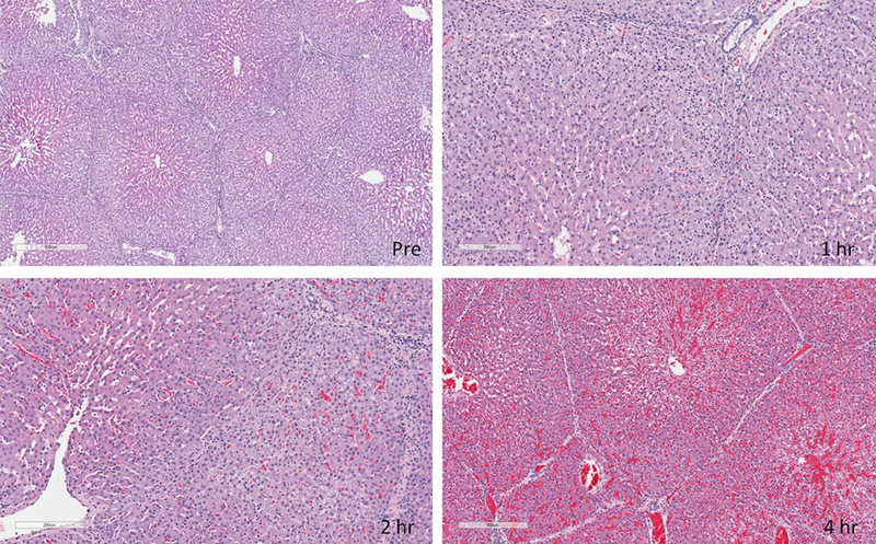
Figure 8: H&E staining of liver samples.
Pre-perfusion biopsies showed normal liver architecture in all experiments. Progressive periportal vacuolization extending towards the central veins and edema was noted starting at 1 hour and progressing through perfusion. Sinusoidal spaces became less clear starting around 2 hours. At about 4 hours there were areas of red blood cell congestion and hemorrhage.

