Abstract
Background and study aims There are limited published data on endoscopic removal of colorectal polyps by endoscopic mucosal resection (EMR) and endoscopic mucosal dissection (ESD) in patients with inflammatory bowel disease (IBD).
Patients and methods We performed a retrospective review of patients with colonic IBD and colonic polyps >10mm who underwent EMR and/or ESD at our institution between January 1, 2012 and June 31, 2016.
Results Ninety-seven patients with pathology-confirmed IBD (median disease duration 16 years) were included. Mild or moderate active colitis (in background biopsies) was seen in 85 %. Of the total 124 polyps, location was ascending colon in 44 %, transverse in 15 % and sigmoid in 18.5 %; of the total, 55 % were < 20 mm and 45 % were ≥20mm in maximal diameter. Using the Paris classification, 56 % of polyps were polypoid sessile (Is) polyps, while 38 % were non-polypoid (IIa, IIb, IIc). EMR was used in 118 polyps, three required ESD, and three by combined EMR-ESD. Seventy-two percent were resected en-bloc; 28 % underwent piecemeal resection. Histology included low-grade dysplasia in 75, serrated adenoma in 31, and tubular adenoma in 14 polyps. Chromoendoscopy was used in 33 (26.6 %). Adverse events occurred in three patients. Colectomy was performed in 11 patients within 12 months. Recurrence was seen in 20 polyps, 11 of which were successfully resected en-bloc using EMR. Polyps ≥ 20 mm and polyps treated with APC were found to have a statistically significantly higher risk of recurrence.
Conclusion This study demonstrates the efficacy and safety of endoscopic resection of large polyps in patients with IBD, making them effective alternatives to colectomy.
Introduction
Patients with inflammatory bowel disease (IBD) are at increased risk of colorectal dysplasia with further risk of transforming into colorectal cancer 1 . Chronic colonic inflammation in both ulcerative colitis (UC) and Crohn’s disease (CD) increases risk of dysplasia 2 .
Endoscopic mucosal resection (EMR) is currently used routinely for treatment of sporadic colorectal dysplasia and polyps, including small and large (> 20 mm) polyps 3 . EMR uses submucosal injection of fluid to separate the superficial mucosal layer (containing the dysplastic lesion) from the underlying muscle layer, after which the lesion can be removed with an electrosurgical snare 4 . EMR as a procedure is considered comparatively safe, simple, adaptable and easier to master for a less-experienced endoscopist compared to endoscopic submucosal dissection (ESD) 5 .
ESD was developed in the early 2000 s as a new resection method based on EMR 6 . ESD is used most often for polyps >20 mm or in conjunction with EMR when EMR is unsuccessful in completely excising the polyp or dysplastic lesion 7 . ESD in comparison to EMR has a higher likelihood of complete resection of the lesion and hence, can provide en bloc specimens which can be used for reliable pathological examination 8 . ESD offers the possibility of achieving en bloc resections regardless of lesion size but theoretically is limited by presence of submucosal fibrosis 9 .
Traditionally, presence of dysplasia in the colon in patients with IBD has been an indication for colectomy. However, more recent guidelines such as the SCENIC guidelines suggest that patients with effective endoscopic resection of dysplastic polyps can continue endoscopic surveillance rather than proceed to colectomy 10 11 . The key condition is feasibility of complete endoscopic resection, which depends on characteristics of the dysplastic polyp. There are very limited data in the literature on outcomes of EMR of large polyps in patients with IBD 11 .
In our study, we aimed to describe our experience with EMR and ESD of polyps larger than 10 mm in patients with colitis due to IBD at Mayo Clinic. We also sought to further describe recurrence in patients who had undergone an EMR or ESD previously.
Patients and methods
Ethical considerations
This study was approved by the Mayo Clinic Institutional Review Board. Per Minnesota state law, patients who had withdrawn authorization for their medical records to be reviewed for research purposes were not included in the study. This is a referral center for patients from all across the United States and outside of the country for their medical care.
Patient selection
This study was a retrospective chart review of patients with IBD involving the colon and colonic polyps > 10 mm who underwent colonoscopy with ESD and/or EMR at our institution between January 1, 2012 and June 30, 2016.
We used a medical record filter from Mayo Clinic called Advanced Cohort Explorer (ACE). ACE is a data exploring software in which Mayo Clinic medical record numbers can be entered and matched to the International statistical Classification of Diseases (ICD) codes for specific diseases and to different variables, such as in our study to polyps of > 10 mm. We initially had a list of patients who had polyps > 10 mm; these patients were matched to ICD codes for CD and UC (CD – 550.X and UC – 556.X) on at least three different billing encounters to minimize error. Diagnosis of IBD was confirmed by pathology specimens obtained (random background biopsies) during index and prior colonoscopies. These had to be consistent with active or quiescent IBD involving the colonic segment where the polyp was resected. Once we had a list of patients with IBD and polyps, we abstracted data for different variables for these patients. Study data were collected and managed using Research Electronic Data Capture (Redcap) hosted by Mayo Clinic 12 .
Endoscopic resection techniques
All EMR and ESD cases were performed by specially trained endoscopists with large-volume experience in advanced resection techniques. EMR was performed using standard or stiff snares, either en bloc or piecemeal (described at endoscopy), following submucosal fluid injection of a mixture of saline, methylene blue and dilute epinephrine (1:100,000). The hot biopsy forceps avulsion technique (Endocut I, setting 3, ERBE VIO 300 electrosurgical generator) was used for tissue removal when submucosal fibrosis and non-lifting prevented ensnarement of polyp tissue. Alternatively, argon plasma coagulation was used to ablate residual polyp tissue in some cases. Choice of endoscopic resection technique was dependant on the endoscopist performing the procedure, and not per any protocol. ESD was performed in standard fashion with marking of the lesion border by thermal coagulation dots followed by intermittent submucosal fluid injection using a mixture of hydroxypropyl methylcellulose with dilute epinephrine and methylene blue. A circumferential incision was performed using an electrosurgical knife (Dual and/or Hook knives, Olympus Corp., Japan) followed by submucosal dissection. When severe fibrosis precluded dissection, an attempt was made to snare resect the partially dissected lesion en bloc or in a piecemeal fashion, with use of the hot biopsy forceps avulsion or APC technique for residual non-lifting polyp tissue ( Fig. 1 ).
Fig. 1.
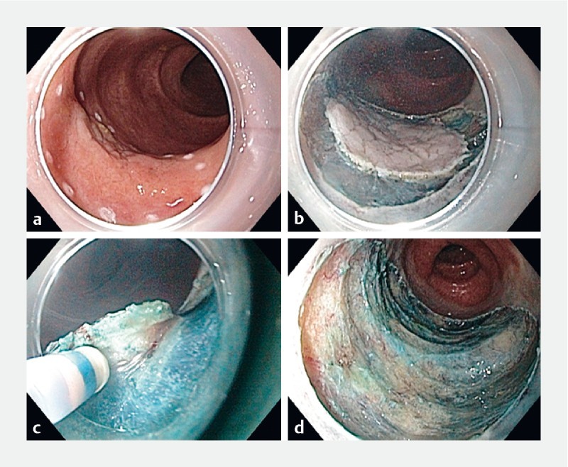
ESD of Paris Type IIa/b lesion. a Thermal marking of periphery of lesion. b Circumferential submucosal incision. c Submucosal dissection through fibrosis. d Post-resection defect
Data collection
Medical records were reviewed by a single reviewer (SY) – any doubts or discrepancies regarding IBD status were clarified by EVL, and any questions regarding the polyps or actual procedure were clarified by the other authors (LWKS and NCP). The variables collected were gender, date of IBD diagnosis, duration of IBD at time of procedure, type of IBD (CD/UC/indeterminate), involvement of small bowel, severity at time of procedure (mild/mod/severe), date of procedure, use of chromoendoscopy, number of polyps resected > 10 mm, location of these polyps, size of polyp in mm, scarring associated with lesion as mentioned in endoscopy report, morphology (Paris classification), ESD/EMR performed, previous ESD/EMR, resectioning technique (en-bloc vs piecemeal), type of snare used [standard/small (Sensation, Boston Scientific, Massachusetts, United States) , Histolock (US Endoscopy, Ohio, United States), Spiral (SnareMaster, Olympus, Pennsylvania, United States), Traxtion (US Endoscopy, Ohio, United States), AcuSnare (Cook Endoscopy, North Carolina, United States), or Crescent (Olympus, Pennsylvania, United States)], any additional therapy (argon plasma coagulation [APC], hot biopsy, cold snare, cold biopsy), prophylactic use of clips, histology (tubular adenoma, tubulovillous adenoma, serrated, low-grade dysplasia, high-grade dysplasia, adenocarcinoma), post-procedural adverse events (perforation, bleeding, fever), surgery for colonic resection post polypectomy, follow-up procedures, and polyp recurrence.
Statistical analysis
A descriptive statistical analysis was performed that included the median (range) or mean (standard deviation) for quantitative variables and frequency (%) for discrete variables. We estimated the recurrence rate and survival free of first recurrence using the Kaplan-Meier estimate along with the 95 % confidence interval (CI). All statistical analyses were conducted using SAS version 9.2 for Windows (SAS Institute Inc., Cary, North Carolina, United Staes). P < 0.05 was considered statistically significant.
Results
Patient and IBD characteristics
We identified 97 patients with IBD who were found to have 124 polyps at colonoscopy. Median age at IBD diagnosis was 44 years and at diagnosis of polyps for these patients was 59.1 years (range, 49.2 – 87.7 years). Sixty-three patients had UC (64.9 %), 27 had CD (27.8 %) and seven had indeterminate colitis (7.2 %). All of the patients had background biopsies including the segment of the polyp which confirmed chronic colitis. In these background random biopsies, forty-three patients had mild/or quiescent disease (44.3 %), 40 had moderate disease activity (41.2 %) and 14 had severe disease (14.4 %). At the index colonoscopy, active colitis (Mayo UC score 1 – 3) was seen in 88 patients (77.9 %), the others had prior evidence of colitis ( Table 1 ).
Table 1. Demographic features of patients with IBD who underwent polypectomy.
| Characteristics | N = 97 |
| Median age, years (range) | 59.1 (49.2 – 87.7) |
| Median duration of disease, years (range) | 16 (0 – 60.5) |
| Gender, n (%) | |
| Male | 59 (61.5 %) |
| Female | 37 (38.5 %) |
| Family history of colon cancer | 20 (20.6 %) |
| IBD subtype, n (%) | |
| Crohn’s disease | 27 (27.8 %) |
| Ulcerative colitis | 63 (64.9 %) |
| Indeterminate colitis | 7 (7.2 %) |
| Severity of colitis, n (%) | |
| Mild | 43 (44.3 %) |
| Moderate | 40 (41.2 %) |
| Severe | 14 (14.4 %) |
IBD, inflammatory bowel disease
Polyp characteristics
Of the 124 polyps, 68 (54.8 %) were less than 20 mm and 56 (45.2 %) were 20 mm or greater in diameter. Using the Paris classification, 69 polyps were polypoid sessile (Is) (55.6 %), seven were polypoid pedunculated (Ip) (5.6 %), 45 were non-polypoid superficial elevated (IIa) (36.3 %), and two polyps were non-polypoid flat (IIb). Seventy-five polyps were tubular adenomas with low-grade dysplasia (60.5 %), of which one polyp had both low- and high-grade dysplasia, 14 polyps were tubulovillous adenomas with low-grade dysplasia (11.3 %) and one polyp had both low- and high-grade dysplasia, and 31 polyps were serrated (25 %) ( Table 2 ). There were three adenocarcinomas, of which two were intramucosal and the other was in a pedunculated polyp, and the adenocarcinoma extended to the margin of resection
Table 2. Features of endoscopic resection of colonic polyps by EMR/ESD in patients with IBD.
| Characteristics | |
| Median size of polyp (mm) | 15 (10 – 60) |
|
68 (54.8 %) |
|
56 (45.1 %) |
| Location of polyps, n (%) | |
| Ileo-cecal valve/cecum | 5 (4 %) |
| Ascending colon | 55 (44.4 %) |
| Transverse colon | 19 (15.3 %) |
| Descending colon | 9 (7.3 %) |
| Sigmoid colon | 23 (18.5 %) |
| Rectum | 13 (10.5 %) |
| Morphology (Paris classification), n (%) | |
| Polypoid pedunculated | 7 (5.6 %) |
| Polypoid sessile | 69 (55.6 %) |
| Non polypoid superficial elevated | 45 (36.3 %) |
| Non polypoid flat | 2 (1.6 %) |
| Non polypoid depressed | 1 (0.8 %) |
| Procedure type, n (%) | |
| ESD | 3 (2.4 %) |
| EMR | 118 (95.2 %) |
| ESD and EMR | 3 (2.4 %) |
| Resectioning technique, n (%) | |
| En bloc | 88 (70.9 %) |
| Piecemeal | 36 (29.0 %) |
| Type of snare used, n (%) | |
| Standard/small | 113 (91.1 %) |
| Spiral | 13 (10.2 %) |
| Crescent | 1 (0.8 %) |
| Additional therapy, n (%) | |
| APC | 50 (40.3 %) |
| Hot biopsy avulsion | 9 (7.2 %) |
| Hot snare | 109 (87.9 %) |
| Cold biopsy | 11 (8.8 %) |
| Cold snare | 9 (7.2 %) |
| Polyps needing clips used, n (%) | 65 (52.4 %) |
| Histology, n (%) | |
| Tubular adenoma, low grade dysplasia | 75 (60.5 %) |
| Tubulovillous adenoma, low grade dysplasia | 14 (11.3 %) |
| Serrated | 31 (19.7 %) |
| Adenocarcinoma | 3 (1.9 %) |
| Hyperplastic | 22 (14 %) |
| Surgery within 12 months of polypectomy | 11 (11.3 %) |
| Recurrence, n (%) | |
| 1 recurrence | 20 (16 %) |
| 2 recurrences | 9 (45 %) |
| 3 recurrences | 3 (33.3 %) |
EMR, endoscopic mucosal resection; ESD, endoscopic submucosal dissection; IBD, inflammatory bowel disease; APC, adenomatous polyposis coli
Endoscopic interventions
One hundred eighteen polyps were removed using EMR (95.2%), three polyps needed ESD removal (2.4 %), and three patients were treated initially with an unsuccessful ESD followed by EMR during the same session (2.4 %). All ESD and hybrid ESD/EMR procedures were performed by a single therapeutic endoscopist – LMW. Complete en-bloc resection (defined at endoscopy) was achieved for 88 polyps (70.9 %), while piecemeal resection was performed for 36 polyps (29 %). A standard flexible snare (Sensation, Boston Scientific, Massachusetts, United States) was used to resect 113 polyps (91.1 %), and a stiff Spiral snare (SnareMaster, Olympus, Pennsylvania, United States) in 13 polyps (10.2 %). Prophylactic clips were placed after 65 polypectomies (52.4 %). Eleven patients underwent proctocolectomy within 12 months of polypectomy (11.3%). Of these 11 patients, three had adenocarcinoma, two had high-grade dysplasia, three patients had low-grade dysplasia but also had medically refractory clinical disease activity, and in three other cases the polyp resection was incomplete in the setting of low-grade dysplasia in the unresectable polyp. Ten of these 11 patients had EMR polypectomy, and seven of them had polyps larger than 20 mm. Fig. 2 is a flowchart showing outcomes of polyps including recurrences and colectomy. Adverse events (AEs) included two patients with late bleeding (1 – 2 days post procedure) and one patient with fever, but there were no perforations ( Table 2 ).
Fig. 2.
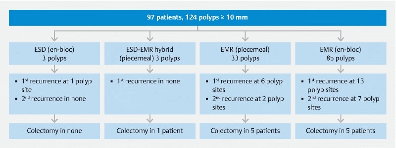
Flowchart of outcomes of polyp resection in terms of recurrence and colectomy.
APC was used in 15 polyps (22.1 %) less than 20 mm compared to 35 polyps (62.5 %) equal or greater than 20 mm ( P > 0.0001). Among the 68 polyps that were less than 20 mm in size, all were resected using EMR and 57 (85.1 %) were resected en-bloc compared to the 56 polyps that were equal to or greater than 2 0 mm, of which only 31 (56.4 %) were resected en-bloc ( P = 0.0004).
Recurrences
Recurrence included any adenomatous tissue found at follow-up colonoscopy at the exact polyp site or within the segment of the colon that had the index polypectomy. Of the initial 20 recurrences, another nine recurred a second time (45 %), and three polyps recurred for a third time ( Table 3 ). Recurrences occurred in six polyps less than 20 mm compared with 14 polyps equal or greater than 20 mm. Among the first recurrences, all polyps were removed using EMR technique. Fig. 3 shows images of some of these recurrent polyps.
Table 3. Features of recurrence in patients with endoscopic resection of colonic polyps by EMR/ESD in patients with IBD.
| Characteristics | Recurrence 1 (20) | Recurrence 2 (9) | Recurrence 3 (3) |
| Index polyp size (mm) | |||
| < 20 | 6 (30.0 %) | 2 (22.2 %) | 0 (0.0 %) |
| ≥ 20 | 14 (70.0 %) | 7 (77.8 %) | 3 (100.0 %) |
| Median size of recurrent polyp (mm) | 6.5 | 5 | 3.5 |
| Resection technique of index polyp | |||
| ESD only | 1 | ||
| EMR only | 19 | ||
| ESD + EMR | 0 | ||
| Location of polyps, n (%) | Top of Form | ||
| Ascending colon | 10 (50 %) | ||
| Transverse colon | 4 (20 %) | ||
| Sigmoid colon | 4 (20 %) | ||
| Rectum | 2 (10 %) | ||
| Morphology (Paris classification), n (%) | |||
| Polypoid sessile | 15 (75.0 %) | 7 (77.8 %) | 3 (100.0 %) |
| Non polypoid flat | 5 (25.0 %) | 2 (22.2 %) | 0 (0.0 %) |
| Resectioning technique; polyps ≥ 20 mm, n (%) | |||
| En bloc | 13 (92.9 %) | 1 | |
| Piecemeal | 1 (7.1 %) | ||
| Type of snare used, n (%) | |||
| Standard/small | 10 (83.3 %) | 5 | |
| Histolock | 1 (9.2 %) | ||
| Spiral | 1 (9.2 %) | ||
| Additional therapy, n (%) | |||
| APC | 5 (16.7 %) | 3 (23.1 %) | 1 (25 %) |
| Hot biopsy | 2 (6.7 %) | ||
| Hot snare | 10 (33.3 %) | 4 (30.8 %) | |
| Cold biopsy | 5 (16.7 %) | 4 (30.8 %) | 2 (50 %) |
| Cold snare | 8 (26.7 %) | 2 (15.4 %) | 1 (25 %) |
| Polyps needing clips used, n (%) | 5 | ||
| Histology, n (%) | |||
| Tubular adenoma, low grade dysplasia | 9 (45 %) | 6 (66.6 %) | 1 (33 %) |
| Tubulovillous adenoma, low grade dysplasia | 5 (25 %) | ||
| Serrated | 5 (25 %) | 1 (11.1 %) | 1 (33 %) |
| Adenocarcinoma | |||
| Hyperplastic | 1 (2.5 %) | 2 (22.2 %) | 1 (33 %) |
EMR, endoscopic mucosal resection; ESD, endoscopic submucosal dissection; IBD, inflammatory bowel disease; APC, adenomatous polyposis coli
Fig. 3.
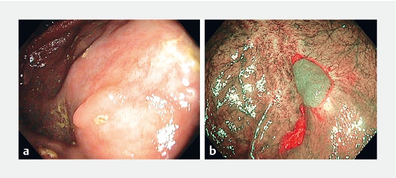
Recurrent polyps at previous polypectomy sites.
Recurrence-free survival
Survival free of recurrence at the end of the first year from polypectomy was 83.3 % (95 % CI, 76 %-92 %), slightly decreased to 76.5 % (66 % – 87 %) in the second year, and remained unchanged in the third year 76.5 % (66 % – 87 %).
The 2-year estimates for recurrence free survival in polyps sized < 20 mm and ≥ 20 mm were 91.2 % and 56.0 %, respectively. A polyp size < 20 mm relative to a polyp sized ≥ 20 mm was at a significantly increased risk of recurrence, P = 0.006, with a hazard ratio of 3.8, 95 % CI 1.4 – 10.0 ( Fig. 4 ).
Fig. 4.
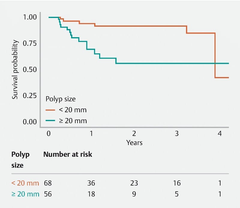
Survival free of recurrence in IBD patients having undergone ESD/EMR classified by polyp size.
When comparing polyp morphology, the 2-year recurrence-free survival was 72.8 % for polypoid lesions (Paris 1 p and 1 s) compared to 82.2 % for non-polypoid lesions (Paris 2a, 2b, and 2c) with a hazard ratio of 1.8 (95 % CI 0.7 – 5.1) ( Fig. 5 ).
Fig. 5.
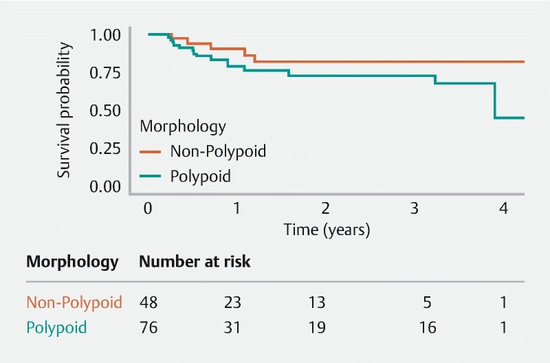
Survival free of recurrence in IBD patients having undergone ESD/EMR classified by polyp morphology (Paris classification).
In the 50 polyps where APC was used during polypectomy, 2-year survival free of recurrence was 61 % compared to 86 % in the 74 polyps where APC was not used. A polyp where APC was used had an increased risk of recurrence compared to a polyp where it was not used with a HR of 2.798 ( P = 0.0250).
The 2-year survival free of recurrence was 74.6 % for en-bloc resected polyps compared to 82.4 % for polyps resected piecemeal (HR 0.9, P = 0.83); and was 72.8 % for polypoid lesions compared to 82.2 % for non-polypoid lesions (HR 1.8, P = 0.23).
Discussion
In recent years, with the advent of better imaging and newer resectioning techniques, the sphere of endoscopic resection is slowly closing the gap to surgery. Guidelines published by the American Society for Gastrointestinal Endoscopy in the last few years, based on the SCENIC consensus statement, recommend endoscopic resection of polypoid dysplasia in patients with chronic colitis, followed by endoscopic surveillance 11 13 . For non-polypoid endoscopically visible dysplasia, resection is still suggested too. However, the data on success and recurrence rates after endoscopic resection of polyps larger than 1 cm remain limited 14 15 16 17 18 . In our cohort of patients, we focused on use of EMR and ESD for large polyps in patients with IBD. Overall, our study concluded that ESD and EMR are effective and safe therapies for polyps > 10 mm.
Polyp resection in patients with colitis is challenging because of the submucosal fibrosis often present, especially in healed colitis. Ability to obtain adequate submucosal lifting is also challenged. However, use of stiffer snares and avulsion technique following some submucosal injection have aided resection. In patients with active colitis, identification of lesion margins can also be difficult due to surrounding inflammation. In these cases, use of narrow-band imaging or contrast chromoscopy can be helpful to delineate the lesion.
Iacopini and colleagues have described their outcome with resection of 10 polyps in patients with quiescent chronic colitis; eight of these patients underwent en-bloc ESD 14 . Median polyp sizes were 33 mm, and most were in the left colon with almost universal submucosal fibrosis. They demonstrated curative resection in 70 %, but noted 55 % recurrence of dysplastic tissue at a median follow-up of 24 months. More recently, there have been more data on the feasibility of ESD for removal of large polyps in colitis. Suzuki et al reported an en-bloc resection in 91 % of 32 dysplastic lesions with a median diameter of 33mm 18 . Submucosal fibrosis and adipose deposition were observed in 31 (97 %) and 13 lesions (41 %). Recurrence was seen in only one patient after median follow-up of 33 months. Kinoshita et al described universal en-bloc resection and 71 % R0 resection in 25 dysplastic lesions with a mean size of 34.9mm 17 . During a 21-month follow-up, no local recurrence was noted.
In the first cited study 14 , all of the patients were in complete histologic remission at the time of their ESD. This is unusual in common practice, where polyps are often seen in a background of active inflammation. In our cohort, 44 % of patients had mild/quiescent disease, but the majority had active inflammation which more accurately reflects routine practice. Submucosal fibrosis was not universally documented in our cases, but in all three hybrid EMR-ESD cases, the ESD had to be converted to EMR technique because submucosal fibrosis prevented adequate submucosal dissection. Also, a larger number of the polyps in our study were in the right colon, where potentially more adenomatous polyps may be seen both in the IBD and non-IBD population.
Another important study by Smith et al described efficacy of ESD-assisted EMR in resection of 67 large polyps, median size ranging from 12 to 30 mm, in patients with chronic colitis 15 . En bloc resection was achieved in 78 % cases, without any invasive adenocarcinomas. With a median follow-up of 1.5 years, recurrent disease was seen in 7 %, of whom all but one of which were endoscopically resected again. EMR is a much more widely used endoscopic technique compared to ESD, and therefore our study is more widely applicable to gastroenterologists in practice.
One of the potential limitations of our study is that we included all polyps > 10mm – both polypoid and flat. However, 38% of the polyps were non-polypoid and therefore, clinically very relevant. Also, 45 % were ≥ 20 mm, and even though recurrences were significantly more common in this size compared to smaller polyps, they were also endoscopically treatable. Currently available data on the efficacy of endoscopic resection of non-polypoid polyps are scarce. This study shows that even polyps larger than 2 cm in size and non-polypoid in morphology can be safely and effectively resected using either EMR or ESD.
Recurrences were noted to occur significantly more often in polyps treated with APC, but the caveat is that APC was used more often in polyps greater than 20 mm, which is an independent risk factor for recurrence. Interestingly, piecemeal resection was not shown to be associated with increased risk of recurrences.
It is well known that patients with chronic colitis with dysplastic lesions are at high risk for developing further dysplastic lesions 19 20 . In our study, we had a relatively low rate of recurrence of dysplasia, with a recurrence-free survival at 1 year of 83.3 %. We included polyps as recurrent if they were in the same colonic segment as the initial polyps and not necessarily present on or adjacent to the tattoo or scar tissue (because not all polyps had clear site demarcation). An important feature of our study is the fact that almost all dysplasia recurrence could be resected endoscopically. This underscores the current guidelines which recommend close follow-up surveillance after endoscopic resection of polyps 11 .
Our study had certain limitations. It was a retrospective review of data at a tertiary center. However, our practice is such that almost all our patients get their follow-up and surveillance procedures performed at our institution, making the follow-up data more meaningful. The procedures were performed by multiple gastroenterologists with variable levels of skill, but this represents a majority of gastroenterology practices. We had very few ESDs in this dataset, so a direct comparison between ESD and EMR is not possible.
Conclusion
In conclusion, we present here the largest retrospective review examining efficacy, safety, and outcome of endoscopic resection of large polyps greater than 1 cm in the IBD population. We show that endoscopic resection of polyps in IBD is a feasible and curative process which may help patients avoid proctocolectomy. This study is important because there isn’t a large body of literature on this subject in this specific population. The results of our study serve to affirm and support current guidelines on the endoscopic management of dysplasia in patients with IBD.
Acknowledgement
The authors thank Teresa Nolte, Gastroenterology Tech Support, for helping acquire patient and procedural details.
Footnotes
Competing interests Dr. Loftus is a consultant for AbbVie, Takeda, Janssen, UCB, Amgen, Pfizer, Celgene, Celltrion, Eli Lilly, Napo Pharma and receives research suppor from AbbVie, Takeda, Janssen, UCB, Amgen, Pfizer, Celgene, Genentech, Receptos, MedImmune, Allergan, Seres Therapeutics, and Robarts Clinical Trials.
References
- 1.Rutter M D, Riddell R H. Colorectal dysplasia in inflammatory bowel disease: a clinicopathologic perspective. Clin Gastroenterol Hepatol. 2014;12:359–367. doi: 10.1016/j.cgh.2013.05.033. [DOI] [PubMed] [Google Scholar]
- 2.Fort Gasia M, Ghosh S, Iacucci M. Colorectal polyps in ulcerative colitis and Crohnʼs colitis. Minerva Gastroenterol Dietol. 2015;61:215–222. [PubMed] [Google Scholar]
- 3.Sakamoto T, Matsuda T, Nakajima T et al. Efficacy of endoscopic mucosal resection with circumferential incision for patients with large colorectal tumors. Clin Gastroenterol Hepatol. 2012;10:22–26. doi: 10.1016/j.cgh.2011.10.007. [DOI] [PubMed] [Google Scholar]
- 4.Consolo P, Luigiano C, Strangio G et al. Efficacy, risk factors and complications of endoscopic polypectomy: ten year experience at a single center. World J Gastroenterol. 2008;14:2364–2369. doi: 10.3748/wjg.14.2364. [DOI] [PMC free article] [PubMed] [Google Scholar]
- 5.Kaimakliotis P Z, Chandrasekhara V. Endoscopic mucosal resection and endoscopic submucosal dissection of epithelial neoplasia of the colon. Expert Rev Gastroenterol Hepatol. 2014;8:521–531. doi: 10.1586/17474124.2014.902305. [DOI] [PubMed] [Google Scholar]
- 6.Cai S L, Shi Q, Chen T et al. Endoscopic resection of tumors in the lower digestive tract. World J Gastrointest Endosc. 2015;7:1238–1242. doi: 10.4253/wjge.v7.i17.1238. [DOI] [PMC free article] [PubMed] [Google Scholar]
- 7.Noshirwani K C, van Stolk R U, Rybicki L A et al. Adenoma size and number are predictive of adenoma recurrence: implications for surveillance colonoscopy. Gastrointest Endosc. 2000;51:433–437. doi: 10.1016/s0016-5107(00)70444-5. [DOI] [PubMed] [Google Scholar]
- 8.Wang J, Zhang X H, Ge J et al. Endoscopic submucosal dissection vs endoscopic mucosal resection for colorectal tumors: a meta-analysis. World J Gastroenterol. 2014;20:8282–8287. doi: 10.3748/wjg.v20.i25.8282. [DOI] [PMC free article] [PubMed] [Google Scholar]
- 9.Nakajima T, Saito Y, Tanaka S et al. Current status of endoscopic resection strategy for large, early colorectal neoplasia in Japan. Surg Endosc. 2013;27:3262–3270. doi: 10.1007/s00464-013-2903-x. [DOI] [PubMed] [Google Scholar]
- 10.Annese V, Daperno M, Rutter M D et al. European evidence based consensus for endoscopy in inflammatory bowel disease. J Crohns Colitis. 2013;7:982–1018. doi: 10.1016/j.crohns.2013.09.016. [DOI] [PubMed] [Google Scholar]
- 11.Laine L, Kaltenbach T, Barkun A et al. SCENIC international consensus statement on surveillance and management of dysplasia in inflammatory bowel disease. Gastroenterology. 2015;148:639–651 e628. doi: 10.1053/j.gastro.2015.01.031. [DOI] [PubMed] [Google Scholar]
- 12.Harris P A, Taylor R, Thielke R et al. Research electronic data capture (REDCap) – a metadata-driven methodology and workflow process for providing translational research informatics support. J Biomed Inform. 2009;42:377–381. doi: 10.1016/j.jbi.2008.08.010. [DOI] [PMC free article] [PubMed] [Google Scholar]
- 13.American Society for Gastrointestinal Endoscopy Standards of Practice C . Shergill A K, Lightdale J R et al. The role of endoscopy in inflammatory bowel disease. Gastrointest Endosc. 2015;81:1101–1121 e1101-1113. doi: 10.1016/j.gie.2014.10.030. [DOI] [PubMed] [Google Scholar]
- 14.Iacopini F, Saito Y, Yamada M et al. Curative endoscopic submucosal dissection of large nonpolypoid superficial neoplasms in ulcerative colitis (with videos) Gastrointest Endosc. 2015;82:734–738. doi: 10.1016/j.gie.2015.02.052. [DOI] [PubMed] [Google Scholar]
- 15.Smith L A, Baraza W, Tiffin N et al. Endoscopic resection of adenoma-like mass in chronic ulcerative colitis using a combined endoscopic mucosal resection and cap assisted submucosal dissection technique. Inflamm Bowel Dis. 2008;14:1380–1386. doi: 10.1002/ibd.20497. [DOI] [PubMed] [Google Scholar]
- 16.Gulati S, Emmanuel A, Burt M et al. Outcomes of endoscopic resections of large laterally spreading colorectal lesions in inflammatory bowel disease: a single United Kingdom center experience. Inflamm Bowel Dis. 2018;24:1196–1203. doi: 10.1093/ibd/izx113. [DOI] [PubMed] [Google Scholar]
- 17.Kinoshita S, Uraoka T, Nishizawa T et al. The role of colorectal endoscopic submucosal dissection in patients with ulcerative colitis. Gastrointest Endosc. 2018;87:1079–1084. doi: 10.1016/j.gie.2017.10.035. [DOI] [PubMed] [Google Scholar]
- 18.Suzuki N, Toyonaga T, East J E. Endoscopic submucosal dissection of colitis-related dysplasia. Endoscopy. 2017;49:1237–1242. doi: 10.1055/s-0043-114410. [DOI] [PubMed] [Google Scholar]
- 19.Fumery M, Dulai P S, Gupta S et al. Incidence, risk factors, and outcomes of colorectal cancer in patients with ulcerative colitis with low-grade dysplasia: a systematic review and meta-analysis. Clin Gastroenterol Hepatol. 2017;15:665–674 e665. doi: 10.1016/j.cgh.2016.11.025. [DOI] [PMC free article] [PubMed] [Google Scholar]
- 20.Wanders L K, Dekker E, Pullens B et al. Cancer risk after resection of polypoid dysplasia in patients with longstanding ulcerative colitis: a meta-analysis. Clin Gastroenterol Hepatol. 2014;12:756–764. doi: 10.1016/j.cgh.2013.07.024. [DOI] [PubMed] [Google Scholar]


