Abstract
Purpose
Beckwith–Wiedemann syndrome (BWS) is a developmental disorder caused by dysregulation of the imprinted gene cluster of chromosome 11p15.5 and often associated with loss of methylation (LOM) of the imprinting center 2 (IC2) located in KCNQ1 intron 10. To unravel the etiological mechanisms underlying these epimutations, we searched for genetic variants associated with IC2 LOM.
Methods
We looked for cases showing the clinical features of both BWS and long QT syndrome (LQTS), which is often associated with KCNQ1 variants. Pathogenic variants were identified by genomic analysis and targeted sequencing. Functional experiments were performed to link these pathogenic variants to the imprinting defect.
Results
We found three rare cases in which complete IC2 LOM is associated with maternal transmission of KCNQ1 variants, two of which were demonstrated to affect KCNQ1 transcription upstream of IC2. As a consequence of KCNQ1 haploinsufficiency, these variants also cause LQTS on both maternal and paternal transmission.
Conclusion
These results are consistent with the hypothesis that, similar to what has been demonstrated in mouse, lack of transcription across IC2 results in failure of methylation establishment in the female germline and BWS later in development, and also suggest a new link between LQTS and BWS that is important for genetic counseling.
Keywords: Beckwith–Wiedemann syndrome, imprinting disorders, genomic imprinting, long QT syndrome, DNA methylation
INTRODUCTION
KCNQ1 (potassium voltage-gated channel subfamily Q member 1), the major gene involved in long QT syndrome (LQTS), is part of the imprinted gene cluster that is associated with Beckwith–Wiedemann syndrome (BWS, OMIM 130650) at chromosome 11p15.5.
LQTS (prevalence 1:2000 [ref.1]) is a cardiac arrhythmia characterized by prolongation of the electrocardiographic (ECG) QT interval and high risk of syncope, seizures, and sudden death. The genetic form of this disease is associated with more than 700 variants in 13 genes encoding ion channel subunits.2,3 Pathogenic KCNQ1 variants (LQT1, OMIM 192500) represent 35% of LQTS defects and are generally inherited as isolated cardiac defects within dominant pedigrees (also known as Romano–Ward syndrome), or in combination with other clinical features (e.g., deafness) as an autosomal recessive trait (also known as Jervell and Lange–Nielsen syndrome).
BWS (prevalence 1:10,500) is a congenital genomic imprinting disorder, characterized by variable presence of macroglossia, abdominal wall defects, lateralized overgrowth, enlarged abdominal organs, and increased risk of developing embryonal tumors.4,5 Heterogeneous molecular alterations affect expression of an imprinted gene cluster located at 11p15.5. The cluster is divided in two domains controlled by separate imprinting control regions (ICRs). The ICR of the centromeric domain, known as KCNQ1OT1:transcription start site (TSS) differentially methylated region (DMR), also known and herein indicated as imprinting center 2 (IC2), is located within KCNQ1 intron 10. IC2 corresponds to the promoter of the long noncoding RNA KCNQ1OT1 and is methylated and inactive on the maternal chromosome. On the paternal chromosome, KCNQ1OT1 is transcribed and represses in cis the flanking imprinted genes, including the growth inhibitor CDKN1C, which is normally transcribed from the maternal allele. In 50% of the BWS patients, loss of methylation (LOM) of IC2 leads to biallelic expression of KCNQ1OT1 and biallelic silencing of CDKN1C. One-third of these cases also show methylation abnormalities at other ICRs (multilocus imprinting disturbances, MLID). Further molecular lesions of BWS are gain of methylation of the H19/IGF2: intergenic (IG) DMR (5–10%), 11p15 paternal uniparental isodisomy (20%), loss-of-function pathogenic variants of CDKN1C (5%), and 11p15.5 copy-number variants (CNVs, 1–4%) (ref. 6). Clinical features, risk of recurrence, and frequency and histotype of associated tumors differ significantly among the main molecular classes of BWS.7
The KCNQ1 gene is 404 kb long, organized in 19 exons, and harbors two major transcript isoforms arising from alternative use of promoters: isoform 1 from exon 1a and isoform 2 from exon 1b. Both isoforms are expressed in heart from both parental alleles. While isoform 2 is heart-specific, isoform 1 is expressed in several other tissues and imprinted with the paternal allele silenced during embryogenesis,8,9 but expressed from both parental alleles in postnatal tissues including peripheral blood cells.10
Mouse studies indicate that transcription across ICRs is a prerequisite for establishing methylation imprints in the maternal germline.11,12 Because IC2 lies within the transcriptional unit of the KCNQ1 gene, we hypothesized that maternally inherited pathogenic variants leading to defects of KCNQ1 transcription might also result in loss of maternal IC2 methylation in BWS. We also expect that such pathogenic variants cause KCNQ1 haploinsufficiency and possibly prolonged ECG QTc values and LQTS. To identify such rare cases, we looked for BWS patients with ECG alterations and IC2 LOM. We identified three patients who carried maternally inherited pathogenic variants of KCNQ1. Complete absence of methylation at the IC2 region was present in all three cases and defective transcription of the maternal KCNQ1 allele could be demonstrated in two of them. These results indicate that genetic variants interfering with transcription across IC2 are associated with dramatic methylation loss at this region, which is suggestive of defective establishment of imprint methylation in oocytes and may evolve into BWS later in embryo development.
MATERIALS AND METHODS
Clinical reports
The proband of family 1 was a Dutch girl born from healthy and unrelated parents. Her mother became pregnant at 40 years of age. Amniocentesis was performed at 17 weeks of gestation because of advanced age and ultrasound abnormalities. The fetus showed features typical of BWS, omphalocele, organomegaly, macroglossia, and polyhydramnios.5 The karyotype showed a balanced paternally derived Robertsonian translocation 45,XX,der(13;14)(q10;q10). Alpha-fetoprotein (AFP) was normal (17.6 μg/ml). She was born prematurely at 33 + 2/7 weeks of gestation with a birth weight of 2565 grams (90th percentile) and length at the 97th percentile. Clinical diagnosis of BWS was obtained at birth based on the presence of hypoglycemia, macrosomia, macroglossia, nevus flammeus, ear creases and pits, and organomegalia.5 Electrocardiography showed a prolonged QTc interval of 471 ms indicative for LQTS.13–15 Molecular diagnosis was obtained by multiplex ligation-dependent probe amplification (MLPA) (assays ME-030 and P144 for BWS and LQTS, respectively). The familial Robertsonian balanced translocation segregated in this family over five generations and did not have any effect on the phenotype. At age 18 months she had developed celiac disease. At 4.5 years, electrocardiography still showed a prolonged QTc interval (449 ms). At the clinical follow-up at 10 years, besides the BWS characteristics, LQTS, and celiac disease, she had developed mild bilateral hearing loss.
The proband of family 2 was an American girl born from unrelated parents. BWS was clinically diagnosed at birth at the University of Florida Hospital, based on the presence of omphalocele, macroglossia, and ear pits.5 Molecular diagnosis of BWS with IC2 LOM was obtained by Southern blot analysis (Mayo Clinic, Rochester, MN, USA, data not shown). She also had a history of learning delay and a low-frequency seizure disorder. At the age of 9 years, an ECG, performed following an episode of chest pain, revealed prolonged QTc (492 ms), and LQTS was diagnosed. Asymptomatic LQTS was diagnosed by ECG in her mother (QTc = 473), who did not show any sign of BWS. No BWS sign and normal QTc values were found in her half-sister, maternal grandmother and maternal grandfather. The maternal aunt, not available to clinical and molecular analyses, had several miscarriages. The first fetus, which was aborted at the 20th week, had large omphalocele, a clinical sign commonly found in BWS associated with molecular defects of the centromeric 11p15.5 domain. Her only full-term pregnancy was of two male twins. Of these, one infant was stillborn at scheduled Cesarean section, while his brother had a heart defect that required surgery shortly after birth.
The BWS features of family 3, in which segregates a 160-kb duplication within KCNQ1, were previously reported.16 Two further family members, healthy cousins of the proband who were not born at the time of the first study, were added to the present study. The proband was subjected to ECG within a screening for LQTS of BWS patients (see below in “ECG to BWS patients with IC2 LOM” section). Her QTc value was of 478 ms at 7.8 years of age, which is diagnostic of LQTS (Table S1).13–15 Following this finding, ECG was offered to all other carriers of the duplication of this family. All the individuals examined for ECG showed borderline prolonged QTc values (mother: 460 ms at 35 years, maternal uncle: 447 ms at 37 years, cousin: 440 ms at 2 years).
All genetic analyses in this paper were performed after written informed consent was obtained from the patients or patients’ parents. All research work has been carried out in accordance with the ethical principles and the legislation of Italy, the Netherlands, and the United States. The study was approved by the Ethical Committee of the University of Campania “Luigi Vanvitelli” (approval number 1135, 13 October 2016).
ECG to BWS patients with IC2 LOM
Twenty-four patients with clinical and molecular diagnosis of BWS with IC2 LOM5 were enrolled in an ECG screening. The patients, aged between 1 month and 24 years, were subjected to clinical visit, baseline ECG with QTc measurement, and family history collection. QTc interval was calculated using Bazett formula.13 QT values are reported in Table S1.
RESULTS
Because the centromeric ICR of the BWS locus is located in intron 10 of the KCNQ1 gene, which is also associated with LQTS, we wondered if abnormal QTc values in BWS patients with IC2 LOM could reveal the presence of pathogenic variants interfering with the imprinted methylation of this region. Thus, we looked for the presence of features of LQTS in BWS patients with IC2 LOM. Three cases were identified and are described in the following sections.
LQTS and BWS with complete IC2 LOM associated with a transcription defect of KCNQ1
Unusually low (<5%) IC2 methylation levels were detected in the family 1 proband (Fig. 1a) by methylation-specific MLPA (MS-MLPA) (ME-030 kit for BWS/Silver–Russell syndrome [SRS]) (Fig. 1b) and confirmed by pyrosequencing (Fig. 1c), indicating absence of the epigenetic mosaicism (10–35% methylation) found in most BWS patients. Methylation levels comparable with values of healthy individuals were found at IC1 and five other ICRs in the proband, and at IC1 and IC2 in her parents (Fig. 1c and Table S4). MLPA (P144 kit for LQTS) demonstrated a de novo heterozygous deletion of KCNQ1 exons 1a–1b in the proband (II-1) (Fig. 1d). By typing two single-nucleotide variants (SNVs; SNV2 and SNV3) falling in the intron between exon 1a and exon 1b, we demonstrated that the deletion arose on the maternal chromosome (Fig. 1e).
Fig. 1.
Characterization of a 120–kb deletion abolishing the KCNQ1 promoter. a Pedigree of family 1. The symbol partitioned in two halves with different filling indicate the proband (II-1) affected by Beckwith–Wiedemann syndrome (BWS) and long QT syndrome 1 (LQTS1). b DNA methylation analysis of IC2 and IC1 by methylation-specific MLPA (MS-MLPA). The histogram shows DNA methylation levels of the proband (II-1) and controls. Numbers indicate the length (nt) of the probes according to the manufacturer's instructions (ME-030, MRC-Holland); 355 is a digestion control probe. c DNA methylation analysis of IC2 and IC1 by pyrosequencing. Each dot represents the methylation value of a single CpG. Ctrl 1 and Ctrl 2 are unrelated healthy individuals; BWS 1 and BWS 2, two further BWS patients with IC2 loss of methylation (LOM). d Copy-number (CN) analysis of KCNQ1 exons in the trio by MLPA. The histograms represent the normalized CN detected with 18 MLPA probes (SALSA MLPA 144 Long QT probemix version A2) hybridizing within the KCNQ1 exons. Exon numbering is according to Lee et al.8 e Segregation in the trio of two single-nucleotide variants (SNVs) (SNV2: rs2023818 and SNV3: rs800336) located in the deleted region, as determined by Sanger sequencing, and demonstrating the maternal origin of the deletion. A schematic diagram of the KCNQ1 locus is reported. KCNQ1 (represented by a thin black bar) and KCNQ1OT1 (represented by a thicker blue bar) are transcribed on opposite strands. The pink rectangle highlights the region involved in the deletion. The vertical red lines indicate the location of SNVs 2 and 3 typed for segregation analysis and SNVs 10 and 11 reported in 1f. f Allele-specific expression analysis obtained by typing SNV10: rs2283168 A/G and SNV11: rs2283169 on total RNA from blood leukocytes of the proband. SNV10 and SNV11 are located between exon 1 and exon 2, downstream of the deletion and are present in heterozygosity in the proband. The nucleotides of the single-nucleotide polymorphisms (SNPs) determined by Sanger sequencing in DNA and RNA are reported below each electropherogram. Note the monoallelic expression of KCNQ1.
A comparative genomic hybridization (CGH)-based chromosome array analysis confirmed the presence of the deletion in heterozygosity and demonstrated that it was 120 kb long and included the first two KCNQ1 exons, the KCNQ1 promoter, and other three genes (TRPM5, TSSC4, and CD81). The telomeric breakpoint was mapped between 2,383,764 and 2,397,123, and the centromeric breakpoint between 2,510,194 and 2,532,843 of chromosome 11, about 30 kb downstream of KCNQ1 exon 1b (reference genome: hg19. Figure S2). By sequencing the entire coding region, no additional pathogenic variants were detected, indicating KCNQ1 haploinsufficiency as the most probable mechanism of LQTS in the proband.
To investigate the effect of the deletion on KCNQ1 transcription, allele-specific expression analysis was performed on total RNA from peripheral blood leukocytes of the proband. Consistent with a previous report,10 we observed biallelic KCNQ1 expression in three age-matched healthy individuals (Figure S3). Transcription was derived from promoter 1a, while promoter 1b–specific transcript was not detected in blood (data not shown). Significantly, KCNQ1 was expressed from only one allele in the proband, consistent with the loss of its promoters on the maternal chromosome (Fig. 1f). These data demonstrate that a deletion removing both main promoters and affecting the expression of KCNQ1 is associated with absence of IC2 methylation on the maternal chromosome.
LQTS and BWS with complete IC2 LOM associated with a splice-site variant and defective elongation of KCNQ1 transcript
Similar to family 1, low IC2 methylation level (<5%) was found in the proband (III-1) of family 2 (Fig. 2b). In contrast, methylation values comparable with healthy controls were found at IC1 and five other ICRs in the proband and at both IC1 and IC2 in her mother (II-2) and maternal grandparents (I-1 and I-2; see Fig. 2b and Table S4). The maternal aunt (II-3) and her children (III-3 and III-4) were unavailable for molecular analysis. The molecular cause of LQTS was investigated by SNV-based chromosomal microarray (CMA) and targeted sequencing. No clinically relevant CNV was detected in the proband (data not shown). However, a novel SNV was identified in heterozygosity at the 5’ donor site of the first intron of KCNQ1 (KCNQ1-ENST00000155840: c.386+1G>C [IVS1+1G>C]) (Fig. 2c). This variant was absent in all available SNV databases (https://www.ncbi.nlm.nih.gov/projects/SNP; http://www.internationalgenome.org; http://gnomad.broadinstitute.org; http://exac.broadinstitute.org) and predicted to be potentially damaging by Human Splicing Finder (http://www.umd.be/HSF3/). The variant was also found in heterozygosity in the proband's mother (II-2) and maternal grandfather (I-1), but absent in her half-sister (III-2) and maternal grandmother (I-2) (Fig. 2c).
Fig. 2.
Clinical and molecular features associated with a splice-site variant in the 5’ donor site of KCNQ1 first intron. a Pedigree of the family. The proband (III-1) affected by Beckwith–Wiedemann syndrome (BWS) and long QT syndrome 1 (LQTS1) is indicated as in Fig. 1. The symbol with stripes indicates the LQTS1 condition affecting the proband’s mother (II-2), who is a carrier of the splice-site variant. The symbol with a dot in the middle indicates the silent carrier of the splice-site variant. Black triangle: male fetus with large omphalocele, demised in utero at 5 months of gestation. White triangles: spontaneous miscarriages. III-4: Stillborn (SB) twin. Asterisks: individuals unavailable for molecular analysis. b Methylation analysis of IC2 and IC1, as determined by pyrosequencing. Each dot represents the methylation value of a CpG. Ctrl: unrelated healthy individual. c Electropherogram showing the novel variant (KCNQ1-ENST00000155840: c.386+1G>C [IVS1+1G>C], GRCh37/hg19 chr11: 2,466,715) located at the donor splice site of the first intron of KCNQ1. The splice-site variant was found in heterozygosity in III-1, II-2, and I-1, but was absent in III-2 and I-2.
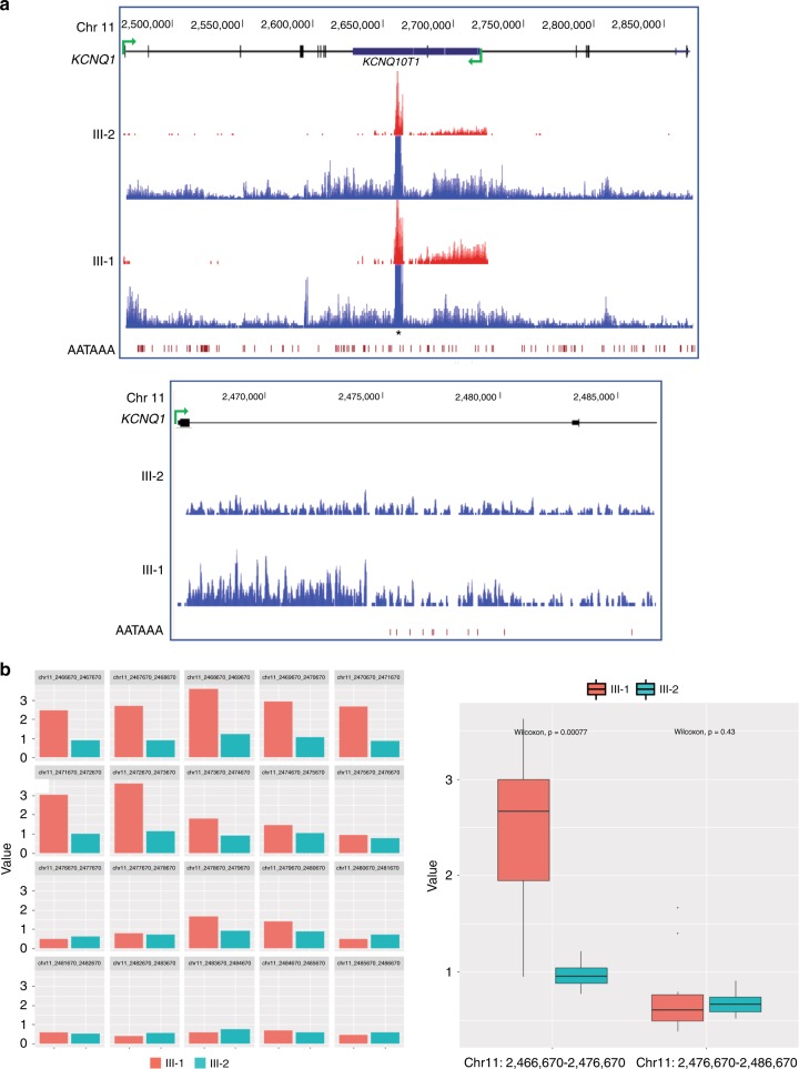
To investigate the functional effect of the splice-site variant, we determined the transcription profile of KCNQ1 on total RNA from blood leukocytes of the proband (III-1) and her half-sister (III-2) by genome RNA sequencing. This analysis indicated that, although the overall expression of KCNQ1 was lower in the proband (III-1) than in her half-sister (Figure S5), the RNA level was not homogeneously distributed along the KCNQ1 gene in III-1, with the most 5’ 10 kb about two to threefold more abundant than the rest of the gene, while RNA level was homogeneously distributed in III-2 (Fig. 3a, b). Interestingly, a cluster of poly-A addition sites is present immediately downstream of the position of KCNQ1 intron 1, in which the RNA level declines, suggesting a premature termination of transcription in III-1 (Fig. 3a). Finally, consistent with IC2 LOM, KCNQ1OT1 RNA level was increased in the proband with respect to her sister (Fig. 3a).
Fig. 3.
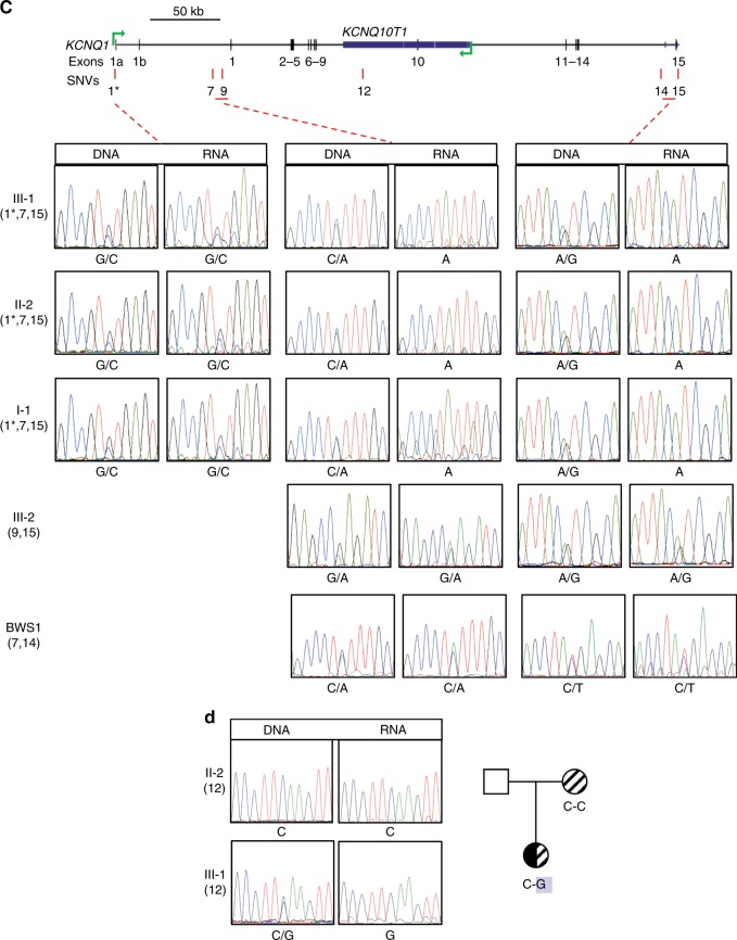
Effect of the splice-site variant on KCNQ1 expression. (a) Transcription profile of KCNQ1 in III-1 and III-2 obtained by RNAseq on total RNA from blood leukocytes. Top: screenshot from University of California–Santa Cruz (UCSC) Genome Browser showing the RNA transcribed from the positive (blue) and negative (red) strands of the KCNQ1 locus. A track showing the location of AATAAA poly-A addition sites is reported below the RNA tracks. The peak of reads on both strands in the middle of the gene (indicated by an asterisk) corresponds to an L1 repeat. Bottom: zoom in of the first 5’ 20 kb of KCNQ1. (b) Observed increased expression of the most 5’ 10 kb of KCNQ1 in the proband (III-1) compared with her sister (III-2). Expression profile of the most 5’ 20 kb (2466670-2486670) represented as normalized read count partitioned in windows of 1 kb (bar plots on the left) or normalized read count distribution in two windows of 10 kb (box plots on the right) of III-1 and III-2. Y-axis values in bar plots are absolute counts in millions normalized by the number of uniquely mapped reads. Box plots show a statistically significant enrichment of the coverage in the first 10 kb (2466670-2476670) of III-1 compared with III-2, but similar coverages in the following 10 kb (2476670-2486670). (c) Allele-specific expression analysis of KCNQ1 in III-1, II-2, I-1, and III-2 of family 2 and an unrelated Beckwith–Wiedemann syndrome (BWS) case with IC2 loss of methylation (LOM), analyzed as in Fig. 1f. A schematic diagram of KCNQ1 is reported at the top to indicate the positions of the analyzed single-nucleotide variants (SNVs) (red vertical lines). SNV codes are listed in Table S2. The electropherograms are organized in three panels corresponding to SNVs located in different parts of the KCNQ1 gene. The numbers in parentheses at the left of the electropherograms represent the SNVs analyzed in each individual. The splice-site variant described in Fig. 2c is indicated by an asterisk. (d) Allele-specific expression analysis of KCNQ1 to determine the parental origin of the transcribed allele in III-1. The typed SNV (SNV12 = rs7942590 G/C) falls in the part of the KCNQ1 gene that is expressed from only one allele. The boxed G represents the only allele present in the RNA of III-1.
To confirm the hypothesis of defective KCNQ1 elongation in the proband, allele-specific expression of KCNQ1 was analyzed in total RNA from peripheral blood leukocytes, by typing SNVs present in heterozygosity at different positions of the primary transcript (Fig. 3c). By typing the intron 1 splice-site variant (SNV1), we found biallelic KCNQ1 expression in the proband, as well as in II-2 and I-1 (Fig. 3c, left panels). In contrast, transcription from only one allele was found in the same individuals, by typing SNVs in the 3’ part of intron 1 (SNV7, middle panels) and in the 3’ untranslated region (UTR) (SNV15, right panels) of the KCNQ1 gene. III-2 has not inherited the splice-site variant and therefore was not informative at this position. However, she showed biallelic KCNQ1 expression in both intron 1 (SNV9, middle panel) and 3’ UTR (SNV15, right panel). Similarly, KCNQ1 was transcribed from both parental alleles in intron 1 (SNVs 6–9, middle panel of Fig. 3c and left panel of Figure S3) and intron 14 (SNVs 13–14, right panels of Fig. 3c and Figure S3) in three age-matched healthy individuals (Ctrl 1–3) and four further BWS patients with IC2 LOM (BWS 1–4). To determine the parental origin of the full-length KCNQ1 RNA in the individuals carrying the splice-site variant, we typed a SNV (SNV12) of intron 9 that is present in heterozygosity in the proband and in homozygosity in her mother (II-2). Only the G allele that is absent in the mother was amplified from III-2 RNA, indicating that the splice pathogenic variant is associated with the defective transcript (Fig. 3d).
To better define the position of the KCNQ1 gene in which expression shifts from two alleles to one allele in the individuals carrying the splice-site variant, we typed two further SNVs (4 and 5) that were located in the 5’ part of intron 1 (Figure S4). Both SNV4 and SNV5 were informative in the maternal grandfather (I-1). Expression was found biallelic at SNV4 but monoallelic at SNV5 in this individual. Only one allele was transcribed at SNV5 also in the mother (II-2) and proband (III-1), while both parental alleles were expressed in two informative healthy individuals (Ctrl 4 and 5). Consistent with the RNAseq data, these results demonstrated that expression shifts from two alleles to one allele between 6.6 (SNV4, chr11: 2473311) and 26 kb (SNV5, chr11: 2492629) downstream of the splice-site variant (Figure S4).
Overall, these data demonstrate defective expression of the KCNQ1 transcript downstream of the splice-site variant and complete absence of methylation at IC2 on the maternal chromosome, suggesting that premature transcription termination upstream of IC2 did not allow to properly establish the methylation imprint in the germline of the proband’s mother leading to an imprinting error in the embryo.
LQTS in a BWS family with a 160-kb KCNQ1 duplication and complete IC2 LOM
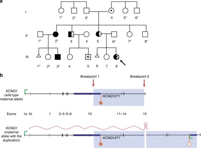
To identify asymptomatic cases of LQTS in an Italian BWS cohort, ECG was evaluated in 24 patients with IC2 LOM. A prolonged QTc value diagnostic of LQTS was found in only one patient (patient 24 in Table S1). The proband (III-8 in Fig. 4a) was part of a previously described family, including several other individuals with BWS features, and in which segregates a 160-kb KCNQ1 duplication (Fig. 4a, b, and Chiesa et al.16). Two apparently healthy children (III-4 and III-5) of II-3, not born at the time of the first study, were added to the present study. The duplication was confirmed in III-5, but excluded in III-4 (Figure S6). Further clinical examination of the family showed borderline prolonged QTc values in all maternal and paternal duplication carriers who accepted the ECG exam including III-5 (Fig. 4a). This demonstrates that LQTS cosegregates with the duplication on both maternal and paternal transmission, while BWS does it only on maternal transmission. Importantly, the original study also showed that maternal transmission of the duplication was associated with complete loss of DNA methylation of the duplicated IC2, leading to activation of a truncated form of KCNQ1OT1 and silencing of CDKN1C.16 To test if the methylation defect was extended to other ICRs or restricted to IC2 in this family, five loci that are frequently affected in MLID17 were analyzed. The methylation values of all tested ICRs were comparable with the values of healthy individuals, in II-3, II-5, III-5, and III-8, indicating that the epigenetic defect was restricted to IC2 (Table S4).
Fig. 4.
Segregation of long QT syndrome (LQTS) and Beckwith–Wiedemann syndrome (BWS) phenotypes in a family with a 160-kb KCNQ1 duplication. a Pedigree of family 3, modified from Chiesa et al.16 The proband (III-8) affected by both BWS and LQTS1 is indicated as in Fig. 1. The gray stripes in II-3, II-5, and III-5 indicate borderline prolonged QTc values. Asterisks: family members unavailable for molecular analysis. b Schematic representation of the 160-kb KCNQ1 duplication. The light blue rectangle highlights the region involved in the duplication. Breakpoint 1 is telomeric; breakpoint 2, centromeric. The position of the inverted duplication in the centromeric breakpoint and the the stem-and-loop structure in the KCNQ1 transcript are proposed to explain the molecular data.
These results demonstrate that a pathogenic variant within KCNQ1 segregates with BWS and complete IC2 LOM on maternal transmission and with LQTS on both maternal and paternal transmission.
DISCUSSION
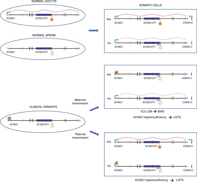
In rare cases, DNA methylation defects in BWS occur secondary to genetic variants acting in cis or in trans and are associated with higher recurrence risk.17–20 The identification of these genetic variants is therefore important for genetic counseling. However, identifying variants associated with IC2 LOM in cis is challenging, because of the large extension of the centromeric 11p15.5 imprinted domain. By looking for the presence of LQTS features within three BWS cohorts, we identified three cases in which complete IC2 LOM occurs secondary to cis-acting DNA variants in the KCNQ1 gene. In all three cases, the genetic variants cosegregate with BWS on maternal transmission and in at least two cases were demonstrated to affect KCNQ1 transcription upstream of IC2, suggesting that lack of transcription across this ICR leads to LOM, likely because of defective de novo methylation in oocytes (Fig. 5).
Fig. 5.
Model of how a single genetic KCNQ1 variant may determine both long QT syndrome (LQTS) and Beckwith–Wiedemann syndrome (BWS). In the female germline, the KCNQ1 promoter drives a transcript across IC2, which is required for de novo methylation of this imprinting control region (ICR). Differential IC2 methylation regulates KCNQ1OT1 and CDKN1C imprinting in somatic cells. Genetic variants causing loss or premature termination of KCNQ1 transcription result in (1) BWS due to defective IC2 methylation establishment, biallelic KCNQ1OT1 expression, and CDKN1C silencing, and LQTS due to KCNQ1 haploinsufficiency, on maternal transmission; (2) LQTS due to KCNQ1 haploinsufficiency on paternal transmission. Active promoters and transcription orientation are indicated by bent green arrows. Methylated IC2 is indicated by filled lollipop, unmethylated IC2 by open lollipop. The KCNQ1 transcript is depicted as a pink curved line. The dashed curved line indicates the paternal KCNQ1 transcript that is expressed in heart and adult tissues and imprinted in embryo. Genetic variants causing loss of KCNQ1 transcription are indicated by a red cross. The KCNQ1 gene is represented nonimprinted in somatic cells, according to the results obtained in blood leukocytes.
LQTS and BWS with complete IC2 LOM associated with a transcription defect of KCNQ1
The pathogenic variant found in the proband of family 1 removes the promoter and causes loss of both main KCNQ1 transcript isoforms on the maternal chromosome. The complete absence of IC2 methylation in the proband indicates that this epimutation arose as a consequence of defective imprint establishment in the maternal germline. Consistent with the role of Kcnq1 transcription in de novo IC2 methylation demonstrated in mouse,11 it is possible that this imprinting defect derived from lack of KCNQ1 transcription in the oocyte. Loss of expression of one allele also results in KCNQ1 haploinsufficiency and this is the likely cause of LQTS in this individual. A deletion of similar extension on chromosome 11p15 but associated with more complex rearrangements has been recently described in a family with multiple offspring with large omphalocele and complete IC2 LOM, which was incompatible with life.21 Similarly to our case, this epimutation could be originated as a consequence of defective KCNQ1 transcription and imprint establishment.
LQTS and BWS with complete IC2 LOM associated with a splice-site variant and premature termination of KCNQ1 transcript
Although several KCNQ1 splice-site variants have been identified in LQTS22,23 (https://www.ensembl.org/Homo_sapiens/Transcript/ProtVariations?db=core;g=ENSG00000053918;r=11:2444684-2849110;t=ENST00000155840), our case 2 splicing defect is the only one, to our knowledge, that has ever been reported as associated with BWS. Often, abnormal splicing leads to premature translation termination and messenger RNA (mRNA) degradation by nonsense-mediated decay.24 However, in the case of this splice-site variant, the twofold reduction and the shift from two alleles to one allele in KCNQ1 expression between the variant and a cluster of poly-A sites in intron 1 suggests a rather different mechanism, in which premature transcription termination occurs in the KCNQ1 gene.
It is known that pre-mRNA's processing of eukaryotic genes including splicing occurs cotranscriptionally.25 Several studies have demonstrated that the splicing apparatus stimulates transcription elongation through direct interaction with elongation factors, suggesting that checkpoint mechanisms ensure that only spliced transcripts are efficiently elongated.26–28 Consistent with this hypothesis, knockdown of U1 small nuclear ribonucleoprotein (snRNP) results, in addition to splicing inhibition, in premature cleavage of pre-mRNAs at cryptic polyadenylation signals in introns located near the transcription start site of large genes.29–31 Intriguingly, in our case, the KCNQ1 splice-site variant alters the 5’ splice site that normally pairs with the U1 snRNP, suggesting that weakening this binding causes termination, cleavage, and polyadenylation of the promoter 1a–derived transcript upstream of IC2. Because 1a is an active KCNQ1 promoter in most fetal tissues,8 we expect that the exon 1a–derived transcript is prematurely terminated in oocytes and this possibly results in failure of IC2 methylation establishment in the maternal germline. Further experiments, such as 3’-RACE, may be performed in the future to more precisely determine the transcription termination of KCNQ1 relative to the cryptic poly-A addition site in intron 1, in the present case.
In family 2, the BWS phenotype is associated with the splice-site variant on maternal transmission, while LQTS occurs on both maternal and paternal transmission. This is consistent with the maternal methylation of IC2 and the lack of imprinting of KCNQ1 in heart. KCNQ1 haploinsufficiency is expected from incomplete elongation of the transcript caused by the splice-site variant. Consistent with our findings, exon 1a pathogenic variants have been reported associated with LQTS.22,32 The absence of BWS features including normal IC2 methylation in the maternal grandfather suggests that the variant is present on his paternal chromosome, while the absence of LQTS in this individual is probably explained by incomplete penetrance of the variant.33 The multiple miscarriages, including one fetus with a large omphalocele, are reminiscent of what was described in a family with KCNQ1 deletion,21 and suggest that the proband’s aunt is a silent carrier of the splice-site variant and that the phenotypes of the progeny are the result of combined KCNQ1 haploinsufficiency and IC2 imprinting defect.
LQTS and BWS with complete IC2 LOM associated with a 160-kb KCNQ1 duplication
The proband of family 3 scored positive for prolonged QTc value in an ECG screening offered to all BWS patients with IC2 LOM of an Italian cohort (Table S1). Following this result, three further members of this family were found to have prolonged QTc value. All these individuals carry a previously described 160-kb internal KCNQ1 duplication segregating with BWS with IC2 LOM on maternal transmission.16 We now find that this pathogenic variant also causes overt LQTS or prolonged QTc value upon either maternal and paternal transmission. Unfortunately, the lack of informative SNVs in the 3’ part of KCNQ1 prevented investigation of allelic expression in this family. However, we originally demonstrated that one of the duplicated copies of IC2 is completely nonmethylated on the maternal chromosome and that the duplication is inverted in cis (Fig. 4b and Chiesa et al.16). Here, we propose that KCNQ1 transcription is arrested before reaching the duplicated IC2, because of the formation of a stem-and-loop secondary structure of the transcript caused by the inverted duplication and this in turn causes a failure of imprint establishment in the maternal germline, similarly to families 1 and 2 (Fig. 4). Indeed, stem-and-loop-dependent mechanisms of transcription termination have been described in mammals.34 Further studies are needed, however, to directly demonstrate the role of this secondary structure in transcription termination of KCNQ1 in the present case. Interestingly, a case of BWS associated with a maternally inherited KCNQ1 exon1c-1 duplication and complete IC2 LOM, which could also be caused by premature transcription termination, has been reported by Demars et al.35
This is the first case of LQTS associated with a KCNQ1 duplication. The cardiac phenotype may be caused by either (1) truncated KCNQ1 protein lacking the amino acids encoded by last exon; or (2) degradation of the transcript lacking the normal poly-A signal. Consistent with the first mechanism, a truncating pathogenic variant close to the last KCNQ1 exon has been reported associated with LQTS36 (https://www.ensembl.org/Homo_sapiens/Transcript/ProtVariations?db=core;g=ENSG00000053918;r=11:2444684-2849110;t=ENST00000155840).
LQTS in BWS patients with IC2 LOM
Congenital disorders of the heart rhythm, such as LQTS, have been rarely reported in BWS and generally associated with maternal chromosome 11p15 rearrangements.37,38 In one of these cases,38 the proband carries a translocation with a breakpoint between KCNQ1 exons 2 and 10 and shows IC2 LOM, which could indeed be caused by defective transcription across IC2. In this study, we identified three cases with combined BWS and LQTS related to three different genetic/genomic lesions leading to a common epigenotype derangement. Although further studies are needed to clarify the exact prevalence, the ECG screening performed in the Italian cohort indicates a low incidence of LQTS among the BWS patients with IC2 LOM, confirming the relatively rare overlap of these two diseases. The BWS guidelines recommend that physicians should be aware of the increased prevalence of cardiac anomalies in children with BWS and indicate annual evaluation and electrocardiogram in patients with genomic rearrangements involving the IC2 region. However, because of the incomplete penetrance of KCNQ1 variants and the associated high risk of major cardiac events in adult age,23,33 an ECG (and if positive further KCNQ1 analysis) should be performed in all BWS patients with IC2 LOM.
In summary, this study proposes a new molecular mechanism, in which two diseases, LQTS and BWS, share the same genetic defect.39,40 We demonstrate that, by affecting KCNQ1 transcription, genetic variants may result in KCNQ1 haploinsufficiency and LQTS, and at the same time we suggest these lesions may interfere with de novo IC2 methylation in the oocyte and cause BWS later in development. As consequence of the differential imprinting status of KCNQ1OT1 and KCNQ1, BWS occurs only on maternal transmission of the variants while no parent-of-origin-dependent effect is observed with LQTS (Fig. 5).
Supplementary information
Acknowledgements
We are very grateful to the patients and their families for their collaboration. This work was supported by Associazione Italiana Ricerca sul Cancro (IG 2016 N.18671 to A.R.), Telethon-Italia (GGP15131 to A.R.), MIUR-CNR Flag Project Epigen (to A.R.), EU-FP7-ITN INGENIUM 290123 (to A.R.), and MIUR-PRIN 2015 JHLY35 (to A.R. and F.C.).
Disclosure
The authors declare no conflict of interest.
Footnotes
Publisher’s note: Springer Nature remains neutral with regard to jurisdictional claims in published maps and institutional affiliations.
Contributor Information
Andrea Riccio, Email: andrea.riccio@unicampania.it.
Flavia Cerrato, Email: flavia.cerrato@unicampania.it.
SUPPLEMENTARY INFORMATION
The online version of this article (10.1038/s41436-018-0416-7) contains supplementary material, which is available to authorized users.
References
- 1.Schwartz PJ, Stramba-Badiale M, Crotti L, et al. Prevalence of the congenital long-QT syndrome. Circulation. 2009;120:1761–1767. doi: 10.1161/CIRCULATIONAHA.109.863209. [DOI] [PMC free article] [PubMed] [Google Scholar]
- 2.Hedley PL, Jørgensen P, Schlamowitz S, et al. The genetic basis of long QT and short QT syndromes: a mutation update. Hum Mutat. 2009;30:486–511. doi: 10.1002/humu.21106. [DOI] [PubMed] [Google Scholar]
- 3.Tester DJ, Ackerman MJ. Genetics of long QT syndrome. Methodist Debakey Cardiovasc J. 2014;10:29–33. doi: 10.14797/mdcj-10-1-29. [DOI] [PMC free article] [PubMed] [Google Scholar]
- 4.Mussa A, Russo S, De Crescenzo A, et al. Prevalence of Beckwith-Wiedemann syndrome in North West of Italy. Am J Med Genet A. 2013;161:2481–2486. doi: 10.1002/ajmg.a.36080. [DOI] [PubMed] [Google Scholar]
- 5.Brioude F, Kalish JM, Mussa A, et al. Expert consensus document: clinical and molecular diagnosis, screening and management of Beckwith-Wiedemann syndrome: an international consensus statement. Nat Rev Endocrinol. 2018;14:229–249. doi: 10.1038/nrendo.2017.166. [DOI] [PMC free article] [PubMed] [Google Scholar]
- 6.Baskin B, Choufani S, Chen YA, et al. High frequency of copy number variations (CNVs) in the chromosome 11p15 region in patients with Beckwith-Wiedemann syndrome. Hum Genet. 2014;133:321–330. doi: 10.1007/s00439-013-1379-z. [DOI] [PubMed] [Google Scholar]
- 7.Mussa A, Russo S, Larizza L, et al. (Epi)genotype-phenotype correlations in Beckwith-Wiedemann syndrome: a paradigm for genomic medicine. Clin Genet. 2016;89:403–415. doi: 10.1111/cge.12635. [DOI] [PubMed] [Google Scholar]
- 8.Lee MP, Hu RJ, Johnson LA, et al. Human KVLQT1 gene shows tissue-specific imprinting and encompasses Beckwith-Wiedemann syndrome chromosomal rearrangements. Nat Genet. 1997;15:181–185. doi: 10.1038/ng0297-181. [DOI] [PubMed] [Google Scholar]
- 9.Neyroud N, Richard P, Vignier N, et al. Genomic organization of the KCNQ1 K+channel gene and identification of C-terminal mutations in the long-QT syndrome. Circ Res. 1999;84:290–297. doi: 10.1161/01.RES.84.3.290. [DOI] [PubMed] [Google Scholar]
- 10.Baran Y, Subramaniam M, Biton A, et al. The landscape of genomic imprinting across diverse adult human tissues. Genome Res. 2015;25:927–936. doi: 10.1101/gr.192278.115. [DOI] [PMC free article] [PubMed] [Google Scholar]
- 11.Singh VB, Sribenja S, Wilson KE, et al. Blocked transcription through KvDMR1 results in absence of methylation and gene silencing resembling Beckwith-Wiedemann syndrome. Development. 2017;144:1820–1830. doi: 10.1242/dev.145136. [DOI] [PMC free article] [PubMed] [Google Scholar]
- 12.Chotalia M, Smallwood SA, Ruf N, et al. Transcription is required for establishment of germline methylation marks at imprinted genes. Genes Dev. 2009;23:105–117. doi: 10.1101/gad.495809. [DOI] [PMC free article] [PubMed] [Google Scholar]
- 13.Goldenberg I, Moss AJ, Zareba W. QT interval: how to measure it and what is “normal”. J Cardiovasc Electrophysiol. 2006;17:333–336. doi: 10.1111/j.1540-8167.2006.00408.x. [DOI] [PubMed] [Google Scholar]
- 14.Goldenberg I, Horr S, Moss AJ, et al. Risk for life-threatening cardiac events in patients with genotype-confirmed long-QT syndrome and normal-range corrected QT intervals. J Am Coll Cardiol. 2011;57:51–59. doi: 10.1016/j.jacc.2010.07.038. [DOI] [PMC free article] [PubMed] [Google Scholar]
- 15.Schwartz PJ, Garson AJ, Paul T, et al. Guidelines for the interpretation of the neonatal electrocardiogram. A task force of the European Society of Cardiology. Eur Heart J. 2002;23:1329–1344. doi: 10.1053/euhj.2002.3274. [DOI] [PubMed] [Google Scholar]
- 16.Chiesa N, De Crescenzo A, Mishra K, et al. The KCNQ1OT1 imprinting control region and non-coding RNA: new properties derived from the study of Beckwith-Wiedemann syndrome and Silver-Russell syndrome cases. Hum Mol Genet. 2012;21:10–25. doi: 10.1093/hmg/ddr419. [DOI] [PMC free article] [PubMed] [Google Scholar]
- 17.Docherty LE, Rezwan FI, Poole RL, et al. Mutations in NLRP5 are associated with reproductive wastage and multilocus imprinting disorders in humans. Nat Commun. 2015;6:8086. doi: 10.1038/ncomms9086. [DOI] [PMC free article] [PubMed] [Google Scholar]
- 18.Sparago A, Russo S, Cerrato F, et al. Mechanisms causing imprinting defects in familial Beckwith-Wiedemann syndrome with Wilms’ tumour. Hum Mol Genet. 2007;16:254–264. doi: 10.1093/hmg/ddl448. [DOI] [PubMed] [Google Scholar]
- 19.Sparago A, Cerrato F, Riccio A. Is ZFP57 binding to H19/IGF2:IG-DMR affected in Silver-Russell syndrome? Clin Epigenetics. 2018;10:23. doi: 10.1186/s13148-018-0454-7. [DOI] [PMC free article] [PubMed] [Google Scholar]
- 20.Abi Habib W, Azzi S, Brioude F, et al. Extensive investigation of the IGF2/H19 imprinting control region reveals novel OCT4/SOX2 binding site defects associated with specific methylation patterns in Beckwith-Wiedemann syndrome. Hum Mol Genet. 2014;23:5763–5773. doi: 10.1093/hmg/ddu290. [DOI] [PubMed] [Google Scholar]
- 21.Beygo J, Joksic I, Strom TM, et al. A maternal deletion upstream of the imprint control region 2 in 11p15 causes loss of methylation and familial Beckwith-Wiedemann syndrome. Eur J Hum Genet. 2016;24:1280–1286. doi: 10.1038/ejhg.2016.3. [DOI] [PMC free article] [PubMed] [Google Scholar]
- 22.Kapa S, Tester DJ, Salisbury BA, et al. Genetic testing for long-QT syndrome: distinguishing pathogenic mutations from benign variants. Circulation. 2009;120:1752–1760. doi: 10.1161/CIRCULATIONAHA.109.863076. [DOI] [PMC free article] [PubMed] [Google Scholar]
- 23.Napolitano C, Priori SG, Schwartz PJ, et al. Genetic testing in the long QT syndrome: development and validation of an efficient approach to genotyping in clinical practice. JAMA. 2005;294:2975–2980. doi: 10.1001/jama.294.23.2975. [DOI] [PubMed] [Google Scholar]
- 24.Schweingruber C, Rufener SC, Zünd D, et al. Nonsense-mediated mRNA decay—mechanisms of substrate mRNA recognition and degradation in mammalian cells. Biochim Biophys Acta. 2013;1829:612–623. doi: 10.1016/j.bbagrm.2013.02.005. [DOI] [PubMed] [Google Scholar]
- 25.Bentley DL. Coupling mRNA processing with transcription in time and space. Nat Rev Genet. 2014;15:163–175. doi: 10.1038/nrg3662. [DOI] [PMC free article] [PubMed] [Google Scholar]
- 26.De Almeida SF, Carmo-Fonseca M. Cotranscriptional RNA checkpoints. Epigenomics. 2010;2:449–455. doi: 10.2217/epi.10.21. [DOI] [PubMed] [Google Scholar]
- 27.Fong YWand Zhou Q. Stimulatory effect of splicing factors on transcriptional elongation. Nature. 2001;414:929–933. doi: 10.1038/414929a. [DOI] [PubMed] [Google Scholar]
- 28.Kameoka S, Duque P, Konarska MM. p54(nrb) associates with the 5´ splice site within large transcription/splicing complexes. EMBO J. 2004;23:1782–1791. doi: 10.1038/sj.emboj.7600187. [DOI] [PMC free article] [PubMed] [Google Scholar]
- 29.Kaida D, Berg MG, Younis I, et al. U1 snRNP protects pre-mRNAs from premature cleavage and polyadenylation. Nature. 2010;468:664–668. doi: 10.1038/nature09479. [DOI] [PMC free article] [PubMed] [Google Scholar]
- 30.Berg MG, Singh LN, Younis I, Liu Q, Pinto AM, Kaida D, Zhang Z, Cho S, Sherrill-Mix S, Wan L, et al. U1 snRNP determines mRNA length and regulates isoform expression. Cell. 2012;150:53–64. doi: 10.1016/j.cell.2012.05.029. [DOI] [PMC free article] [PubMed] [Google Scholar]
- 31.Oh JM, Di C, Venters CC, et al. U1 snRNP telescripting regulates a size-function-stratified human genome. Nat Struct Mol Biol. 2017;24:993–999. doi: 10.1038/nsmb.3473. [DOI] [PMC free article] [PubMed] [Google Scholar]
- 32.Motoi N, Marehiko U, Ryota E, et al. A novel KCNQ1 nonsense variant in the isoform-specific first exon causes both Jervell and Lange-Nielsen syndrome 1 and long QT syndrome 1: a case report. BMC Med Genet. 2017;18:66. doi: 10.1186/s12881-017-0430-7. [DOI] [PMC free article] [PubMed] [Google Scholar]
- 33.Priori SG, Schwartz PJ, Napolitano C, et al. Risk stratification in the long-QT syndrome. N Engl J Med. 2003;348:1866–1874. doi: 10.1056/NEJMoa022147. [DOI] [PubMed] [Google Scholar]
- 34.Marzluff WF, Koreski KP. Birth and death of histone mRNAs. Trends Genet. 2017;33:745–759. doi: 10.1016/j.tig.2017.07.014. [DOI] [PMC free article] [PubMed] [Google Scholar]
- 35.Demars J, Rossignol S, Netchine I, et al. New insights into the pathogenesis of Beckwith-Wiedemann and Silver-Russell syndromes: contribution of small copy number variations to 11p15 imprinting defects. Hum Mutat. 2011;32:1171–1182. doi: 10.1002/humu.21558. [DOI] [PubMed] [Google Scholar]
- 36.Kapplinger JD, Tester DJ, Salisbury BA, et al. Spectrum and prevalence of mutations from the first 2,500 consecutive unrelated patients referred for the FAMILION long QT syndrome genetic test. Heart Rhythm. 2009;6:1297–1303. doi: 10.1016/j.hrthm.2009.05.021. [DOI] [PMC free article] [PubMed] [Google Scholar]
- 37.Gurrieri F, Zollino M, Oliva A, et al. Mild Beckwith-Wiedemann and severe long-QT syndrome due to deletion of the imprinting center 2 on chromosome 11p. Eur J Hum Genet. 2013;21:965–969. doi: 10.1038/ejhg.2012.280. [DOI] [PMC free article] [PubMed] [Google Scholar]
- 38.Kaltenbach S, Capri Y, Rossignol S, et al. Beckwith-Wiedemann syndrome and long QT syndrome due to familial-balanced translocation t(11;17)(p15.5; q21.3) involving the KCNQ1 gene. Clin Genet. 2013;84:78–81. doi: 10.1111/cge.12038. [DOI] [PubMed] [Google Scholar]
- 39.Boonen SE, Freschi A, Christensen R, et al. Two maternal duplications involving the CDKN1C gene are associated with contrasting growth phenotypes. Clin Epigenetics. 2016;8:69. doi: 10.1186/s13148-016-0236-z. [DOI] [PMC free article] [PubMed] [Google Scholar]
- 40.Tarazona S, Furió-Tarí P, Turrà D, et al. Data quality aware analysis of differential expression in RNA-seq with NOISeq R/Bioc package. Nucleic Acids Res. 2015;43:e140. doi: 10.1093/nar/gkv711. [DOI] [PMC free article] [PubMed] [Google Scholar]
Associated Data
This section collects any data citations, data availability statements, or supplementary materials included in this article.


