Short abstract
Body mass index is a common and well-known measure in daily life. A body mass index higher than 25 is assumed to be an indicator for overweight and obesity and a high amount of total body fat. But body mass index overestimates body fat in subjects with high muscle mass and underestimates it in persons with a low lean body mass, especially in elderly and diseased persons. In the present study, we investigate the performance of the body mass index as a measure of body fatness and its ability to distinguish between well-trained and untrained subjects. Twenty-one well-trained male members of a police task force named “Cobra” and 38 non-active controls, matched by age, weight and height were participants of the study. The age range of these subjects was between 30 and 45 years. Subcutaneous adipose tissue thicknesses and body fat distributions were measured non-invasively by an optical device named the “Lipometer.” Statistics were performed with SPSS. We found that the body mass index did not show a difference between the two groups, whereas all Lipometer results were able to discriminate significantly between the trained and untrained subjects. Furthermore, the receiver operating characteristic curve analysis was calculated and all Lipometer measurements provided significant results up to a correct classification of all subjects of 86.4%, which was for the lateral thigh body site. In conclusion, the body mass index was not able to recognize the difference between trained and untrained participants, while body fat distribution measured with the Lipometer was able to distinguish more clearly the large body fat differences between these two groups.
Impact statement
Body mass index (BMI) is a common measure of body fatness but overestimates body fat in subjects with high muscle mass. We have developed previously a device named “Lipometer,” an alternative way to measure body fatness. We show herein that the Lipometer is able to distinguish more clearly (than the BMI) the large body fat differences between well-trained and untrained subjects. Thus, the Lipometer is superior to BMI with respect to body fat measurements.
Keywords: Body mass index, Lipometer, Cobra, subcutaneous fat, obesity, body fat measurement
Introduction
Higher living standards and subsequently increased access to relatively cheap high-density foods are accompanied by a higher proportion of overweight people in contemporary society. Along with an increase in caloric consumption, a general lack of physical activity also contributes to obesity and chronic diseases like the metabolic syndrome and cardiovascular diseases, which can affect people’s quality of life. To detect and measure obesity and overweight in human populations, different methods have been suggested. In 1985, Garrow established the body mass index (BMI, kg/m2) as a measure of weight classification (underweight through to morbid obesity), and nowadays, the BMI is used ubiquitously in clinical and sport practice to determine optimal body weight.1 Because of its simplicity, the BMI has become the most commonly used measurement of fatness and obesity. For example, searching for papers in the Web of Science (Core collection) using the terms “body mass index” as keywords we retrieved substantially more results (about 180,000 hits) compared with other fatness and obesity measurement methods, including Dual X-Ray Absorptiometry (DXA) (approximately 10,000 hits), Skinfold Calipers (5000 hits), and Lipometer (50 hits).
Given its popularity, it seems that many researchers have accepted the BMI as a relatively accurate measure of body fatness. However, previous research indicates that the association between BMI and levels of body fat is not strong and that BMI is unable to distinguish between tissue type (adipose versus non-adipose) or indicate fat distribution throughout the body.2 It is understood that total body fat is an important risk factor for a number of diseases; however, studies have also shown that central adiposity (an indicator of visceral fat) is a greater risk factor for obesity-related disorders compared with total body fatness.3 Indeed, a number of diseases including type 2 diabetes,4,5 arteriosclerosis,6 coronary heart diseases7 and polycystic ovary syndrome8 are associated with a typical body fat distribution with thicker adipose tissue layers at the trunks of the patients (apple-like body fat distribution), which is ignored by the BMI measurement. Furthermore, the use of the BMI is regarded critically in athletic populations performing strength training.9 For example, body builders with a high amount of muscle mass can have a BMI of >30 kg/m2, but a total body fat of about 6%.10
The optical device named the “Lipometer,” patented and developed at the Medical University of Graz, enables a non-invasive, precise, and quick measurement of the thickness of the subcutaneous adipose tissue (SAT) layers at any site of the human body.11,12 Lipometer measurements at 15 well-defined body sites from neck to calf show the individual body fat distribution of a subject, the so-called “subcutaneous adipose tissue topography” (SAT-Top).13 Previously, a comparison between BMI and the topography of subcutaneous adipose tissue layers in young athletes (about 20–30 years old) and age-matched peers suggested that subcutaneous fat patterns were a better screening tool to characterize fatness in physically active people of this age class compared with the BMI.14,15
In this study, we investigate for the first time the role of BMI versus Lipometer-indicated fat patterns in a higher age class of 30 to 45-year-old men. We present a comparison of well-trained men employed at a special Austrian police task force called “Cobra” with an untrained control group of comparable age, height, and weight. Our hypothesis is that the BMI will not be a good screening tool of body fatness for these subjects: BMI will be unable to distinguish between the two groups, thereby treating very well trained men with healthy fat mass the same as non-trained men with substantially higher and unhealthy fat mass.
Subjects and methods
Subjects
Twenty-one male members of a well-trained police unit named “Cobra” and 38 non-active controls of comparable age, height, and weight were recruited by personal invitation to participate in this cross-sectional study. The participants were between the ages of 30 and 45 years and were measured in light clothing without shoes. A portable calibrated stadiometer (SECA®-220, Hamburg, Germany) was used to measure standing height to the nearest 0.1 cm, and body mass was determined to the nearest 0.01 kg using calibrated scales (Soehnle® 7700, Murrhardt, Germany). Finally, the BMI was calculated for all subjects (BMI = body mass (kg)/height (m)2). The participants of the study were thoroughly informed about the measurements and gave their written informed consent. The study protocol was designed under the Code of Ethics according to the Declaration of Helsinki and approved by the Ethic Committee of the Medical University of Graz (NCT01474629).
COBRA men
The special forces Cobra unit is one of the most effective anti-terrorism and violence suppression forces in the world. Cobra squad members need to have high levels of mental resilience and physiological fitness to undergo the demanding training routines that involve up to 6 h of heavy exercise on at least five days per week. Long endurance runs with interval training, swimming climbing, and weapons training are practiced daily. The members of this task force require high levels of mental toughness along with their high levels of physical fitness because of the mental and physical stress involved in subduing criminals under high danger equipped with heavy protective clothing.
Male control group
Thirty-eight men aged between 30 and 45 years were recruited via advertisements at health fairs. The men were currently taking no medication, were non-smokers and untrained (performing not more than 1 h of exercise per week).
Measurement of SAT-Top
Subcutaneous adipose tissue thickness in mm was measured by the patented optical device Lipometer (EU patent no. 0516251) on 15 anatomically well-defined body sites from neck to calf on the right side of the subject’s body,13,16 while the subjects were in an upright standing position. All measurements were performed once by a qualified technician. Consequently, a detailed SAT profile of the human body was obtained. The optical measurement process of the Lipometer device has been described previously.16,17
Statistics
IBM SPSS Statistics 24 (IBM, New York, USA) was applied for statistical calculations. To get a measure of regional body fat mass, additional compartment variables were calculated from the individual Lipometer site thicknesses for arms (sum of biceps and triceps), trunk (sum of neck, upper back, front chest, lateral chest), abdomen (sum of upper abdomen, lower abdomen, lower back, hip), and legs (sum of front thigh, lateral thigh, rear thigh, inner thigh, calf). To give information about the total amount of subcutaneous fat, the sum of all 15 SAT thicknesses was calculated as “Total SAT.” Body fat percentage (BF %) was obtained from an equation published by Toivo Jürimäe et al.11 using DXA as a prediction method. The normal distribution of the variables was tested using the Kolmogorov-Smirnov and the Shapiro-Wilk tests. Differences in the distributions of variables between Cobra men and control males were tested by a Student’s t-test for two independent samples (in case of normally distributed variables) and by a Mann-Whitney U-test for two independent samples (if variables were not normally distributed). To investigate the discrimination power of each Lipometer thickness value, receiver operating characteristic (ROC) curve analysis was applied, providing sensitivity, specificity, an area index, the optimal cut-off value, and the correctly classified cases.14,18
Results
Significant deviation from normal distribution suggested to present descriptive statistics as median, minimum, and maximum. Descriptive statistics of 21 trained Cobra men and 38 untrained controls are presented in Table 1.
Table 1.
Descriptive statistics (median (minimum–maximum)) of the two male groups matched by age, height, and weight.
| Personal | Controls | COBRA men | Difference | Significancea |
|---|---|---|---|---|
| parameters | (N = 38) | (N = 21) | ||
| Age (y) | 36.7 (29.8–45.2) | 38.3 (30.0–45.1) | +4.4% | n.s.b,c |
| Height (m) | 1.83 (1.73–1.91) | 1.85 (1.73–1.92) | +1.1% | n.s.c |
| Weight (kg) | 81.3 (65.0–94.0) | 84.0 (66.0–97.0) | +3.3% | n.s.c |
| BMI (kg/m2) | 24.6 (21.4–29.0) | 24.3 (19.7–28.4) | –1.2% | n.s.c |
| SAT-Top (mm)d | ||||
| Neck | 8.6 (3.0–15.7) | 5.9 (2.7–11.4) | –31.4% | p = 0.001c |
| Triceps | 6.0 (1.9–11.7) | 4.5 (1.8–12.1) | –25.0% | p = 0.039 |
| Biceps | 4.0 (1.6–9.3) | 2.5 (1.6–6.2) | –37.5% | p < 0.001 |
| Upper back | 5.7 (2.2–10.4) | 3.5 (1.9–10.1) | –38.6% | p = 0.002 |
| Front chest | 7.6 (1.7–14.7) | 3.5 (1.7–9.5) | –53.9% | p < 0.001 |
| Lateral chest | 9.8 (1.7–16.7) | 3.9 (2.0–9.9) | –60.2% | p < 0.001 |
| Upper abdomen | 12.3 (2.6–22.1) | 6.2 (1.8–13.7) | –49.6% | p < 0.001c |
| Lower abdomen | 11.5 (2.4–21.5) | 6.1 (2.8–10.6) | –47.0% | p < 0.001c |
| Lower back | 9.9 (3.1–18.3) | 5.4 (2.7–8.8) | –45.5% | p < 0.001c |
| Hip | 12.5 (2.1–19.4) | 7.1 (2.3–13.3) | –45.5% | p < 0.001c |
| Front thigh | 5.7 (1.7–11.2) | 3.5 (1.6–9.0) | –38.6% | p < 0.001 |
| Lateral thigh | 4.6 (1.8–12.2) | 2.0 (1.5–4.0) | –56.5% | p < 0.001 |
| Rear thigh | 3.6 (1.3–10.6) | 2.3 (1.2–4.7) | –36.1% | p < 0.001 |
| Inner thigh | 8.1 (1.2–18.3) | 5.0 (1.7–10.5) | –38.3% | p < 0.001c |
| Calf | 3.2 (1.2–7.6) | 2.0 (1.2–3.8) | –37.5% | p = 0.001 |
| Compartments (mm) | ||||
| Trunke | 32.9 (8.6–52.0) | 15.5 (8.7–33.8) | –52.9% | p < 0.001c |
| Armsf | 10.3 (3.7–18.6) | 6.2 (3.9–17.0) | –39.8% | p = 0.004 |
| Abdomeng | 47.8 (10.6–69.5) | 25.8 (10.1–42.3) | –46.0% | p < 0.001 |
| Legsh | 26.5 (9.0–59.9) | 15.1 (8.3–28.0) | –43.0% | p < 0.001 |
| Total SATi | 120.2 (33.6–179.2) | 66.5 (34.8–116.0) | –44.7% | p < 0.001c |
| BF %j | 26.5 (12.2–38.2) | 18.6 (12.8–28.1) | –29.8% | p < 0.001c |
BMI: body mass index; SAT-Top: subcutaneous adipose tissue topography; SAT: subcutaneous adipose tissue; BF: body fat.
aBy Mann-Whitney U-test.
bNot significant (p > 0.05).
cBy t-test for independent samples.
dSAT thickness of 15 body sites in mm.
eTrunk = neck + upper back + front chest + lateral chest.
fArms = triceps + biceps.
gAbdomen = upper abdomen+lower abdomen + lower back + hip.
hLegs = front thigh + lateral thigh + rear thigh + inner thigh + calf.
iTotal SAT = sum of all 15 body sites.
jBF % = body fat percentage obtained from an equation using DXA.11
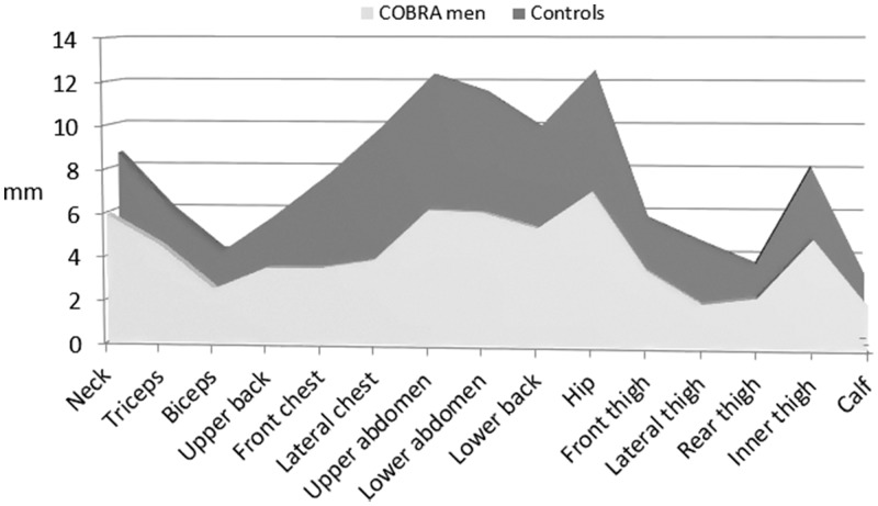
While BMI was not significantly different for the two groups, all Lipometer results were able to discriminate significantly between the trained (Cobra) and the untrained (Control) subjects. Lipometer measurements are presented in Table 1 at three levels: as SAT thicknesses at 15 body sites, as four compartment variables and as the single value Total SAT. The trained athletes of the task force Cobra showed significantly thinner adipose tissue layers on every measured body site from neck to calf compared with the untrained controls. The difference was between –25% on the triceps (p = 0.039) up to –60% (p < 0.001) on the lateral chest. Figure 1 shows the body fat profiles of the two groups.
Figure 1.
SAT-Top plot comparing the subcutaneous body fat patterns of Cobra men and their male controls. The medians of the 15 top-down sorted body sites show the SAT differences between the two male subject groups.
The highest absolute SAT difference was 6.1 mm found at the body site upper abdomen. Generally, high absolute SAT differences occurred in the middle of the profiles between the front chest body site and the hip body site (Figure 1). Similar to the individual Lipometer measurement thicknesses, the four compartment thicknesses of the Cobra men were significantly lower than the thicknesses in the control men (Table 1). Among all compartment variables, the abdomen provided the highest absolute SAT difference of –22.0 mm between the two groups (Table 1, Figure 1). Finally, Cobra men showed about 45% lower Total SAT (p < 0.001) and about 30% lower BF % (p < 0.001) compared with their untrained control group, which corresponds to absolute value of –53.7 mm Total SAT and –7.9% BF % (Table 1).
To obtain information about the discrimination power of each variable ROC curve analysis was calculated, providing the area index, sensitivity, specificity, the optimal cut-off value, and the correctly classified cases (Table 2).
Table 2.
Results obtained from ROC curve analysis for age, height, weight, BMI, 15 specified SAT-Top body sites, four compartments, and total SAT of 21 COBRA men and 38 male controls.
| Personal | Area indexa | p | Optimal cut-offb | Sensitivity | Specificity | Correctly classified cases | |
|---|---|---|---|---|---|---|---|
| parameters | H0:small | H0:large | (mm) | (%) | (%) | ||
| Age (y) | – | 0.551 | n.s.c | ||||
| Height (m) | – | 0.609 | n.s. | ||||
| Weight (kg) | – | 0.533 | n.s. | ||||
| BMI (kg/m2) | 0.561 | – | n.s. | 25.04 | 76.2 | 42.1 | 54.2% (32 of 59) |
| SAT-Topd | |||||||
| Neck | 0.721 | – | 0.05 | 9.80 | 90.5 | 47.4 | 62.7% (37 of 59) |
| Triceps | 0.664 | – | 0.039 | 4.90 | 66.7 | 76.3 | 72.9% (43 of 59) |
| Biceps | 0.824 | – | <0.001 | 2.85 | 76.2 | 84.2 | 81.4% (48 of 59) |
| Upper back | 0.748 | – | 0.002 | 4.60 | 71.4 | 76.3 | 74.6% (44 of 59) |
| Front chest | 0.777 | – | <0.001 | 5.10 | 76.2 | 71.1 | 72.9% (43 of 59) |
| Lateral chest | 0.820 | – | <0.001 | 7.55 | 85.7 | 68.4 | 74.6% (44 of 59) |
| Upper abdomen | 0.860 | – | <0.001 | 9.20 | 90.5 | 78.9 | 83.1% (49 of 59) |
| Lower abdomen | 0.853 | – | <0.001 | 9.85 | 95.2 | 71.1 | 79.7% (47 of 59) |
| Lower back | 0.828 | – | <0.001 | 8.50 | 95.2 | 65.8 | 76.3% (45 of59) |
| Hip | 0.820 | – | <0.001 | 9.45 | 90.5 | 76.3 | 81.4% (48 of 59) |
| Front thigh | 0.786 | – | <0.001 | 4.95 | 81.0 | 71.1 | 74.6% (44 of 59) |
| Lateral thigh | 0.902 | – | <0.001 | 2.70 | 81.0 | 89.5 | 86.4% (51 of 59) |
| Rear thigh | 0.776 | – | <0.001 | 3.05 | 81.0 | 65.8 | 71.2% (42 of 59) |
| Inner thigh | 0.785 | – | <0.001 | 5.90 | 71.4 | 84.2 | 79.7% (47 of 59) |
| Calf | 0.754 | – | 0.001 | 2.05 | 52.4 | 89.5 | 76.3% (45 of 59) |
| Compartments | |||||||
| Trunke | 0.798 | – | <0.001 | 25.55 | 76.2 | 71.1 | 72.9% (43 of 59) |
| Armsf | 0.726 | – | 0.004 | 7.15 | 66.7 | 89.5 | 81.4% (48 of 59) |
| Abdomeng | 0.869 | – | <0.001 | 35.90 | 90.5 | 84.2 | 86.4% (51 of 59) |
| Legsh | 0.845 | – | <0.001 | 17.35 | 71.4 | 86.8 | 81.4% (48 of 59) |
| Total SATi | 0.863 | – | <0.001 | 86.40 | 85.7 | 84.2 | 84.7% (50 of 59) |
| BF %j | 0.777 | – | <0.001 | 23.86% | 85.7 | 65.8 | 72.9% (43 of 59) |
BMI: body mass index; SAT-Top: subcutaneous adipose tissue topography; SAT: subcutaneous adipose tissue; BF: body fat.
aThere are two possible hypotheses (H0): that either small/large values provide stronger evidence for positivity.
bOptimal cut-off value estimated by Youden-Index.19
cNot significant (p > 0.05).
dSAT thickness of 15 body sites in mm.
eTrunk = neck + upper back + front chest + lateral chest.19
fArms = triceps+biceps.
gAbdomen = upper abdomen + lower abdomen + lower back + hip.
hLegs = front thigh + lateral thigh + rear thigh + inner thigh + calf.
iTotal SAT = sum of all 15 body sites.
jBF % = body fat percentage obtained from an equation using DXA.11
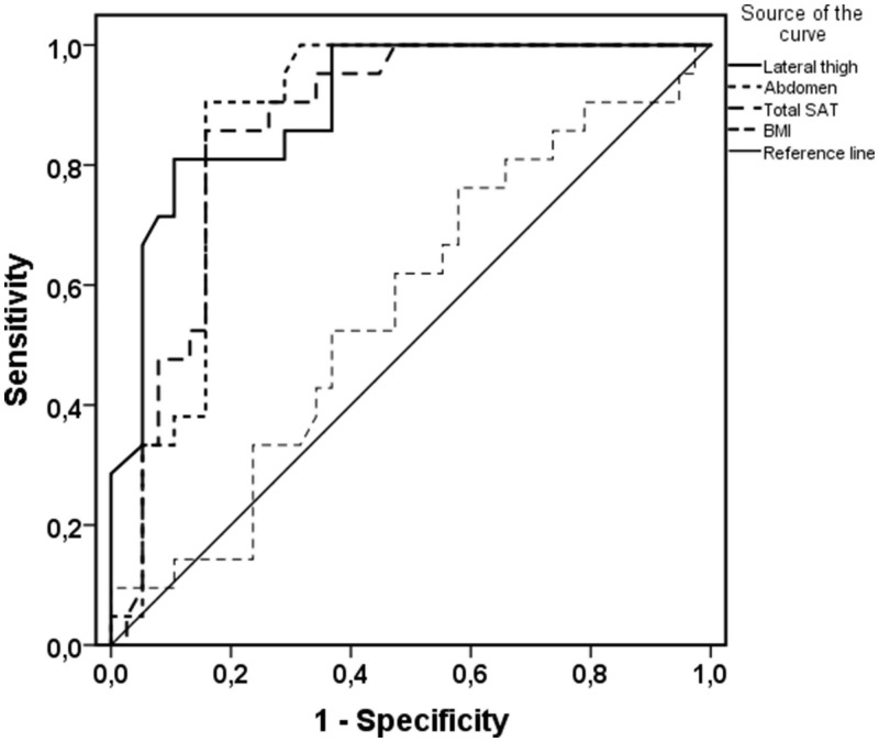
No significant result was obtained for the BMI, indicating that the BMI is not able to discriminate between the trained and the untrained group. On the other hand, all Lipometer measurements provided significant ROC results up to the highest area index of 0.9 for the body site lateral thigh. Figure 2 shows the ROC curves for the lateral thigh body site, the abdominal compartment variable, total SAT, and the BMI.
Figure 2.
ROC curves for lateral thigh, abdomen, total SAT and BMI. The curve describes the association between the sensitivity and the specificity at different cut-off values. ROC curves that approach the upper leftmost corner indicate high discrimination power.
SAT: subcutaneous adipose tissue.
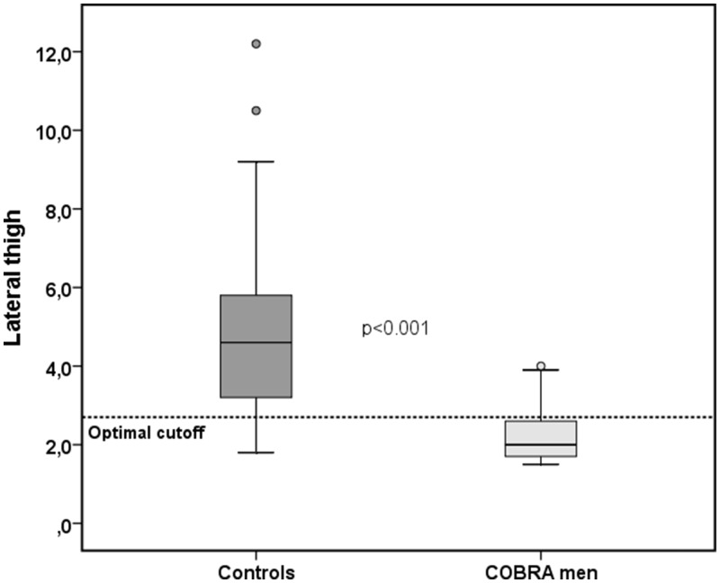
Using 2.7 mm as the optimal cutoff, the Lipometer measurement results at the lateral thigh are able to correctly classify 51 of the 59 subjects (86.4%) as Cobra men or untrained controls (Table 2). Figure 3 shows the corresponding boxplot including the line for the optimal cutoff at 2.7 mm.
Figure 3.
Box plot at the body site lateral thigh for Cobra men and male controls. The lateral thigh provides the highest discrimination power of all 15 body sites. A dotted horizontal line shows the optimal cut-off value for the two subject groups.
A similar classification result (86.4%) was obtained for the abdomen compartment values (Table 2, Figure 2). Finally, Total SAT provided a significant area index of 0.86 and was able to correctly classify 50 of the 59 subjects (84.7%) as either Cobra or controls (Table 2). Overall, we obtained high discrimination results for the Lipometer measurements on the 15 body sites, for the compartment variables, for the Total SAT value and for the BF %, but not for BMI.
Discussion
Our findings show that regardless of having similar BMI values, men involved in heavy training routines (Cobra group) had significantly lower subcutaneous fat patterns in all body areas compared with inactive men. The well-trained men showed approximately 45% lower Total SAT thickness compared to untrained control men suggesting well-trained men have substantially lower amounts of subcutaneous fat. Moreover, while the BMI is a commonly used indicator of body fat in humans, our results clearly show that is not always the case, particularly in a more physically active athletic population.
Despite the shortcomings of the BMI measure found in this study and previously reported,14 it continues to be recommended, that individuals should know if their BMI sits within the healthy range (BMI: 18–25 kg/m2).20 A BMI higher than 25 is assumed to be an indicator for overweight and obesity with a high amount of total body fat. However, total body fat in individuals can only be precisely measured using expensive methods such as MRI and computed tomography, while other methods like bioelectric impedance and DXA indicate “indirectly” a measure for total body fat. In any case, the BMI is an inaccurate measure of body composition because age, sex, muscle mass, and body fat distribution are not taken into consideration.21
In those sports disciplines where speed is the characteristic determinant (e.g. to win a foot race), a BMI between 17 and 20 has been suggested to produce the optimal performance.20 Long distance runners who train regularly for competition are naturally very lean with a low amount of body fat. On the other hand, Santos et al. published a broad study with about 800 male and female athletes involved in 21 different sports.22 Body composition was measured with DXA, and reference percentiles were calculated. Athletes showed a higher fat-free mass than untrained persons, and the BMI was not useful at describing the body composition of these trained athletes.
A further study investigating BMI appropriateness involved a police agency in the USA who tested 1941 police officers (1826 men and 114 women) concerning their body fat percentages and fitness levels.23 This study also confirmed that the prevalence of overall fitness decreased linearly with the increase of body fat in men and women independent of age and rank, but not with the increase of BMI.
The results of the current study are similar to an earlier study comparing young athletes and non-athletic controls.14 While the power to discriminate between athletes and untrained subjects was very poor using the BMI, subcutaneous adipose tissue thicknesses measured by the Lipometer provided high discrimination power and was able to classify correctly 90.6% of the male subjects only by the neck body site and 88.1% of the female subjects only by the upper back body site.
The present study is a follow-up of this earlier study. The study participants in the present study were in a higher age group with a mean age of 38 years, but results were similar to the earlier study. Age and height were matched with the control group, and the high performance training showed a maximum difference between these two groups: On one hand, the men of the task force Cobra with a high training load of about 30 h per week and on the other hand the relatively untrained controls. As we expected, the BMI measure was unable to distinguish the high training efforts of the Cobra men and showed no significant difference between the two groups (Table 1), while all 15 Lipometer body site thicknesses, the four-compartment thicknesses, and the Total SAT were significantly different between the groups. Notably, Total SAT provided an absolute difference of 53.7 mm of subcutaneous body fat (120.2 mm versus 66.5 mm; Table 1) between the two subject groups, whereas the BMI was unable to discriminate this difference. Therefore, we conclude that the BMI is not an appropriate measure for body fat determination for these groups.
In addition to the statistical tests, ROC curve analysis was applied to calculate the discrimination power of each single variable (Table 2). Again all Lipometer SAT thicknesses showed significant discrimination results up to a correct classification of 86.4% (51 of 59 subjects were correctly classified as untrained or Cobra men) only by the SAT thicknesses at the body site “lateral thigh.” This result is very similar compared with the result of the previous study of young male athletes,14 which were about 15 years younger and showed a correct classification result of 90.6%. On the other hand, the BMI did not provide significant ROC curve results and, consequently, showed no discrimination power.
BMI underestimates overweight and obesity in untrained individuals and overestimates excess adipose tissue in trained individuals with high muscle mass and more lean body mass.24 Our dataset confirms that statement: BMI classified six Cobra men as “overweight” (BMI≥25) and 15 Cobra men as “normal” (BMI < 25). Sixteen controls were classified as “overweight” and 22 controls as “normal.” In both groups, there were no “underweight” subjects (BMI < 18.5) and no “obese” subjects (BMI > 30). The Total SAT median of the Cobra-“normal”-group was 58.7 mm, the Cobra-“overweight”-group showed 83.0 mm, 108.2 mm was obtained from the Control-“normal”-group, and finally the Control-“overweight”-group provided 133.6 mm. Notably, the Cobra-“overweight”-group showed a lower Total SAT median compared with the Control-“normal”-group. This is an example that the BMI overestimates the body fat situation of the Cobra-“overweight”-group, classifying them as “overweight.” On the other hand, the Control-“normal”-group might be (partly) underestimated by the BMI when classified as “normal.” Therefore, to get accurate measures of body fat and body composition, other ways of thinking and more refined measurement methods are demanded.
In this study, we present for the first time data on the BMI and Lipometer body fat measures in trained and untrained men of a higher age group. While BMI was unable to distinguish fat levels between the two groups, the Lipometer method showed distinctive differences and therefore was able to account for the fat levels which were probably related to the physical activity differences in the groups.
A limitation of this study is that we did not use the Gold standard method for measuring body fat (e.g. DXA); instead, we used the Lipometer, which is quicker, less expensive, more portable, and has no radiation associated with its use. Moreover, previous studies have reported high correlations between DXA and Lipometer body fat measurements.11,25,26 For example, Jürimäe et al.11 reported a correlation coefficient of r = 0.88 in males and r = 0.91 in females for body fat measurements between DXA and Lipometer devices. In their study, Jürimäe et al.11 used a stepwise regression analysis to produce an equation to predict body fat percentage from Lipometer data. We used the same equation in the current study (BF % = 1.308 neck + 0.638 hip + 6.971) to produce the BF % values which were able to distinguish fat levels between Cobra men and untrained controls (Table 1, Table 2).
ACKNOWLEDGMENTS
We wish to thank the Cobra task force Graz for their kind cooperation.
Authors’ contribution
ET analyzed data; GC wrote the article; ML, MH, and RM performed experiments and interpreted results; MJH and RH reviewed the article.
DECLARATION OF CONFLICTING INTERESTS
The author(s) declared no potential conflicts of interest with respect to the research, authorship, and/or publication of this article.
FUNDING
The author(s) received no financial support for the research, authorship, and/or publication of this article.
References
- 1.Garrow JS, Webster JD. Quetelet’s index (W/H2) as a measure of fatness. Int J Obes 1985; 9:147–53 [PubMed] [Google Scholar]
- 2.Meeuwsen S, Horgan GW, Elia M. The relationship between BMI and percent body fat, measured by bioelectrical impedance, in a large adult sample is curvilinear and influenced by age and sex. Clin Nutr 2010; 29:560–6 [DOI] [PubMed] [Google Scholar]
- 3.Fox CS, Massaro JM, Hoffmann U, Pou KM, Maurovich-Horvat P, Liu CY, Vasan RS, Murabito JM, Meigs JB, Cupples LA, D'Agostino RB, Sr, O'Donnell CJ. Abdominal visceral and subcutaneous adipose tissue compartments: association with metabolic risk factors in the Framingham Heart Study. Circulation 2007; 116:39–48 [DOI] [PubMed] [Google Scholar]
- 4.Horejsi R, Möller R, Pieber TR, Wallner S, Sudi K, Reibnegger G, Tafeit E. Differences of subcutaneous adipose tissue topography between type-2 diabetic men and healthy controls. Exp Biol Med (Maywood) 2002; 227:794–8 [DOI] [PubMed] [Google Scholar]
- 5.Tafeit E, Möller R, Sudi K, Reibnegger G. The determination of three subcutaneous adipose tissue compartments in non-insulin-dependent diabetes mellitus women with artificial neural networks and factor analysis. Artif Intell Med 1999; 17:181–93 [DOI] [PubMed] [Google Scholar]
- 6.Fantuzzi G, Mazzone T. Adipose Tissue and atherosclerosis: exploring the connection. Arterioscler Thromb Vasc Biol 2007; 27:996–1003 [DOI] [PubMed] [Google Scholar]
- 7.Wallner SJ, Horejsi R, Zweiker R, Watzinger N, Möller R, Schnedl WJ, Schauenstein K, Tafeit E. ROC analysis of subcutaneous adipose tissue topography (SAT-Top) in female coronary heart disease patients and healthy controls. J Physiol Anthropol 2008; 27:185–91 [DOI] [PubMed] [Google Scholar]
- 8.Tafeit E, Möller R, Rackl S, Giuliani A, Urdl W, Freytag U, Crailsheim K, Sudi K, Horejsi R. Subcutaneous adipose tissue pattern in lean and obese women with polycystic ovary syndrome. Exp Biol Med (Maywood) 2003; 228:710–6 [DOI] [PubMed] [Google Scholar]
- 9.Rothman KJ. BMI-related errors in the measurement of obesity. Int J Obes ( Obes) 2008; 32:S56–9 [DOI] [PubMed] [Google Scholar]
- 10.Bazzarre TL, Kleiner SM, Litchford MD. Nutrient intake, body fat, and lipid profiles of competitive male and female bodybuilders. J Am Coll Nutr 1990; 9:136–42 [DOI] [PubMed] [Google Scholar]
- 11.Jürimäe T, Sudi K, Jürimäe J, Payerl D, Möller R, Tafeit E. Validity of optical device lipometer and bioelectric impedance analysis for body fat assessment in men and women. Coll Antropol 2005; 29:499–502 [PubMed] [Google Scholar]
- 12.Tafeit E, Möller R, Sudi K, Reibnegger G. Artificial neural networks as a method to improve the precision of subcutaneous adipose tissue thickness by means of the optical device lipometer. Comput Biol Med 2000; 30:355–65 [DOI] [PubMed] [Google Scholar]
- 13.Tafeit E, Möller R, Jurimae T, Sudi K, Wallner SJ. Subcutaneous adipose tissue topography (SAT-Top) development in children and young adults. Coll Antropol 2007; 31:395–402 [PubMed] [Google Scholar]
- 14.Kruschitz R, Wallner-Liebmann SJ, Hamlin MJ, Moser M, Ludvik B, Schnedl WJ, Tafeit E. Detecting body fat – a weighty problem BMI versus subcutaneous fat patterns in athletes and non-athletes. PLoS One 2013; 8:e72002. [DOI] [PMC free article] [PubMed] [Google Scholar]
- 15.Wallner-Liebmann SJ, Kruschitz R, Hübler K, Hamlin MJ, Schnedl WJ, Moser M, Tafeit E. A measure of obesity: BMI versus subcutaneous fat patterns in young athletes and nonathletes. Coll Antropol 2013; 37:351–7 [PubMed] [Google Scholar]
- 16.Moeller R, Horejsi R, Pilz S, Lang N, Sargsyan K, Dimitrova R, Tafeit E, Giuliani A, Almer G, Mangge H. Evaluation of risk profiles by subcutaneous adipose tissue topography in obese juveniles. Obesity (Silver Spring) 2007; 15:1319–24 [DOI] [PubMed] [Google Scholar]
- 17.Sudi K, Jürimäe J, Payerl D, Pihl E, Möller R, Tafeit E, Jürimäe T. Relationship between subcutaneous fatness and leptin in male athletes. Med Sci Sports Exerc 2001; 33:1324–9 [DOI] [PubMed] [Google Scholar]
- 18.Tafeit E, Möller R, Sudi K, Reibnegger G. ROC and CART analysis of subcutaneous adipose tissue topography (SAT-Top) in type-2 diabetic women and healthy females. Am J Hum Biol 2000; 12:388–94 [DOI] [PubMed] [Google Scholar]
- 19.Youden WJ. Index for rating diagnostic tests. Cancer. 1950;3 :32--5 [DOI] [PubMed] [Google Scholar]
- 20.Sedeaut A, Marc A, Marck A, Dor F, Schipman J, Dorsay M, Haida A, Berthelot G, Toussaint JF. BMI, a performance parameter for speed improvement. PLoS One 2014; 9:e90183. [DOI] [PMC free article] [PubMed] [Google Scholar]
- 21.Rothman KJ. BMI-related errors in the measurement of obesity. Int J Obes (Lond) 2008; 32:56–69 [DOI] [PubMed] [Google Scholar]
- 22.Santos DA, Dawson JA, Matias CN, Rocha PM, Minderico CS, Alison DB, Sardinha LB, Silva A. Reference values for body composition and anthropometric measurements in athletes. PLoS One 2014; 9:e97846. [DOI] [PMC free article] [PubMed] [Google Scholar]
- 23.Violanti JM, Ma CC, Fekedulegn D, Andrew ME, Gu JK, Hartley TA, Charles LE, Burchfield CM. Associations between body fat percentage and fitness among police officers: a statewide study. Saf Health Work 2017; 8:36–41 [DOI] [PMC free article] [PubMed] [Google Scholar]
- 24.Shah NR, Braverman ER. Measuring adiposity in patients: the utility of body mass index (BMI), percent body fat, and leptin. PLoS One 2012; 7:e33308. [DOI] [PMC free article] [PubMed] [Google Scholar]
- 25.Jürimäe T, Jürimäe J, Wallner SJ, Lipp RW, Schnedl WJ, Möller R, Tafeit E. Relationships between body fat measured by DXA and subcutaneous adipose tissue thickness measured by lipometer in adults. J Physiol Anthropol 2007; 26:513–6 [DOI] [PubMed] [Google Scholar]
- 26.Tafeit E, Greilberger J, Cvirn G, Lipp RW, Schnedl WJ, Jürimäe T, Jürimäe J, Wallner-Liebmann SJ. Estimating DXA total body fat percentage by lipometer subcutaneous adipose tissue thicknesses. Coll Antropol 2009; 33:391–6 [PubMed] [Google Scholar]


