Abstract
Many diseases affect multiple tissues and/or organ systems, or affect tissues that are broadly distributed. For these diseases, an effective gene therapy will require systemic delivery of the therapeutic vector to all affected locations. Adeno-associated virus (AAV) has been used as a gene therapy vector for decades in preclinical studies and human trials. These studies have shown outstanding safety and efficacy of the AAV vector for gene therapy. Recent studies have revealed yet another unique feature of the AAV vector. Specifically, AAV can lead to bodywide gene transfer following a single intravascular injection. Here we describe the protocols for effective systemic delivery of AAV in both neonatal and adult mice and dogs. We also share lessons we learned from systemic gene therapy in the murine and canine models of Duchenne muscular dystrophy.
Keywords: DMD, Systemic delivery, AAV, Neonatal mice, Neonatal dogs, Adult mice, Adult dogs
1. Introduction
Based on the delivery method, gene therapy can be divided into local therapy and systemic therapy. Local gene therapy refers to direct injection of a therapeutic vector to the diseased tissue in situ. Such an approach works extremely well for diseases that affect a confined region such as congenital blindness caused by mutations in the retinal gene. In this case, a simple one-time subretinal injection of an adeno-associated virus (AAV) vector can result in marvelous vision improvement [1]. As a matter of fact, a gene therapy drug (Luxturna) for treating an inherited form of blindness received Food and Drug Administration (FDA) approval on December 19, 2017, for commercial use [2]. However, many diseases affect multiple body systems or affect tissues that are widely distributed. An effective therapy for these diseases will require systemic delivery. In this case, a therapeutic vector is injected into the circulation to allow it to spread throughout the whole body. Systemic delivery is much more challenging because the vector has to escape from the vasculature and still maintain its infectivity to transduce the target cells. Systemic delivery is also more risky due to the administration of a huge quantity of the vector (≥1014 particles/kg) over a short period of time and inevitable spreading of the vector to untoward tissues.
A variety of viral and nonviral vector systems have been explored for systemic delivery (reviewed in Ref. [3]). Unfortunately, most of them have failed to result in robust, persistent bodywide gene transfer following intravascular delivery. Currently, AAV is the only vector that can lead to whole body systemic transduction at a level that can meet the needs of clinical gene therapy in human patients [4]. The exact molecular mechanisms underlying AAV-mediated systemic gene transfer remain to be elucidated. However, a number of factors may contribute to systemic AAV delivery, such as the small size of the viral particle (AAV is one of the smallest viruses), the reported transcytosis of certain AAV serotypes, and the unique viral capsid structure of certain AAV serotypes. The first successful systemic delivery in rodent (normal and diseased mice) was reported in 2004 by the Chamberlain lab [5]. The first systemic delivery in large mammals was performed in neonatal dogs in 2008 by the Duan lab [6]. The first systemic delivery in an adult diseased large mammal was performed in the canine model of Duchenne muscular dystrophy (DMD) in 2015 by the Duan lab [7]. The first systemic delivery in human patients was performed by the Mendell lab in 2017 for treating type I spinal muscular atrophy [4].
Over the last 11 years, we have conducted extensive preclinical systemic AAV delivery in normal mice and dogs, and murine and canine DMD models [3, 6–18]. In these studies, we tested both reporter and therapeutic vectors and explored a variety of AAV serotypes. Here we describe detailed protocols we have developed for systemic AAV delivery in mice and dogs.
2. Materials
2.1. Systemic Delivery of AAV to Neonatal Mice (Fig. 1a)
Fig. 1.
Systemic injection in newborn mice via the facial vein. (a) Material required for the procedure: (1) loop headband magnifier, (2) Hamilton syringe with attached tubing and needle, (3) food dye, (4) Wee Sight transilluminator. (b) A newborn pup taped to the Wee Sight illuminator. (c) Position of the facial vein (arrow). (d) A photograph showing the placement of the injection needle in the facial vein. (e) Representative photographs showing an un-injected puppy (top) and an injected puppy (bottom)
Personal protective equipment (PPE) including disposable gowns and/or dedicated scrubs, head cover, shoe covers, surgical mask, and gloves.
30G Hamilton syringe (Hamilton, Reno, NV, USA).
PE10 tubing (Hamilton, Reno, NV, USA).
Wee Sight transilluminator vein finder (Philips, Omaha, NE, USA).
Food dye (Wilton, Darien, IL, USA) sterilized with 0.2 μm filter before use.
Wet ice bucket.
Sterile gauge sponges (2 in. × 2 in.).
Medical tape (3 M, Maplewood, MN, USA).
Optivisor adjustable headband magnifier with a 2.5 × lens (Donegan Optical Company Inc., Lenexa, KS, USA).
Recombinant AAV vector (see Note 1).
2.2. Systemic Delivery of AAV to Adult Mice
Personal protective equipment (PPE) including disposable gowns and/or dedicated scrubs, head cover, shoe covers, surgical mask, and gloves.
Flat bottom rodent restrainer (Plas Labs Inc., Lansing, MI, USA).
BD Insulin syringe with a capacity of 100 μL and a 30G × 0.5 in. needle.
Thermophore heating pad (Medwing, Columbia, SC, USA). Webcol alcohol prep pad (Covidien, Dublin, Republic of Ireland).
Digital scale.
Biosafety hood.
Recombinant AAV vector (see Note 1).
2.3. Systemic Delivery of an AAV Vector to Neonatal Dogs (Fig. 2a)
Fig. 2.
Systemic injection of neonatal dog puppies via the jugular vein. (a) Materials required for the procedure(1). a butterfly needle attached to the infusion line, (2) a Luer lock connector, (3) the 3-mL syringe containing the AAV vector, (4) the prefilled saline syringe, (5), alcohol wipe, (6) gauze sponge, (7) a ChloraPrep one step. (b) A photograph showing the position of the puppy for injection. (c) A photograph showing the placement of the injection needle in the jugular vein
Personal protective equipment (PPE) including disposable gowns and/or dedicated scrubs, head cover, shoe covers, surgical mask, and gloves.
MyWeigh SCM2600BLACK iBalance tabletop digital scale (MyWeigh, Phoenix, Arizona, USA).
Digital thermometer (ReliOn Inc., Spokane Valley, WA, USA).
Pediatric stethoscope (3 M Littmann, St. Paul, MN, USA).
Masimo Rad-57 handheld pulse oximeter (DRE, Louisville, KY, USA).
Thermal blanket (100% cotton) (Elite Home Products Inc., Saddle Brook, NJ, USA).
Animal fine hair clipper.
Infusion set (23G × 3/4 in. butterfly needle attached to a 7.5” infusion line with Luer lock connector) (Greiner Bio-One, Kremsmünster, Austria).
Syringe with Luer-Lock Tip (3 mL) (Becton Dickinson company, Franklin Lakes, NJ, USA).
BD PosiFlush Pre-filed saline (0.9% sodium chloride) syringe for intravenous (IV) injection (Becton Dickinson company, Franklin Lakes, NJ, USA).
Sterile gauge sponges (2 in. × 2 in.).
ChloraPrep one step (2% Chlorhexidine gluconate and 70% Isopropyl alcohol) (CareFusion, Leawood, KS, USA).
Recombinant AAV vector (see Note 1).
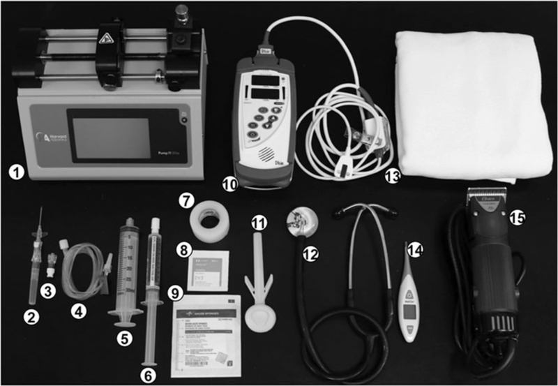
2.4. Systemic Delivery of AAV to Young/Adult Dogs (Fig. 3)
Fig. 3.
Materials required for the systemic injection of young/adult dog via the cephalic vein (1) Infusion pump,(2) shielded IV catheter, (3) Luer lock, (4) connector tubing, (5) 30 mL syringe, (6) prefilled saline syringe,(7) medical tape, (8) alcohol wipe, (9) gauze sponge, (10) handheld pulse oximeter, (11) ChloraPrep one step,(12) stethoscope, (13) thermal blanket, (14) digital thermometer, (15) animal hair clipper
Personal protective equipment (PPE) including disposable gowns and/or dedicated scrubs, head cover, shoe covers, surgical mask, and gloves.
Digital vet scale (DRE, Louisville, KY, USA).
Digital thermometer (ReliOn Inc., Spokane Valley, WA, USA).
Stethoscope (3 M Littmann, St. Paul, MN, USA).
Handheld pulse oximeter (Masimo Rad-57) (DRE, Louisville, KY, USA).
Thermal blanket (100% cotton) (Elite Home Products Inc., Saddle Brook, NJ, USA).
Animal work table.
Animal hair clipper equipped with a size 50 blade (Oster professional product, Sunbeam products, Inc., Boca Raton, FL, USA).
Shielded I.V. Catheter (22G × 1.00 in. needle, or 20G × 1.00 in. needle) (Becton Dickinson company, Franklin Lakes, NJ, USA).
SmartSite Needle free valve (CareFusion, San Diego, CA, USA).
Micro-Bore IV Extension set (A 60 in. tube line with 0.3 mL volume capacity, and contains Luer lock connectors as well as a slide clamp) (NeoChild, Oklahoma City, OK, USA).
Medical Tape.
Vet wrap (3 M, Maplewood, MN, USA).
Webcol Alcohol Prep pad (Covidien, Dublin, Republic of Ireland).
Syringe with Luer-Lock Tip (30 mL) (Becton Dickinson Company, Franklin Lakes, NJ, USA).
BD PosiFlush Pre-filed saline (0.9% Sodium chloride) syringe for IV injection (Becton Dickinson Company, Franklin Lakes, NJ, USA).
Infusion pump (KD Scientific Inc., Holliston, MA, USA).
Sterile gauge sponges (4 in. × 4 in.) (Govidien, Mansfield, MA, USA).
ChloraPrep one step (2% Chlorhexidine gluconate and 70% Isopropyl alcohol) (CareFusion, San Diego, CA, USA).
Recombinant AAV vector (see Note 1).
3. Methods
3.1. Systemic Delivery of AAV to Neonatal Mice
Follow the institutional guidelines for animal care and handling. Obtain the approval from institutional animal care and use committee (ACUC). Wear all PPE before working or handling animals. Use aseptic techniques while preparing the AAV vector for injection and during the injection procedure.
Keep the neonates in a pre-warmed cage by keeping a heat pad beneath the cage. Separate the dam from the pups (see Note 2).
Mix the food dye with stock AAV virus at a 1:100 dilution (see Note 3).
Fill the syringe with the AAV vector. Get rid of air bubbles (see Note 4).
Anesthetize the animal using a thermal shock by wrapping the puppy with a piece of gauze and keeping on ice for few seconds (see Note 5).
Secure the neonate body on its side on the Wee Sight transilluminator by placing a gauze pad on the skin and a tape over the gauze (see Note 6) (Fig. 1b).
Use the magnifier to visualize the facial vein (Fig. 1c).
Slowly insert the needle into the vein and infuse the AAV vector (see Note 7) (Fig. 1d). Once all the solution is injected, keep the needle in the vein for additional 15 s (see Note 8).
After removing the needle, gently pressure the injection site to avoid bleeding.
Allow 2–3 min for the neonate to recover, and after it is conscious, place the neonate back in the cage. If necessary the animal can be warmed up by keeping on the investigator’s hands or a pre-warmed gauze pad.
Monitor the neonates for signs of distress.
Return the injected pup to the mother after the pup becomes conscious and restores ambulation.
3.2. Systemic Delivery of AAV to Adult Mice
Follow the institutional guidelines for animal care and handling. Obtain the approval from institutional animal care and use committee (IACUC). Wear all PPE before working or handling animals. Use aseptic techniques while preparing the AAV vector for injection and during the injection procedure.
Weigh the mouse and calculate the volume of the AAV vector needed to reach a certain viral genome (vg) particles/body weight (see Note 9).
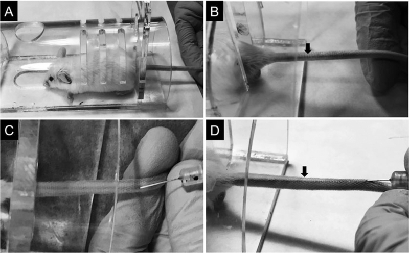
Position the mouse in the rodent restrainer (Fig. 4a).
Gently warm the tail using a heating pad at its lowest setting to allow vasodilation.
Hold the distal end of the tail with the nondominant hand.
Slightly rotate the tail to allow clear visualization of the lateral tail vein (Fig. 4b).
Clean the injection site with alcohol pad.
Insert the needle to the tail vein by holding the tail and needle positioning parallel to the tail (see Note 10) (Fig. 4c, d).
Inject the AAV vector at a steady rate of ~1 mL/min.
Once the AAV vector is injected, carefully remove the needle and apply pressure to the injection site to avoid bleeding (see Note 11).
Transfer the mouse to the cage and observe the mouse for at least 4 h.
Fig. 4.
Systemic injection in adult mice via the tail vein. (a) An adult mouse inside the rodent restrainer. (b) A photograph showing the prominent lateral tail vein (arrow). (c) A photograph showing the placement of the injection needle in the tail vein. (d) A photograph showing the color of the tail becomes dark with a successful injection of the AAV. The color change is due to the food dye in the AAV solution
3.3. Systemic Delivery of AAV to Neonatal Dogs
Follow the institutional guidelines for animal care and handling. Obtain the approval from institutional animal care and use committee (ACUC). Wear all PPE before working or handling animals. Use aseptic techniques while preparing the AAV vector for injection and during the injection procedure.
Monitor the newborn puppy for the body weight and responsiveness (see Note 12). Supplement the puppy with formula as needed (see Note 13). Consult with the veterinary doctor immediately for unexpected changes such as sudden drop of the body weight or temperature.
Weight the puppy to determine the injection volume. A normal neonatal puppy can tolerate up to 25 μL liquid/gram body weight (see Note 14).
Measure the vital signs (heart rate, respiration rate, rectal body temperature, and blood oxygen saturation) (see Note 15).
Gently shave the neck hair on the side to allow better observation of the jugular vein for injection.
Connect the saline prefilled BD PosiFlush syringe with the IV extension line and attach them to the infusion set. All parts are connected through Luer lock fitting. Prefill the IV extension line and the infusion set with saline to remove air from the lines. Lock the sliding clamp on the extension line. Draw the exact volume of AAV vector (calculated based on body weight and desired dose) into the 3 mL syringe and remove any air bubbles form the syringe. Replace the BD PosiFlush syringe with the AAV syringe. Check the extension line and infusion set for any air bubble.
Perform injection procedure with the help of three investigators. The first investigator places a preheated thermal blanket on the animal working table, positions the puppy supine on the blanket, then gently restrains the forelimbs using both hands (Fig. 2b). The second investigator restrains the head and scrubs the ventral side of the neck with the ChloraPrep, then gently inserts the needle in the jugular vein (Fig. 2c). Once the needle enters the vein, blood will appear in the needle hub. The third investigator unlocks the sliding clamp on the extension line and pulls the plunger back to visualize blood in the infusion line. This will confirm that the needle is correctly placed inside the vein. Inject the AAV vector at the rate of 2 mL/min, then flush the infusion line with saline [12].
While pressing the entry site of the needle with a finger, retract the butterfly from the jugular vein. Continue to pressure the injection site to avoid bleeding.
Remeasure the vital signs.
Return the puppy back to the dam and closely monitor the puppy for the following 24 h. Consult with veterinary doctor immediately with any concerns.
3.4. Systemic Delivery of AAV to Young/Adult Dogs
Follow the institutional guidelines for animal care and handling. Obtain the approval from institutional animal care and use committee (ACUC). Wear all PPE before working or handling animals. Use aseptic techniques while preparing the AAV vector for injection and during the injection procedure.
Weigh the dog to determine the injection volume. Up to6.2 mL/kg body weight (6.24 × 10¹⁴ viral genome particles/kg body weight) can be tolerated in young adult dystrophic dogs [7].
Measure the vital signs (heart rate, respiration rate, rectal body temperature, and blood oxygen saturation).
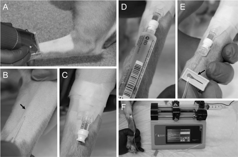
Shave the cranial side of the lower forelimb (Fig. 5a) to allow better observation of the cephalic vein (Fig. 5b) during the injection procedure (see Note 16).
Place a thermal blanket on the animal working table. Place the dog on the table in the sternal recumbency position with the lower forelimb extended. Occlude the cephalic vein at the elbow with your thumb or using a tourniquet by one investigator.
Catheterize the cephalic vein by another investigator. Scrub the cranial aspect of the lower forelimb with ChloraPrep. Pull the lower forelimb skin distally to stabilize the vein. Slowly insert the IV catheter needle (bevel up) through the skin and into the vein (Fig. 5c). Blood will appear in the catheter when the needle punctures the vein. Gently slide the catheter into the vein while retracting the needle out. At this time, blood will flow out form the catheter. Quickly occlude the vein at the catheter tip and attach the smart site needle free valve to the catheter. Secure the catheter with tape and warp it with vet wrap. Flush the catheter with saline to ensure there is no leak.
Connect the saline prefilled BD PosiFlush syringe with the IV extension line using the Luer lock fitting (Fig. 5d). Prefill the IV extension line with saline to remove air. Lock the sliding clamp on the extension line (Fig. 5e). Draw the exact volume of AAV vector into the 30 mL syringe. Remove any air bubbles form the syringe. Replace the BD PosiFlush syringe with the AAV syringe. Check the extension line for any air bubble. Secure the syringe to the infusion pump and set the infusion rate to 2 mL/min (Fig. 5f).
Attach the IV extension line to the valve and unlock the sliding clamp. Start the infusion and monitor the dog’s vital signs closely. Once the infusion is done, flush infusion line with saline and disconnect the extension line from the catheter. While pressing the entry site of the needle with your finger, retract the catheter from the cephalic vein. Continue to pressure the puncture site to avoid bleeding.
Continue measuring the vital signs for 5, 10, 30, and 60-min post injection and examine the dog for any adverse reaction.
Return the dog to its housing and continue monitoring the dog for 72 h. Consult with the veterinary doctor immediately with any concerns.
Fig. 5.
Systemic injection of young/adult dog via the cephalic vein. (a) Shaving of the cranial aspect of the forelimb. (b) A photograph showing the placement of the needle in the cephalic vein. Arrow, the cephalic vein. (c) A photograph showing the catheterization of the cephalic vein. (d) A photograph showing the AAV containing syringe during the injection. (e) A photograph showing the catheter inserted with the extension line. Arrow, the sliding clamp. (f) A photograph showing injection of AAV using the infusion pump
4. Notes
Many different methods have been developed for the production and purification of recombinant AAV vectors (reviewed in refs. 19, 20). For the protocols described in this chapter, AAV vectors were generated using the transient plasmid transfection method [17]. AAV vector titer was determined using quantitative polymerase chain reaction. AAV vector was dialyzed in HEPES buffer (Sodium chloride 8.7 g, HEPES 4.75 g, 10 N Sodium Hydroxide 1.5 mL, Distilled water up to 1 L, pH adjusted to 7.8). AAV-6 and −8 are the first AAV serotypes shown capable of systemic delivery [5, 21]. Since then, a number of AAV serotypes and engineered AAV capsids were found to have the unique systemic delivery property. Some of these AAV serotypes also demonstrated specific tissue tropism following systemic delivery. For example, intravascular injection of AAV-9 results in preferential transduction of the heart in mice [8]. For the protocols described in this chapter, we have used AAV-1, −6, −8, −9 and their tyrosine mutants [22].
Neonatal facial vein injections need to be carried out between 24 and 48 h after birth. During this time window, the facial vein is clearly visible.
The food dye is added to allow easy visualization and confirmation of injection.
A newborn mouse can tolerate up to 100 μL of volume solution.
Do not leave the neonate on ice for longer than 1 min to avoid complications related to hypothermia.
The newborn mouse should be positioned with its neck turned gently for clear visualization of the facial vein and the head secured by fingers without interfering respiration.
Accumulation of solution at the head or neck area (but not in other parts of the body) suggests injection failure. A successful injection should result in the diffusion of the dye throughout the body. The entire puppy will turn the color of the dye.
This will allow sufficient time for the AAV solution to flow through the PE-10 tubing and it will also prevent any back flow of the solution.
The total AAV vector volume (mL) needed to reach a certain viral dosage (vg/body weight) is calculated using the following formula: Injection volume (mL) = [dosage (vg/body weight) × body weight]/viral titer (vg/mL).
Injection should be done approximately 1 in. distal to the midpoint of the tail. Starting at the far end of the tail to allow second or third injection attempt if the first attempt fails.
If the injection creates a bleb, the injection at that site should be terminated. In the case of an unsuccessful injection, up to three additional injections can be attempted.
The body weight should be monitored every 2–4 h. Bowel movement and/or urination may cause transient body weight fluctuation. Puppies that fail to gain weight should not be used for AAV injection.
The Just Born puppy formula produces best results for nutrition supplementation in our hands.
We have performed neonatal AAV injection in puppies as early as 24 h after the birth. Successful systemic AAV injection can also be achieved in puppies up to 1 week of the age.
Throughout the injection procedure, make sure the puppy is wrapped in a thermal blanket and kept warm. If needed, thermal blanket can be pre-warmed to 37 °C.
Besides the cephalic vein, other veins can also be used such as the jugular vein and saphenous vein. For injection through these veins, shave the related area. For example, for saphenous vein injection, we shave the lateral side of the distal tibia above the tarsus joint.
Acknowledgments
The research on systemic AAV delivery in the Duan lab was supported by the National Institutes of Health (NS-90634, AR-70517 and AR-69085), Department of Defense (MD150133), Jesse’s Journey-The Foundation for Gene and Cell Therapy, Hope for Javier, Jackson Freel DMD Research Fund and Solid Biosciences Inc. The authors thank Keqing Zhang for the assistance in figure preparation.
Footnotes
Disclosure: D.D. is a member of the scientific advisory board, an equity holder of Solid Biosciences, LLC. D.D. is an inventor on a patent licensed to Solid Biosciences, LLC.
References
- 1.Russell S, Bennett J, Wellman JA et al. (2017) Efficacy and safety of voretigene neparvovec (AAV2-hRPE65v2) in patients with RPE65-mediated inherited retinal dystrophy: a randomised, controlled, open-label, phase 3 trial. Lancet 390(10097):849–860. 10.1016/S0140-6736(17)31868-8 [DOI] [PMC free article] [PubMed] [Google Scholar]
- 2.FDA News Release (2017) FDA approves novel gene therapy to treat patients with a rare form of inherited vision loss. https://www.fda.gov/NewsEvents/Newsroom/PressAnnouncements/ucm589467.htm. Accessed 19 Dec 2017 [Google Scholar]
- 3.Duan D (2016) Systemic delivery of adeno-associated viral vectors. Curr Opin Virol 21:16–25. 10.1016/j.coviro.2016.07.006 [DOI] [PMC free article] [PubMed] [Google Scholar]
- 4.Mendell JR, Al-Zaidy S, Shell R et al. (2017) Single-dose gene-replacement therapy for spinal muscular atrophy. N Engl J Med 377(18):1713–1722. 10.1056/NEJMoa1706198 [DOI] [PubMed] [Google Scholar]
- 5.Gregorevic P, Blankinship MJ, Allen JM et al. (2004) Systemic delivery of genes to striated muscles using adeno-associated viral vectors. Nat Med 10(8):828–834 [DOI] [PMC free article] [PubMed] [Google Scholar]
- 6.Yue Y, Ghosh A, Long C et al. (2008) A single intravenous injection of adeno-associated virus serotype-9 leads to whole body skeletal muscle transduction in dogs. Mol Ther 16(12):1944–1952. 10.1038/mt.2008.207 [DOI] [PMC free article] [PubMed] [Google Scholar]
- 7.Yue Y, Pan X, Hakim CH et al. (2015) Safe and bodywide muscle transduction in young adult Duchenne muscular dystrophy dogs with adeno-associated virus. Hum Mol Genet 24(20):5880–5890 [DOI] [PMC free article] [PubMed] [Google Scholar]
- 8.Bostick B, Ghosh A, Yue Y et al. (2007) Systemic AAV-9 transduction in mice is influenced by animal age but not by the route of administration. Gene Ther 14(22):1605–1609 [DOI] [PubMed] [Google Scholar]
- 9.Ghosh A, Yue Y, Long C et al. (2007) Efficient whole-body transduction with trans-splicing adeno-associated viral vectors. Mol Ther 15(4):750–755. 10.1038/sj.mt.6300081 [DOI] [PMC free article] [PubMed] [Google Scholar]
- 10.Bostick B, Yue Y, Lai Y et al. (2008) Adeno-associated virus serotype-9 microdystrophin gene therapy ameliorates electrocardiographic abnormalities in mdx mice. Hum Gene Ther 19(8):851–856. 10.1089/hum.2008.058 [DOI] [PMC free article] [PubMed] [Google Scholar]
- 11.Ghosh A, Yue Y, Shin J-H et al. (2009) Systemic trans-splicing AAV delivery efficiently transduces the heart of adult mdx mouse, a model for Duchenne muscular dystrophy. Hum Gene Ther 20(11):1319–1328 [DOI] [PMC free article] [PubMed] [Google Scholar]
- 12.Yue Y, Shin JH, Duan D (2011) Whole body skeletal muscle transduction in neonatal dogs with AAV-9. Methods Mol Biol 709:313–329. 10.1007/978-1-61737-982-6_21 [DOI] [PMC free article] [PubMed] [Google Scholar]
- 13.Bostick B, Shin JH, Yue Y et al. (2012) AAV micro-dystrophin gene therapy alleviates stress-induced cardiac death but not myocardial fibrosis in >21-m-old mdx mice, an end-stage model of Duchenne muscular dystrophy cardiomyopathy. J Mol Cell Cardiol 53(2):217–222. S0022–2828(12)00179–4 [pii]. 10.1016/j.yjmcc.2012.05.002 [DOI] [PMC free article] [PubMed] [Google Scholar]
- 14.Pan X, Yue Y, Zhang K et al. (2013) Long-term robust myocardial transduction of the dog heart from a peripheral vein by adeno-associated virus serotype-8. Hum Gene Ther 24(6):584–594. 10.1089/hum.2013.044 [DOI] [PMC free article] [PubMed] [Google Scholar]
- 15.Hakim CH, Yue Y, Shin JH et al. (2014) Systemic gene transfer reveals distinctive muscle transduction profile of tyrosine mutant AAV-1, −6, and −9 in neonatal dogs. Mol Ther Methods Clin Dev 1:14002 10.1038/mtm.2014.2 [DOI] [PMC free article] [PubMed] [Google Scholar]
- 16.Zhang Y, Yue Y, Li L et al. (2013) Dual AAV therapy ameliorates exercise-induced muscle injury and functional ischemia in murine models of Duchenne muscular dystrophy. Hum Mol Genet 22(18):3720–3729. 10.1093/hmg/ddt224 [DOI] [PMC free article] [PubMed] [Google Scholar]
- 17.Pan X, Yue Y, Zhang K et al. (2015) AAV-8 is more efficient than AAV-9 in transducing neonatal dog heart. Hum Gene Ther Methods 26(4):54–61 [DOI] [PMC free article] [PubMed] [Google Scholar]
- 18.Hakim CH, Pan X, Kodippili K et al. (2016) Intravenous delivery of a novel micro-dystrophin vector prevented muscle deterioration in young adult canine Duchenne muscular dystrophy dogs. Mol Ther 24(Suppl 1): S198–S199 [Google Scholar]
- 19.Kotin RM, Snyder RO (2017) Manufacturing clinical grade recombinant adeno-associated virus using invertebrate cell lines. Hum Gene Ther 28(4):350–360. 10.1089/hum.2017.042 [DOI] [PubMed] [Google Scholar]
- 20.Clement N, Grieger JC (2016) Manufacturing of recombinant adeno-associated viral vectors for clinical trials. Mol Ther Methods Clin Dev 3:16002 10.1038/mtm.2016.2 [DOI] [PMC free article] [PubMed] [Google Scholar]
- 21.Wang Z, Zhu T, Qiao C et al. (2005) Adeno-associated virus serotype 8 efficiently delivers genes to muscle and heart. Nat Biotechnol 23(3):321–328 [DOI] [PubMed] [Google Scholar]
- 22.Zhong L, Li B, Mah CS et al. (2008) Next generation of adeno-associated virus 2 vectors: point mutations in tyrosines lead to high-efficiency transduction at lower doses. Proc Natl Acad Sci U S A 105(22):7827–7832. 10.1073/pnas.0802866105 [DOI] [PMC free article] [PubMed] [Google Scholar]


