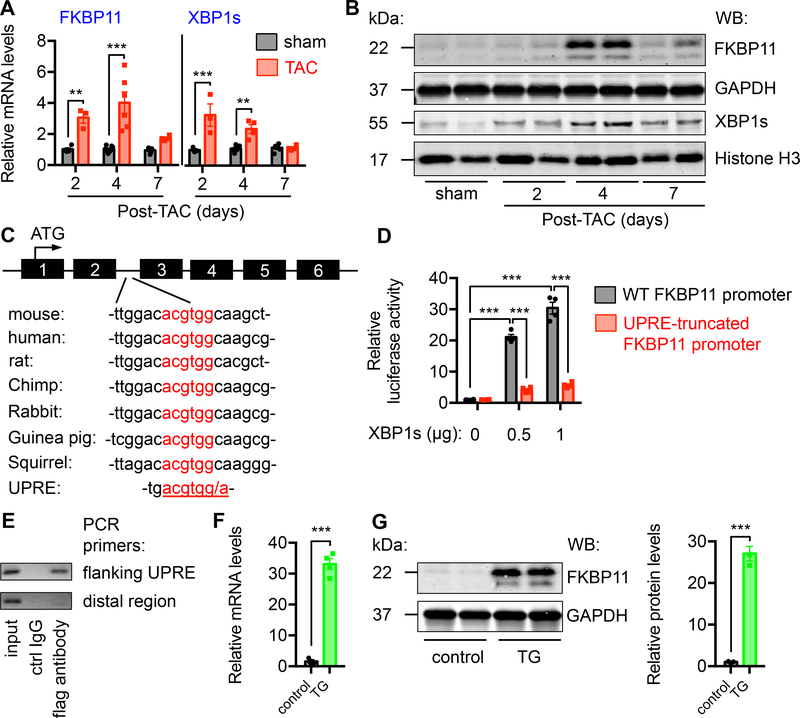
Figure 7. FKBP11 is a direct target of XBP1s.
A. FKBP11 expression was acutely induced by pressure overload in the heart. TAC was conducted in wild type animals. Hearts were harvested at different time points post surgery. Relative FKBP11 mRNA level was determined by quantitative RT-PCR. Note that XBP1s expression was also upregulated by TAC. Two-way ANOVA analysis was conducted, followed by Tukey’s test. N = 3–8.
B. FKBP11 protein level was augmented by TAC in the heart as shown by immunoblotting, along with induction of XBP1s. GAPDH and Histone H3 were used as loading controls for whole cell lysates and nuclear extracts, respectively.
C. A conserved region in the second intron of FKBP11 genome, which resembles the consensus sequence of XBP1s-binding site UPRE (unfolded protein response element). The translational initiation site is in the first exon of FKBP11 genomic DNA.
D. XBP1s expression stimulated FKBP11 promoter activity in a dose-dependent manner. The conserved region of FKBP11 promoter was engineered into a luciferase reporter construct. Co-transfection of a XBP1s-expressing plasmid led to an increase in luciferase activity. Note that a truncated mutant of FKBP11 promoter lacking the UPRE region did not show significant promoter activity. N = 4 per group. Two-way ANOVA analysis was performed, followed by Tukey’s test.
E. Chromatin immunoprecipitation (ChIP) assay was conducted to determine the binding of XBP1s to the FKBP11 promoter. NRVMs were infected by adenovirus expressing flag-tagged XBP1s. ChIP was performed with either control mouse IgG or anti-flag antibody. PCR was conducted using primers spanning the UPRE site. Primers from the distal region were used as a negative control.
F. The mRNA level of FKBP11 was upregulated in the XBP1s transgenic hearts, as determined by quantitative RT-PCR. Student’s t test was performed. N = 3–4.
G. The protein level of FKBP11 was significantly increased in the XBP1s transgenic hearts, as revealed by immunoblotting. GAPDH was used as a loading control. N = 3 for each group. Student’s t test was conducted. **, P<0.01; ***, P<0.001.

