eLife logoLink to eLife
. 2019 Aug 13;8:e40855. doi: 10.7554/eLife.40855

Region-specific myelin differences define behavioral consequences of chronic social defeat stress in mice

Valentina Bonnefil 1, Karen Dietz 2,3, Mario Amatruda 1, Maureen Wentling 1, Antonio V Aubry 4, Jeffrey L Dupree 5, Gary Temple 1, Hye-Jin Park 1, Nesha S Burghardt 4, Patrizia Casaccia 1,2,3, Jia Liu 1,
Editors: Catherine Dulac6, Klaus-Armin Nave7
PMCID: PMC6692108  PMID: 31407664

Abstract

Exposure to stress increases the risk of developing mood disorders. While a subset of individuals displays vulnerability to stress, others remain resilient, but the molecular basis for these behavioral differences is not well understood. Using a model of chronic social defeat stress, we identified region-specific differences in myelination between mice that displayed social avoidance behavior (‘susceptible’) and those who escaped the deleterious effect to stress (‘resilient’). Myelin protein content in the nucleus accumbens was reduced in all mice exposed to stress, whereas decreased myelin thickness and internodal length were detected only in the medial prefrontal cortex (mPFC) of susceptible mice, with fewer mature oligodendrocytes and decreased heterochromatic histone marks. Focal demyelination in the mPFC was sufficient to decrease social preference, which was restored following new myelin formation. Together these data highlight the functional role of mPFC myelination as critical determinant of the avoidance response to traumatic social experiences.

Editorial note: This article has been through an editorial process in which the authors decide how to respond to the issues raised during peer review. The Reviewing Editor's assessment is that all the issues have been addressed (see decision letter).

Research organism: Mouse

eLife digest

High levels of stress do not have the same effect on everybody: some individuals can show resilience and recover quickly, while other struggle to cope. Scientists have started to investigate how these differences may find their origin in biological processes, mainly by focusing on the role of neurons. However, neurons represent only one type of brain cells, and there is increasing evidence that interactions between neuronal and non-neuronal cells play an important role in the response to stress.

Oligodendrocytes are a common type of non-neuronal cells which shield and feed nerve cells. In particular, their membrane constitutes the myelin sheath, a protective coating that insulates neurons and allows them to better communicate with each other using electric signals.

Bonnefil et al. explored whether differences in oligodendrocytes could affect how mice responded to social stress. The rodents were exposed to repeated attacks from an aggressive mouse five minutes a day for ten days. After this period, ‘susceptible’ mice then avoided future contact with any other mice, while resilient animals remained interested in socializing.

Comparing the brain areas of resilient and susceptible mice revealed differences in the oligodendrocytes of the medial prefrontal cortex, the part of the brain that controls emotions and thinking. Susceptible animals had fewer mature oligodendrocytes and their neurons were covered in thinner and shorter segments of myelin sheaths. There was also evidence that, in these animals, the genes that regulate the maturation of oligodendrocytes were more likely to be switched off. Taken together, these results may suggest that, in certain animals, social stress disrupts the genetic program that controls how oligodendrocytes develop, potentially leading to neurons communicating less well.

To explore whether reduced amounts of myelin could be linked to decreased social behavior, Bonnefil et al. then damaged the myelin in the medial prefrontal cortex in another group of rodents. The mice were then less willing to interact with other animals until new sheaths had formed.

The results by Bonnefil et al. undercover how changes in non-neuronal cells can at least in part explain differences in the way individuals respond to stress. Ultimately, this knowledge may be useful to design new strategies to foster resilience.

Introduction

Exposure to stress increases the risk of developing affective disorders such as depression and post-traumatic stress disorder. While stress leads to maladaptive behavioral responses in a subset of humans, others are capable of coping and remain resilient. Differences in the behavioral response to stress can also be detected in experimental mouse models, thereby highlighting the degree of conservation of this response. However, the cellular and molecular basis underlying resilience or susceptibility to negative experiences remains poorly defined.

We and others have previously reported that animal models of psychosocial stressors, such as social isolation (Liu et al., 2012; Liu et al., 2016; Makinodan et al., 2016; Makinodan et al., 2017; Makinodan et al., 2012), chronic social defeat stress (CSDS) (Cathomas et al., 2019; Lehmann et al., 2017), and chronic variable stress (Liu et al., 2018), lead to transcriptional, translational, or ultrastructural changes in oligodendrocytes and myelination. Here we tested the hypothesis that myelinating glia serves a causal role in behavioral susceptibility or resilience following stress exposure. We examined social behaviors, ultrastructural changes in myelination as well as epigenetic modifications in oligodendrocytes in brain regions that have been implicated in depressive-like behavior after a well-established social defeat paradigm (Berton et al., 2006; Golden et al., 2011; Hodes et al., 2014; Krishnan et al., 2007; Vialou et al., 2010). We also provide mechanistic insights into the region-specific differences between the phenotypes, which we attributed to defective oligodendrocyte progenitor differentiation. To provide direct causal evidence, we carried out focal demyelination in the medial prefrontal cortex and showed aversive social behavior in animals undergoing demyelination and a resolution of the behavioral effect consequent to new myelin formation. Together, we suggest the functional role of region-specific myelination in determining depressive-like social behavior.

Results and discussion

Chronic social defeat stress causes region-specific changes in myelination

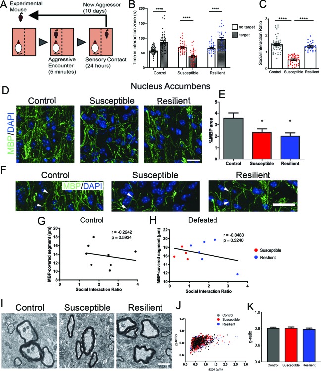
We adopted a mouse model of chronic social defeat stress (CSDS) (Golden et al., 2011), in which mice were exposed to an aggressor challenge for 10 days (Figure 1A) and tested for social behavior afterwards. While some mice showed signs of social withdrawal, characterized by reduced social interaction time when a conspecific mouse is present and reduced social interaction ratio (i.e. susceptible mice), a subset escaped this deleterious consequence (i.e. resilient mice), and were virtually indistinguishable from the control group, which were not exposed to any aggressors (Figure 1B–C).

Figure 1. Effect of aggressive social encounters on myelination in the nucleus accumbens (NAc) of mice which showed two behaviorally distinct phenotypes following chronic social defeat stress (CSDS).

Figure 1.

(A) The experimental paradigm for CSDS. (B–C) Mice susceptible to CSDS spent less time interacting with a conspecific mouse than the control group or resilient mice, as shown in (B) total time spent in the interaction zone when there is a conspecific mouse present and in (C) social interaction ratio defined by time spent in the interaction zone when a conspecific mouse present divided by a conspecific mouse absent. Control, n = 52; susceptible, n = 39; resilient, n = 33; ****, p<0.0001 by one-way ANOVA followed by Tukey’s post hoc test. (D–E) Representative confocal images and quantifications showing immunohistochemistry of myelin basic protein (MBP) counterstained with DAPI. Scale bar = 28 μm. n = 3 mice per group, two 20x images per animal; susceptible vs. control, p=0.0447; resilient vs. control, p=0.0109 by one-way ANOVA followed by Tukey’s post hoc test. (F) Representative confocal images showing MBP-covered myelinated segments. Arrowheads point to one MBP-covered myelinated segment. Scale car = 19 μm. (G–H) Pearson correlation coefficients showed non-significant correlation of MBP-covered segment length and social interaction ratio in control (G) or defeated (H) mice, control, 8 x-y pairs. r = −0.2242, p=0.5932. defeated, 10 x-y pairs, r = −0.3483, p=0.3240. control, n = 8 mice, susceptible, n = 4 mice, resilient, n = 6 mice, 1–2 20x images per mouse; (I) Representative electron microscopy images (scale bar = 1 μm) and (J–K) scatter plot and quantification of g-ratio; control, n = 5 mice; susceptible, n = 7 mice; resilient, n = 5 mice.

Figure 1—source data 1. Source data for social interaction behavior following chronic social defeat stress and myelin content in the nucleus accumbens.
DOI: 10.7554/eLife.40855.004

Next, we sought to determine whether there was any myelination difference between susceptible and resilient mice. We focused our analysis on the nucleus accumbens (NAc) and the medial prefrontal cortex (mPFC), two brain regions shown to play a critical role in determining stress responses (Heshmati et al., 2018; Han and Nestler, 2017) and displaying myelin transcriptional or structural impairment after a stressful experience (Liu et al., 2012; Liu et al., 2016; Makinodan et al., 2016; Lehmann et al., 2017; Liu et al., 2018; Zhang et al., 2016). In the NAc, a significant reduction of myelin basic protein (MBP) was detected in all defeated mice, regardless of their behavioral responses (control, 3.6 ± 0.4%; susceptible, 2.4 ± 0.3%; resilient 2.0 ± 0.3%; Figure 1D–E). However, no significant differences were detected in the length of myelinated segments measured by MBP immunoreactivity (Figure 1F–H) or in myelin thickness (Figure 1I–K) among groups. Pearson coefficients correlation showed no significant correlation between the length of MBP-covered segments and social interaction ratio in either control or defeated group (control, r = −0.2242, p=0.5934, defeated, r = −0.3483, p-0.3240, Figure 1G–H). Altogether, these results suggest that myelination in the NAc uniformly responds to stress and does not distinguish susceptibility and resilience following CSDS.

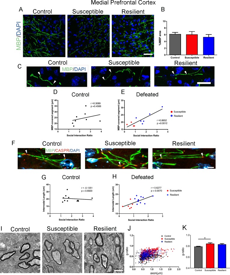
In contrast, the mPFC displayed a unique myelination phenotype following CSDS. While the levels of MBP did not significantly differ between susceptible and resilient mice (control, 6.2 ± 0.5%; susceptible, 6.1 ± 0.9%; resilient, 5.3 ± 0.8%; Figure 2A–B), the length of myelinated segments indicated by MBP immunoreactivity showed a significant positive correlation with social interaction in defeated mice (Figure 2C–E). Importantly, such correlation was not detected in the control (unstressed) group, suggesting that changes in the length of myelinated segments represent an adaptive response to the social defeat stress. To more accurately quantify internodal length, we conducted immunohistochemical analysis using antibodies specific for the contactin-associated protein (Caspr), which marks the paranodal regions (Figure 2F). Also in this case, a significant positive correlation between internodal length and social interaction ratio was detected only in the defeated mice, with shorter internodal lengths identified in susceptible mice (Figure 2G–H). Myelin was also thinner in the susceptible - but not in the resilient – mice, compared to controls (Figure 2I–K). Therefore, region-specific myelination differences in the mPFC could -at least in part- explain the behavioral differences between susceptible and resilient mice in response to stress.

Figure 2. Myelination in the medial prefrontal cortex (mPFC) distinguished resilient from susceptible mice following stress.

Figure 2.

(A–B) Representative confocal images and quantifications of myelin basic protein (MBP) counterstained with DAPI. Scale bar = 30 μm. n = 3 mice per group. Four 20x images taken per mouse (C) Representative confocal images showing MBP-covered myelinated segments. Arrowheads point to one continuous MBP-covered myelinated segment. Scale bar = 17 μm. (D–E) Pearson correlation coefficients showed significant correlation of MBP-covered segment length with social interaction ratio only in defeated (E) mice, but not in control (D), control, 8 x-y pairs, n = 8 mice, defeated 10 x-y pairs, susceptible, n = 4 mice, resilient, n = 6 mice, four 20x images were taken per mouse (F) Representative confocal images showing internodal segment marked by CASPR (Red) and MBP (Green). Arrowheads point to one internode. Scale bar = 5 μm. (G–H) Pearson correlation coefficients showed significant correlation of internodal length with social interaction ratio only in defeated (H) mice, but not in control (G), control, 11 x-y pairs, n = 11 mice, defeated 17 x-y pairs, susceptible, n = 6 mice, resilient, n = 11 mice, four-six 63x images taken per mouse. (I) Representative electron microscopy images, scale bar = 1 μm. (J–K) Scatter plot and quantification of g-ratio in the mPFC; control, n = 5 mice; susceptible, n = 7 mice; resilient, n = 5 mice; susceptible vs. control, p=0.0264 by one-way ANOVA followed by Tukey’s post hoc test.

Figure 2—source data 1. Source Data for myelin content in the medial prefrontal cortex in social defeat mice.
DOI: 10.7554/eLife.40855.006

Different oligodendrocyte populations in the mPFC of susceptible and resilient mice

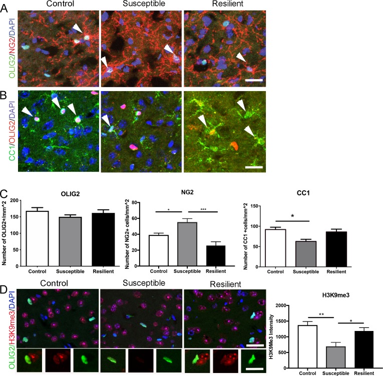
To determine whether reduced myelin content in the mPFC of susceptible mice was limited to the internodal length, we further performed a detailed quantitative immunohistochemical analysis on oligodendrocyte lineage cells. No significant difference in the overall number of OLIG2+ cells was detected (Figure 3A,C), thereby ruling out decreased survival of oligodendrocyte lineage cells in response to stress. However, compared to resilient and controls, the susceptible mice were characterized by a significantly higher number of NG2+ progenitor cells (control, 39.2 ± 2.3 mm−2; susceptible, 55.3 ± 4.4 mm−2; resilient, 25.8 ± 4.8 mm−2; Figure 3A,C), and lower number of CC1+ mature oligodendrocytes (control, 91.9 ± 5.7 mm−2; susceptible, 62.7 ± 5.2 mm−2; resilient, 86.1 ± 5.8 mm−2; Figure 3B–C). Consistent with defective differentiation of NG2+ cells in the mPFC of susceptible mice, a reduction of the histone modification marks associated with differentiation (H3K9me3) was also detected (pixel/area: control, 1373.6 ± 113.3; susceptible, 695.3 ± 127.9; resilient, 1186.0 ± 106.0; Figure 3D). Together these data suggest that social stress might have at least two main effects on oligodendrocyte lineage cells in the mPFC: it promotes myelin remodeling resulting in shorter internodal length and fewer wraps and possibly impairs in the epigenetic program of oligodendrocyte progenitor differentiation, resulting in fewer differentiated oligodendrocytes.

Figure 3. Impaired oligodendrocyte differentiation was associated with reduced repressive histone methylation marks in the mPFC of susceptible mice.

Figure 3.

(A–B) Representative confocal images of cells positive for OLIG2, NG2, and CC1 in the mPFC. DAPI was used as counterstain of nuclei. Scale bar = 25 μm. (C) quantification of OLIG2+ (n = 3 mice per group and 3–4 20x images taken per mouse), NG2+ cells (n = 3 mice per group and four images taken per mouse, *p=0.019, ***p<0.0001 by one-way ANOVA followed by Tukey’s post hoc test) and CC1+ cells (control, n = 8 mice, susceptible, n = 3 mice, resilient n = 7 mice, 3–4 20x images taken per mouse; *p=0.0195 by one-way ANOVA followed by Tukey’s post hoc test). (D) Representative confocal images and quantifications (E) of mean intensity of repressive histone mark H3K9me3 (Red) in OLIG2+ (Green) cells. control, n = 3 mice, susceptible, n = 2 mice, resilient, n = 5 mice, 4 20x images taken per mouse, 50–100 OLIG2+ cells were counted per image **p=0.0038, *p=0.0234 by one-way ANOVA followed by Tukey’s post hoc test. Data are mean ± S.E.M. Scale bar = 20 μm.

Figure 3—source data 1. Source data for the number of oligodendrocyte lineage cells and intensity of a repressive histone mark, H3K9me3, in the medial prefrontal cortex.
DOI: 10.7554/eLife.40855.008

Focal demyelination in the mPFC leads to social avoidance behavior

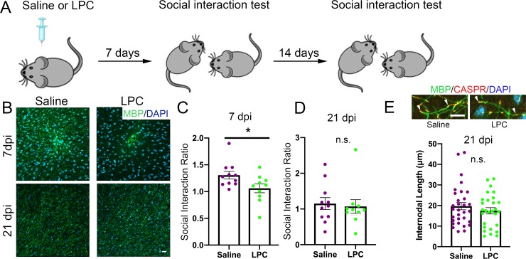
The data above suggested an interesting correlation between myelination in the mPFC and social avoidance behavior in the susceptible mice. To test the causality of this finding, we induced myelin loss by focal injection of lysolecithin (LPC) into the mPFC and asked whether this manipulation would be sufficient to induce behavioral changes. LPC injection is a well characterized model of toxic demyelination, with early myelin loss (detectable one week after injection) followed by spontaneous repair, due to the formation of new myelin by newly differentiated oligodendrocytes (occurring three weeks after injection) (Jeffery and Blakemore, 1995). We reasoned that behavioral differences detected in mice at these two time points after LPC injection would support a causal link between social preference performance and myelin content in the mPFC (Figure 4A). Indeed, the kinetics of demyelination and remyelination after LPC injection was validated by the detection of reduced MBP immunoreactivity at the 7dpi followed by spontaneous recovery of immunoreactivity by 21dpi (Figure 4B). At the early time point (7dpi), LPC-injected mice displayed reduced social preference behavior compared to saline-injected controls (Figure 4C). This difference in social interaction behavior was no longer detectable after 3 weeks (Figure 4D), when myelination recovered to normal level (Figure 4E). Therefore, we conclude that myelin content in the mPFC is a critical determinant of social behavior.

Figure 4. Focal demyelination in the mPFC reduced social preference behavior.

Figure 4.

(A) The experimental paradigm for lysolecithin (LPC) injection and behavioral testing. (B) Representative confocal images showing reduced MBP immunointensity at seven dpi followed by a spontaneous restoration at 21 dpi. (C) Mice received LPC displayed reduced social preference behavior at seven dpi as quantified by social interaction ratio. (D) Restoration of normal social interaction behavior at 21 dpi as quantified by social interaction ratio. Saline, n = 11 mice; LPC, n = 10 mice *, p<0.05 by unpaired t-test. Data are mean ± S.E.M. (E) Representative confocal images of immunohistochemistry of MBP (Green) and CASPR (Red) and scatter plots of internodal length at 21dpi. Counterstained with DAPI. Saline n = 3 mice, LPC, n = 5 mice. 2–4 63x images taken per mouse.

Figure 4—source data 1. Source data for social interaction behavior and internodal length following lysolecithin-induced focal demyelination.
DOI: 10.7554/eLife.40855.010

Altogether, our study reveals region-specific epigenetic dysregulation of oligodendrocyte progenitor differentiation and subsequent defective adult myelination as maladaptive mechanisms occurring only in susceptible mice after exposure to repeated social stress. We have previously reported that myelination defects were detected in socially isolated adult mice, prior to the appearance of social avoidance behavior (Liu et al., 2012). Here, we show that social avoidance behavior can be detected after chronic social defeat stress as well as after focal demyelination in the mPFC, and could therefore be caused by hypomyelination. Furthermore, promoting myelination has been shown to rescue depressive-like behavior in socially isolated mice (Liu et al., 2016). On the same note, normal social behavior was restored following the spontaneously occurring remyelination in LPC-injected mice. However, it is important to note that, social stress did not induce a toxic effect on myelin, whereas LPC did. No cellular toxicity was detected in the mPFC of susceptible mice or in mice undergoing social isolation (Liu et al., 2012). In contrast, we detected fewer mature oligodendrocytes and more progenitors lacking epigenetic marks of differentiation, suggesting an altered epigenetic program. For this reason, we interpret the lower myelin content in the mPFC of susceptible mice as resulting from impaired oligodendrocyte progenitor differentiation, possibly as maladaptive response to social stress. While our data support an inefficient production of new myelin, the detection of a positive correlation between intermodal length and social avoidance behavior, suggests that reorganization of paranodal loops could also be affected. Indeed, shorter internodal length, consequent to impaired myelin formation, has been previously shown to decrease nerve conduction in the optic nerve (Etxeberria et al., 2016). It is, therefore, conceivable that the reduced length of myelinated segments detected in the mPFC of susceptible mice may result in slower conduction and functionally result in the characteristic social avoidance behavior in response to the social stress.

Finally, we suggest that new myelin is formed in the mPFC of resilient mice as an adaptive mechanism to the repeated episodes of aggression. It is conceivable that the formation of new myelin in resilient mice could favor the establishment of neuronal circuits allowing the escape of negative impact following traumatic stress (Krishnan et al., 2007; Fagundes et al., 2013; Fenster et al., 2018; Ménard et al., 2017; Russo et al., 2012), as oligodendrocytes are known to regulate conduction speed and play a crucial role in synchronizing neuronal networks (Saab and Nave, 2017). This explanation is in agreement with the increasing evidence from mice and squirrel monkeys, which suggests stress resilience may arise from active coping strategies, rather than a passive response, defined as lack of adaptive response (Russo et al., 2012; Lyons et al., 2009).

The molecular basis for resilience has been studied extensively in the context of neuronal cells, the immune and neuroendocrine systems (Ménard et al., 2017; Russo et al., 2012). Here we proposed an alternative, although not mutually exclusive explanation involving myelinating glia. One possibility for new myelin formation as a coping strategy is associated with increased neuronal activity in the resilient mice, as reported by a greater degree of FosB, or ΔFosB expression in glutamatergic neurons of mPFC of resilient mice following social defeat (Covington et al., 2010; Lehmann and Herkenham, 2011). Optogenetic stimulation of mPFC has been shown to help resilience phenotype in social defeated mice (Covington et al., 2010). Although not characterized in the previous study (Covington et al., 2010), optogenetic stimulation has been shown to promote oligodendrogliogenesis and new myelin formation (Gibson et al., 2014). An alternative mechanism could involve inflammatory cytokines, such as interleukin-6 (IL-6). IL-6 has been identified as a major cytokine that contributes to the development of depression in human (Dowlati et al., 2010; Erta et al., 2012). In animal models of stress, systemic IL-6, was the only differentially regulated cytokine that distinguished resilient mice from susceptible and control mice (Hodes et al., 2014). Although systemic changes of IL-6 could not account for the region-specific differences in myelination in susceptible and resilient mice, it is known that IL-6 can be produced by neurons, astrocytes, microglia or endothelial cells in the central nervous system (Erta et al., 2012). Several transcriptomic studies suggest that oligodendrocyte progenitors express IL-6 receptors (Zeisel et al., 2015; Zhang et al., 2014). Therefore, it is intriguing to think that IL-6 could be up-regulated in a region-specific pattern with the ability to impact oligodendrocyte progenitor differentiation and new myelin formation in specific regions of the adult brain.

Overall this study extends our knowledge on the functional role of adult myelination by providing a mechanism for adaptation to social stress encounters, which ultimately result in the expression of resilience.

Materials and methods

Key resources table.

Reagent type
(species) or resource
Designation Source or reference Identifiers Additional
information
M. musculus (C57Bl/6J) mouse Jackson Laboratory RRID:IMSR_JAX:000664
M. musculus (CD-1) mouse Charles River RRID:IMSR_CRL:22 Retired breeder
Antibody Mouse monoclonal anti-MBP Covance Cat # SMI99 RRID:AB_2564741 IHC (1:500)
Antibody Rabbit polyclonal anti-Caspr Abcam Cat# ab34151, RRID:AB_869934 IHC (1:100)
Antibody Mouse monoclonal anti-OLIG2 Millipore Cat# MABN50, RRID:AB_10807410 IHC (1:200)
Antibody Rabbit polyclonal anti-OLIG2 Abcam Cat# ab81093, RRID:AB_1640746 IHC (1:200)
Antibody Rabbit anti-H3K9me3 Abcam Cat# ab8898, RRID:AB_306848 IHC (1:100)
Antibody Rabbit polyclonal anti-NG2 Millipore Cat# AB5320, RRID:AB_91789 IHC (1:200)
Antibody Mouse monoclonal anti-APC EMDMillipore Cat# OP80, RRID:AB_2057371 IHC (1:100)
chemical compound, drug DAPI Thermofisher Cat# D1306, RRID:AB_2629482 IHC (1:10000)
chemical compound, drug l-α-lysophosphatidylcholine Sigma-Aldrich Cat# L4129
software, algorithm ImageJ RRID:SCR_003070
software, algorithm Ethovision XT Noldus RRID:SCR_000441
software, algorithm Graphpad Prism 8 RRID:SCR_002798

Animals

All experimental C57Bl/6J male mice (7 weeks) were obtained from the Jackson Laboratory (Bar Harbor, Maine) and allowed one-week acclimation prior to the start of experiment. Retired male CD1 breeders used as the aggressors were obtained from Charles River (Wilmington, Massachusetts). All mice were maintained in a temperature- and humidity-controlled facility on a 12 hr light-dark cycle with food and water ad libitum. All procedures were carried out in accordance with the Institutional Animal Care and Use Committee guidelines of the Icahn School of Medicine at Mount Sinai, Hunter College and Advanced Science Research Center at City University of New York.

Chronic social defeat stress.

Chronic social defeat stress was performed as previously published (Berton et al., 2006; Golden et al., 2011; Krishnan et al., 2007; Vialou et al., 2010; Wilkinson et al., 2009) with slight modification. Briefly, male C57 mice were exposed to a novel aggressive CD1 male mouse for 5 min/day, after which the mice were separated by a Plexiglas barrier that allows for sensory contact without further physical interaction. Control mice were housed two animals/cage under the same conditions as their experimental counterparts but without the presence of an aggressive CD1 mouse. Twenty-four hours after the last of 10 daily defeat or control episodes, mice were evaluated in a social interaction test during the light cycle, as previously described (Liu et al., 2012), then one-way ANOVA tests were performed to assess statistical differences and assess social avoidance. Social interaction ratio was calculated by dividing the time spent in the interaction zone when a conspecific mouse is present by no subject present in the enclosure area. Defeated mice with a social interaction ratio below one are defined as ‘susceptible’, while those with a social interaction ratio above one are defined as ‘resilient’.

Electron microscopy

Mice were processed for standard electron microscopy (EM) analysis as previously described (Liu et al., 2012). Briefly, the mounted section was trimmed to encompass a 1 μm2 region of layers 4–6 of the PFC, thin sectioned at 90 nm, stained with uranyl acetate and lead citrate, and mounted on 200 mesh copper grids. Ten images at 10,000X were collected per mouse using a transmission electron microscope JEOL JEM 1400Plus equipped with a Gatan CCD camera. g-ratios were determined by dividing the diameter of the axon by the diameter of the entire myelinated fiber. ImageJ was used to measure both axon caliber and myelin fiber diameter for a minimum of 100 myelinated axons per mouse. All analyses were performed blind to the experimental conditions. One-way ANOVA tests were performed to assess statistical differences.

Immunohistochemistry

Mice were anesthetized and then perfused, cryopreserved, embedded, and sectioned as previously described (Liu et al., 2012). Immunohistochemistry was performed as previously described (Liu et al., 2012) with primary antibodies against trimethylated histone 3 lysine 9 (H3K9me3, 1:100; ab8898, Abcam), CC1 (1:100; OP80, Calbiochem), myelin basic protein (MBP, 1:500; SMI99, Covance), OLIG2 (1:200, ab81093, Abcam), NG2 (1:200; AB5320, EMD Millipore) or Caspr (1:100, ab34151, Abcam). Stained sections were visualized using confocal microscopy (LSM800 Meta confocal laser scanning microscope, Carl Zeiss Micro-Imaging). For NG2, CC1, OLIG2 cell counts, and H3K9me3 intensity quantifications, 4–6 20x fields were taken per mouse. For MBP-covered segments and internodal length marked by Caspr, 4–6 fields were taken per mouse followed by quantifications using ImageJ. One-way ANOVA tests were performed to assess statistical differences. For correlation of internodal length with social interaction ratio, data normality was determined using D’Agostino and Person test in GraphPad Prism 8. Pearson correlation coefficients were calculated if data passed normality test.

Stereotaxic surgery for lysolecithin injection

While under deep anesthesia induced by inhaled isoflurane, experimental C57BL/6J mice were surgically injected with 1 μl 1% lysolecithin (l-α-lysophosphatidylcholine, Sigma-Aldrich) dissolved in saline, or saline as sham control, bilaterally to the medial prefrontal cortex using a pulled capillary glass pipet at the following stereotaxic coordinates: anterioposterior,+1.5 mm; mediolateral from bregma, 0.5 mm; and dorsoventral-below the surface of the dura, 1.5 mm. The needle was left in place for an additional 2 min to avoid back flow of the lysolecithin or saline. Muscle and skin incisions were sutured with gut and nylon sutures, respectively. To reduce postoperative pain after recovery from anesthesia, animals received a subcutaneous injection of buprenorphine (1.0 mg/kg). Animals were monitored closely following surgery and were tested with social interaction tests at 7- and 21 days post injection.

Acknowledgements

We thank Drs. Eric Nestler, Scott Russo and Rosemary Bagot for the help with animal behavioral assessments. We thank the Epigenetics Core Facility and the Rodent Behavioral Suite at CUNY Advanced Science Research Center for technical help. This work is supported by National Institute of Neurological Disorders and Stroke (2R37NS042925-10, R01NS52738 to PC), NIH-NINDS Center Core Grant 5P30 NS047463, NIH-NCI Cancer Center Grant P30 CAO16059 to JLD, National Institute on Minority Health and Health Disparities (NIMHD) of the NIH (MD007599) to NSB, and City University of New York PSC-CUNY awards to JL. We apologize to our colleagues whose work we did not cite due to limited space.

Funding Statement

The funders had no role in study design, data collection and interpretation, or the decision to submit the work for publication.

Contributor Information

Jia Liu, Email: Jia.Liu@asrc.cuny.edu.

Catherine Dulac, Harvard University, United States.

Klaus-Armin Nave, Max Planck Institute of Experimental Medicine, Germany.

Funding Information

This paper was supported by the following grants:

  • National Institute of Neurological Disorders and Stroke 2R37NS042925-10 to Patrizia Casaccia.

  • National Institute of Neurological Disorders and Stroke R01NS52738 to Patrizia Casaccia.

  • National Institute of Neurological Disorders and Stroke CenterCore Grant 5P30 NS047463 to Jeffrey L Dupree.

  • National Cancer Institute Cancer Center Grant P30 CAO16059 to Jeffrey L Dupree.

  • National Institute on Minority Health and Health Disparities MD007599 to Nesha S Burghardt.

  • City University of New York to Jia Liu.

  • National Institute of Mental Health R21MH114182 to Nesha S Burghardt.

Additional information

Competing interests

No competing interests declared.

Author contributions

Data curation, Formal analysis, Investigation, Methodology, Writing—original draft, Writing—review and editing.

Methodology, Acquisition of data and data analysis.

Methodology, Acquisition of data.

Methodology, Acquisition of data.

Methodology, Acquisition of data.

Data curation, Formal analysis, Methodology.

Methodology, Acquisition of data.

Methodology, Acquisition of data and data analysis.

Supervision, Methodology, Acquisition of data.

Conceptualization, Funding acquisition, Writing—review and editing.

Conceptualization, Data curation, Formal analysis, Supervision, Funding acquisition, Validation, Investigation, Methodology, Writing—original draft, Project administration, Writing—review and editing.

Ethics

Animal experimentation: This study was performed in strict accordance with the recommendations in the Guide for the Care and Use of Laboratory Animals of the National Institutes of Health. All procedures were carried out in accordance with the Institutional Animal Care and Use Committee guidelines of the Icahn School of Medicine at Mount Sinai (Protocol number 08-0676), Hunter College (Protocol number NB-stress 6/18-T3 and NB fear 9/19-02) and Advanced Science Research Center at City University of New York (Protocol number 2018-8).

Additional files

Transparent reporting form
DOI: 10.7554/eLife.40855.011

Data availability

All data generated or analyzed during this study are included in the manuscript. Source data files have been provided.

References

  1. Berton O, McClung CA, Dileone RJ, Krishnan V, Renthal W, Russo SJ, Graham D, Tsankova NM, Bolanos CA, Rios M, Monteggia LM, Self DW, Nestler EJ. Essential role of BDNF in the mesolimbic dopamine pathway in social defeat stress. Science. 2006;311:864–868. doi: 10.1126/science.1120972. [DOI] [PubMed] [Google Scholar]
  2. Cathomas F, Azzinnari D, Bergamini G, Sigrist H, Buerge M, Hoop V, Wicki B, Goetze L, Soares S, Kukelova D, Seifritz E, Goebbels S, Nave KA, Ghandour MS, Seoighe C, Hildebrandt T, Leparc G, Klein H, Stupka E, Hengerer B, Pryce CR. Oligodendrocyte gene expression is reduced by and influences effects of chronic social stress in mice. Genes, Brain and Behavior. 2019;18:e12475. doi: 10.1111/gbb.12475. [DOI] [PubMed] [Google Scholar]
  3. Covington HE, Lobo MK, Maze I, Vialou V, Hyman JM, Zaman S, LaPlant Q, Mouzon E, Ghose S, Tamminga CA, Neve RL, Deisseroth K, Nestler EJ. Antidepressant effect of optogenetic stimulation of the medial prefrontal cortex. Journal of Neuroscience. 2010;30:16082–16090. doi: 10.1523/JNEUROSCI.1731-10.2010. [DOI] [PMC free article] [PubMed] [Google Scholar]
  4. Dowlati Y, Herrmann N, Swardfager W, Liu H, Sham L, Reim EK, Lanctôt KL. A meta-analysis of cytokines in major depression. Biological Psychiatry. 2010;67:446–457. doi: 10.1016/j.biopsych.2009.09.033. [DOI] [PubMed] [Google Scholar]
  5. Erta M, Quintana A, Hidalgo J. Interleukin-6, a major cytokine in the central nervous system. International Journal of Biological Sciences. 2012;8:1254–1266. doi: 10.7150/ijbs.4679. [DOI] [PMC free article] [PubMed] [Google Scholar]
  6. Etxeberria A, Hokanson KC, Dao DQ, Mayoral SR, Mei F, Redmond SA, Ullian EM, Chan JR. Dynamic modulation of myelination in response to visual stimuli alters optic nerve conduction velocity. Journal of Neuroscience. 2016;36:6937–6948. doi: 10.1523/JNEUROSCI.0908-16.2016. [DOI] [PMC free article] [PubMed] [Google Scholar]
  7. Fagundes CP, Glaser R, Hwang BS, Malarkey WB, Kiecolt-Glaser JK. Depressive symptoms enhance stress-induced inflammatory responses. Brain, Behavior, and Immunity. 2013;31:172–176. doi: 10.1016/j.bbi.2012.05.006. [DOI] [PMC free article] [PubMed] [Google Scholar]
  8. Fenster RJ, Lebois LAM, Ressler KJ, Suh J. Brain circuit dysfunction in post-traumatic stress disorder: from mouse to man. Nature Reviews Neuroscience. 2018;19:535–551. doi: 10.1038/s41583-018-0039-7. [DOI] [PMC free article] [PubMed] [Google Scholar]
  9. Gibson EM, Purger D, Mount CW, Goldstein AK, Lin GL, Wood LS, Inema I, Miller SE, Bieri G, Zuchero JB, Barres BA, Woo PJ, Vogel H, Monje M. Neuronal activity promotes oligodendrogenesis and adaptive myelination in the mammalian brain. Science. 2014;344:1252304. doi: 10.1126/science.1252304. [DOI] [PMC free article] [PubMed] [Google Scholar]
  10. Golden SA, Covington HE, Berton O, Russo SJ. A standardized protocol for repeated social defeat stress in mice. Nature Protocols. 2011;6:1183–1191. doi: 10.1038/nprot.2011.361. [DOI] [PMC free article] [PubMed] [Google Scholar]
  11. Han MH, Nestler EJ. Neural substrates of depression and resilience. Neurotherapeutics. 2017;14:677–686. doi: 10.1007/s13311-017-0527-x. [DOI] [PMC free article] [PubMed] [Google Scholar]
  12. Heshmati M, Aleyasin H, Menard C, Christoffel DJ, Flanigan ME, Pfau ML, Hodes GE, Lepack AE, Bicks LK, Takahashi A, Chandra R, Turecki G, Lobo MK, Maze I, Golden SA, Russo SJ. Cell-type-specific role for nucleus accumbens neuroligin-2 in depression and stress susceptibility. PNAS. 2018;115:1111–1116. doi: 10.1073/pnas.1719014115. [DOI] [PMC free article] [PubMed] [Google Scholar]
  13. Hodes GE, Pfau ML, Leboeuf M, Golden SA, Christoffel DJ, Bregman D, Rebusi N, Heshmati M, Aleyasin H, Warren BL, Lebonté B, Horn S, Lapidus KA, Stelzhammer V, Wong EH, Bahn S, Krishnan V, Bolaños-Guzman CA, Murrough JW, Merad M, Russo SJ. Individual differences in the peripheral immune system promote resilience versus susceptibility to social stress. PNAS. 2014;111:16136–16141. doi: 10.1073/pnas.1415191111. [DOI] [PMC free article] [PubMed] [Google Scholar]
  14. Jeffery ND, Blakemore WF. Remyelination of mouse spinal cord axons demyelinated by local injection of lysolecithin. Journal of Neurocytology. 1995;24:775–781. doi: 10.1007/BF01191213. [DOI] [PubMed] [Google Scholar]
  15. Krishnan V, Han MH, Graham DL, Berton O, Renthal W, Russo SJ, Laplant Q, Graham A, Lutter M, Lagace DC, Ghose S, Reister R, Tannous P, Green TA, Neve RL, Chakravarty S, Kumar A, Eisch AJ, Self DW, Lee FS, Tamminga CA, Cooper DC, Gershenfeld HK, Nestler EJ. Molecular adaptations underlying susceptibility and resistance to social defeat in brain reward regions. Cell. 2007;131:391–404. doi: 10.1016/j.cell.2007.09.018. [DOI] [PubMed] [Google Scholar]
  16. Lehmann ML, Weigel TK, Elkahloun AG, Herkenham M. Chronic social defeat reduces myelination in the mouse medial prefrontal cortex. Scientific Reports. 2017;7:46548. doi: 10.1038/srep46548. [DOI] [PMC free article] [PubMed] [Google Scholar]
  17. Lehmann ML, Herkenham M. Environmental enrichment confers stress resiliency to social defeat through an infralimbic cortex-dependent neuroanatomical pathway. Journal of Neuroscience. 2011;31:6159–6173. doi: 10.1523/JNEUROSCI.0577-11.2011. [DOI] [PMC free article] [PubMed] [Google Scholar]
  18. Liu J, Dietz K, DeLoyht JM, Pedre X, Kelkar D, Kaur J, Vialou V, Lobo MK, Dietz DM, Nestler EJ, Dupree J, Casaccia P. Impaired adult myelination in the prefrontal cortex of socially isolated mice. Nature Neuroscience. 2012;15:1621–1623. doi: 10.1038/nn.3263. [DOI] [PMC free article] [PubMed] [Google Scholar]
  19. Liu J, Dupree JL, Gacias M, Frawley R, Sikder T, Naik P, Casaccia P. Clemastine enhances myelination in the prefrontal cortex and rescues behavioral changes in socially isolated mice. The Journal of Neuroscience. 2016;36:957–962. doi: 10.1523/JNEUROSCI.3608-15.2016. [DOI] [PMC free article] [PubMed] [Google Scholar]
  20. Liu J, Dietz K, Hodes GE, Russo SJ, Casaccia P. Widespread transcriptional alternations in oligodendrocytes in the adult mouse brain following chronic stress. Developmental Neurobiology. 2018;78:152–162. doi: 10.1002/dneu.22533. [DOI] [PMC free article] [PubMed] [Google Scholar]
  21. Lyons DM, Parker KJ, Katz M, Schatzberg AF. Developmental cascades linking stress inoculation, arousal regulation, and resilience. Frontiers in Behavioral Neuroscience. 2009;3:32. doi: 10.3389/neuro.08.032.2009. [DOI] [PMC free article] [PubMed] [Google Scholar]
  22. Makinodan M, Rosen KM, Ito S, Corfas G. A critical period for social experience-dependent oligodendrocyte maturation and myelination. Science. 2012;337:1357–1360. doi: 10.1126/science.1220845. [DOI] [PMC free article] [PubMed] [Google Scholar]
  23. Makinodan M, Ikawa D, Miyamoto Y, Yamauchi J, Yamamuro K, Yamashita Y, Toritsuka M, Kimoto S, Okumura K, Yamauchi T, Fukami SI, Yoshino H, Wanaka A, Kishimoto T. Social isolation impairs remyelination in mice through modulation of IL-6. The FASEB Journal. 2016;30:4267–4274. doi: 10.1096/fj.201600537R. [DOI] [PubMed] [Google Scholar]
  24. Makinodan M, Ikawa D, Yamamuro K, Yamashita Y, Toritsuka M, Kimoto S, Yamauchi T, Okumura K, Komori T, Fukami SI, Yoshino H, Kanba S, Wanaka A, Kishimoto T. Effects of the mode of re-socialization after juvenile social isolation on medial prefrontal cortex myelination and function. Scientific Reports. 2017;7:5481. doi: 10.1038/s41598-017-05632-2. [DOI] [PMC free article] [PubMed] [Google Scholar]
  25. Ménard C, Pfau ML, Hodes GE, Russo SJ. Immune and neuroendocrine mechanisms of stress vulnerability and resilience. Neuropsychopharmacology. 2017;42:62–80. doi: 10.1038/npp.2016.90. [DOI] [PMC free article] [PubMed] [Google Scholar]
  26. Russo SJ, Murrough JW, Han MH, Charney DS, Nestler EJ. Neurobiology of resilience. Nature Neuroscience. 2012;15:1475–1484. doi: 10.1038/nn.3234. [DOI] [PMC free article] [PubMed] [Google Scholar]
  27. Saab AS, Nave KA. Myelin dynamics: protecting and shaping neuronal functions. Current Opinion in Neurobiology. 2017;47:104–112. doi: 10.1016/j.conb.2017.09.013. [DOI] [PubMed] [Google Scholar]
  28. Vialou V, Robison AJ, Laplant QC, Covington HE, Dietz DM, Ohnishi YN, Mouzon E, Rush AJ, Watts EL, Wallace DL, Iñiguez SD, Ohnishi YH, Steiner MA, Warren BL, Krishnan V, Bolaños CA, Neve RL, Ghose S, Berton O, Tamminga CA, Nestler EJ. DeltaFosB in brain reward circuits mediates resilience to stress and antidepressant responses. Nature Neuroscience. 2010;13:745–752. doi: 10.1038/nn.2551. [DOI] [PMC free article] [PubMed] [Google Scholar]
  29. Wilkinson MB, Xiao G, Kumar A, LaPlant Q, Renthal W, Sikder D, Kodadek TJ, Nestler EJ. Imipramine treatment and resiliency exhibit similar chromatin regulation in the mouse nucleus accumbens in depression models. Journal of Neuroscience. 2009;29:7820–7832. doi: 10.1523/JNEUROSCI.0932-09.2009. [DOI] [PMC free article] [PubMed] [Google Scholar]
  30. Zeisel A, Muñoz-Manchado AB, Codeluppi S, Lönnerberg P, La Manno G, Juréus A, Marques S, Munguba H, He L, Betsholtz C, Rolny C, Castelo-Branco G, Hjerling-Leffler J, Linnarsson S. Brain structure. Cell types in the mouse cortex and hippocampus revealed by single-cell RNA-seq. Science. 2015;347:1138–1142. doi: 10.1126/science.aaa1934. [DOI] [PubMed] [Google Scholar]
  31. Zhang Y, Chen K, Sloan SA, Bennett ML, Scholze AR, O'Keeffe S, Phatnani HP, Guarnieri P, Caneda C, Ruderisch N, Deng S, Liddelow SA, Zhang C, Daneman R, Maniatis T, Barres BA, Wu JQ. An RNA-sequencing transcriptome and splicing database of Glia, neurons, and vascular cells of the cerebral cortex. Journal of Neuroscience. 2014;34:11929–11947. doi: 10.1523/JNEUROSCI.1860-14.2014. [DOI] [PMC free article] [PubMed] [Google Scholar]
  32. Zhang H, Yan G, Xu H, Fang Z, Zhang J, Zhang J, Wu R, Kong J, Huang Q. The recovery trajectory of adolescent social defeat stress-induced behavioral, (1)H-MRS metabolites and myelin changes in balb/c mice. Scientific Reports. 2016;6:27906. doi: 10.1038/srep27906. [DOI] [PMC free article] [PubMed] [Google Scholar]

Decision letter

Editor: Klaus-Armin Nave1
Reviewed by: Mathias Schmidt

In the interests of transparency, eLife includes the editorial decision letter, peer reviews, and accompanying author responses.

[Editorial note: This article has been through an editorial process in which the authors decide how to respond to the issues raised during peer review. The Reviewing Editor's assessment is that all the issues have been addressed.]

Thank you for submitting your article "Region-specific myelin differences define behavioral consequences of chronic social defeat stress in mice" for consideration by eLife. Your article has been reviewed by three peer reviewers, and the evaluation has been overseen by a Reviewing Editor and Catherine Dulac as the Senior Editor. The following individual involved in review of your submission has agreed to reveal his identity: Mathias Schmidt (Reviewer #3).

The Reviewing Editor has highlighted the concerns that require revision and/or responses, and we have included the separate reviews below for your consideration. If you have any questions, please do not hesitate to contact us.

Summary:

Studying the resilience or susceptibility to stress with a focus on the role of myelin is important and timely, given the recent advances in our understanding myelin plasticity and its impact on higher brain functions. Overall, this is an exciting study demonstrating such a role of myelin in social behaviour and maladaptive social behavior. The authors present evidence that resilience to stress requires maintenance of healthy myelin in the medial prefrontal cortex (mPFC), and that stress susceptible subjects exhibit impaired oligodendrocyte differentiation dynamics following the stressful condition specifically in mPFC.

Major concerns:

The authors interpret the data shown in Figure 1B as proof that exposure to the aggressive mouse changed the behavior of the test mice. However, they do not show that the social behaviors were changed by the experience of aggression since they do not compare the social behavior before and after exposure.

The question emerges whether PFC myelination affects the performance in social interactions and/or is affected itself by social defeat. The authors need to show that there is no correlation between the characteristics of PFC myelin and social interactions in control mice (this appears critical as Liu et al. previously reported a link between PFC myelin and social interactions).

Throughout the text, authors should give exact definition of "n" and state when it is number of mice, number of axons etc. Also, "imaging volumes" should be defined.

Throughout the study, authors use MBP immunohistochemistry alone to measure the myelin internode length. However, MBP alone may not differentiate the difference between an internode and an axon leaving the obrvational field in z-axis. Co-staining with Caspr should clarify that distinction. Rather than re-analyze all of the internode data this way, the authors could demonstrate in a sample group that Caspr co-staining validates the original internode quantifications.

At 21dpi, authors show a recovery of social interaction with the recovery of myelination. Does the recovery of internode length also occur?

The authors should plot individual data points in the bar graphs to better estimate group size and distribution of the data. How exactly were resilient and vulnerable animals defined?

The first experiment included a large number of mice, while all follow-up analyses only used very few animals per group (e.g. 3 or 4). How were these animals selected from the respective groups? What was the social avoidance behavior of the selected mice?

The differences in histone modification are interesting, but only correlative. The authors should avoid claiming a causal relationship with myelination or stress susceptibility.

One is not convinced by the conclusions drawn from the LPC experiment. At 7 days following treatment, the authors observed a reduction in MBP levels. However, MBP levels were not significantly different between stress resilient and susceptible mice. The manipulation does therefore not reflect the stress-induced situation, even though a similar behavioral phenotype was observed.

The authors tend to overstate their conclusions, as all observations are correlational and no experiments were performed that would indicate a causal relationship between the observed differential myelination phenotype of resilient and susceptible animals with their social behavior or epigenetic regulation.

Separate reviews (please respond to each point):

Reviewer #1:

The study by Bonnefil et al. explores "the cellular and molecular basis underlying resilience or susceptibility to negative experiences", focusing on the role of myelin. This is an important and timely question, given the recent advances in the understanding of CNS myelin plasticity and its impact on behavior. However, several of the key experiments have major issues and the results do not convincingly support the author's conclusions.

Main concerns:

The authors use a well-established social defeat paradigm to differentiate between mice that are susceptible and resilient based on how they behave on a social interaction test after exposure to an aggressive mouse. Later, they use this difference to explore the role of myelination on social interactions. I'm concerned with the basic assumptions of this approach. The authors interpret the data shown in Figure 1B as proof that exposure to the aggressive mouse changed the behavior of the test mice. However, there are two significant problems. 1) They do not really show that the social behaviors were changed by the experience of aggression since they do not compare the social behavior before and after exposure. 2) It is not clear that there is a significant difference between the naïve (control) group and all the exposed mice (resilient and susceptible groups together). A lack of difference would suggest that the important question is whether PFC myelination impacts the performance in social interactions rather than have anything to do with the social defeat. The authors need to show there is no correlation between the characteristics of PFC myelin and social interactions in control mice before justifying looking at the effects of the defeat paradigm. This is critical as Liu and Casaccia and others previously reported a link between PFC myelin and social interactions. Moreover, the observation that lysolecithin-induced focal demyelination in the mPFC leads to social avoidance by itself further suggests that the social defeat might be irrelevant.

The characterization of the differences between the susceptible and resilient mice at the cellular levels (Figure 3) has several problems.

1) It is my understanding that Olig2 is a transcription factor and therefore should be detected in the nucleus. However, the olig2 staining seems to extend beyond the nucleus. I would have expected to colocalize with the H3K9me3, which for sure should be nuclear. The authors need to use a nuclear dye to prove that the stainings are in the right cellular compartments.

2) The olig2 staining in panel A is very different from the one shown in panel D both in density and quality, raising doubts about the results.

3) It is not clear that measuring the intensity of the H3K9me3 is the right variable to compare. It seems that% of olig2+ cells with H3K9me3 staining is necessary as well.

4) How can the density of CC1+ and olig2+ cells be the same? it is my understanding that CC1 marks mature oligodendrocytes, while olig2 also labels precursors.

Based on the results shown in Figure 3A, the authors conclude that the exposure doesn't affect survival of cells of the oligodendrocyte lineage, yet they found a reduction in CC1+ cells (3C). Do they propose that mature oligodendrocyte revert to a progenitor state?

In the Introduction (second paragraph) the authors claim that this study provides " mechanistic insights into the region-specific differences between the phenotypes, which we attribute to defective epigenetic regulation of oligodendrocyte progenitor differentiation." Given the issues with Figure 3, I don't believe that the results support this conclusion.

In the third paragraph of the Results section the authors mention that susceptible mice display significantly shorter myelinated segments. How was the internode length measured?

In the Discussion, the authors state that "new myelin is formed in the mPFC of resilient mice as an adaptive mechanism to the repeated episodes of aggression". What data do they use to reach this conclusion ?

Other concerns:

In the Introduction the statement that "multiple" brain regions were evaluated is not appropriate given that only NAc and mPFC were described in the study.

Even though no significant correlation was seen between myelination in the NAc and social interaction in mice after chronic social defeat stress, the manuscript would benefit with a discussion on what mechanisms would possibly underlie the region-specific differences observed in the NAc and mPFC. How is the profile of expression of OLIG2, NG2, CC1 and H3K9me3 in the NAc in susceptible and resilient mice? Is the NAc somehow protected from the effects of defeat stress on myelination?

How was the proper sample size determined in each protocol of the study? The figure legend 1 indicates that 66 controls, 67 susceptible and 50 resilient mice were used in the chronic social defeat stress paradigm. On the other hand, the immunohistochemistry data that strongly support the assumptions of the study were obtained from only 2 to 3 mice per group, according to the Materials and methods section. Such small n for the histology studies does not generate confidence in the results.

All graphs in the results, particularly for the behavior assessment, should show the distribution of each individual sample, as points in the bar graph.

For rigor and reproducibility, the time of the day for the behavioral assessments and the anesthetic used for the stereotaxic surgery need to be added to the Material and methods section.

How did the authors confirm that lysolecithin injection hit the desired site in the mPFC? What is the success rate of the stereotaxic surgery?

Were the same mice tested for social interaction at 7- and 21- days post lysolecithin injection? Is it possible that those mice present any adaptation to the experimental protocol that could bias the results of social interaction?

The images in the Figure 4B do not clearly show an increase of MBP expression in mice 21 days after LPC injection, compared to 7 days post treatment. Higher magnification images might better represent the remyelination process after 21 days.

Because IL-6 circulating levels or mRNA expression were not assessed, the discussion on IL-6 role in resilience and susceptibility to stress seems unwarranted.

Reviewer #2:

This study investigates the relationship between myelin structure and chronic social defeat stress to pinpoint the cellular basis underlying susceptibility or resilience to negative social experiences. A well-established chronic social defeat paradigm is used to identify the different behavioural responses to stress. Two distinct phenotypes emerge as the susceptible mouse group shows social withdrawal and resilient mouse group continues to exhibit normal social interaction.

These two group of mice also show differences in myelin and in oligodendrocyte cell linage differences in the mPFC region but not the NAc. Authors investigate the causal relationship between myelination and behavioural susceptibility to stress using a focal demyelination model. Demyelination of mPFC causes a decrease in the social interaction, similarly to stress exposure, and remyelination rescues that behavioural phenotype.

Overall, this is an exciting study demonstrating the role of myelin in social behaviour and maladaptive social behaviour following stress conditions in functionally relevant brain regions. The authors present evidence that resilience to stress requires maintenance of healthy myelination in mPFC, and that a susceptible subject exhibits impaired oligodendrocyte differentiation dynamics following the stressful condition specifically in mPFC, implicating disordered mPFC myelination in susceptibility to stress-induced social withdrawal. Together, the manuscript represents an important contribution to the field and lends compelling support for the concept that dysmyelination can play important roles in vulnerability to stress, and mood disorders.

I have the following suggestions for improvement to better support the interesting claims:

  • Regarding statistical rigor: Throughout the text, authors should give exact definition of "n" and state when it is number of mice, number of axons etc. Also, "imaging volumes" should be defined.

  • Figure 1A. The social defeat paradigm could be explained better by including the social interaction test that is performed in the end of chronic social defeat setting. This will make it clearer for the reader and it would be more coherent for Figure 4, and also help explain Figure 1B which shows the time spent in interaction zone that is measured during social interaction test.

  • Figure 1B. Including social interaction ratio as well will make it more consistent with the rest of the figure.

  • Including more information on control group animals in the text would make it easier to understand the differences between mice groups. For example, "control group, mice that were not exposed to any aggressor" would be helpful in understanding the comparisons.

  • Throughout the study, authors use MBP immunohistochemistry alone to measure the myelin internode length. However, MBP alone may not differentiate the difference between an internode and an axon going into the field in z-axis. Co-staining with caspr should clarify that distinction. Rather than re-analyze all of the internode data this way, the authors could demonstrate in a sample group that caspr co-staining validates the original internode quantifications.

  • Including a scatter plot (g-ratio vs axon calibre) is a more informative way to present g-ratio data.

  • What does% MBP+ area measurement represent? Unaffected N.Ac. shows reduction in susceptible and resilient mice, but the affected mPFC show no difference for% MBP+ area. Maybe there are fewer myelinated axons in N.Ac for susceptible and resilient mice groups, does the EM data give any suggestion towards this?

  • Figure 4. Regarding social interaction test, presentation of time spent in the interaction zone or distance travelled in the presence and absence of a target mouse would show that subject mouse is fine to leave the interaction zone (i.e. motor abilities). In other words, unpacking the social interaction ratio.

  • Figure 4. At 21dpi, authors show that there is recovery of social interaction with the recovery of myelination in mPFC. Does the recovery of internode length also occur? Or MBP area, to reconcile with the initial mPFC phenotype they show in Figure 2?

Minor Comments:

  • The reduction in H3k9me3 immunostaining in Olig2+ cells in mPFC is consistent with the observed decreased OPC differentiation, but it seems an overstatement to claim "region-specific epigenetic dysregulation of oligodendrocyte differentiation", There is not at present evidence that the lack of mPFC oligodendrogenesis is primarily due to epigenetic dysregulation…the reduced H3K9me3 could alternatively simply reflect the loss of differentiation signal from the environment. Wording should be modified.

  • Regarding wording, some softening of claims should be made in the Discussion. For example, "social avoidance behavior can be detected after chronic social defeat stress as well as after focal demyelination in the mPFC, and is therefore caused by hypomyelination." Would be better reading, "…and therefore can be caused by hypomyelination"

Reviewer #3:

In the current manuscript the authors examine the effects of 10 days of chronic social defeat (CSD), a well-known chronic stress model, on myelin differences in two brain regions, the nucleus accumbens and the medial prefrontal cortex (mPFC). When the CDS animals were stratified by their social avoidance behavior in susceptible (high social avoidance) and resilient (low social avoidance, comparable to un-stressed controls), the authors observed that only in the mPFC susceptible animals were characterized by shorter myelinated segments and decreased myelin thickness. In parallel, susceptible mice had a reduced number of mature oligodendrocytes and decreased H3K9 methylation marks. Finally, the authors chemically and transiently induced demyelination in the mPFC and observed social avoidance behavior only under conditions of demyelination, not following spontaneous remyelination. The data are potentially interesting. At the same time I have a number of questions and concerns, which I believe the authors should address:

a) It would be helpful if the authors would plot individual data points in the bar graphs to better estimate group size and distribution of the data. How exactly where resilient and vulnerable animals defined?

b) The first experiment included a large number of mice, while all follow-up analyses only used very few animals per group (e.g. 3 or 4). How were these animals selected from the respective groups? What was the social avoidance behavior of the selected mice? Specifically I am worried that if only extremes of a group were selected that the results would not be representative of the whole group.

c) For the correlation analyses in Figure 1F and 2F: Are animals from all 3 groups included? The data would be more convincing if a correlation of intermodal length and social interaction ratio would be present within experimental groups or at least within the CSD group.

d) The differences in histone modification are interesting, but only correlative. The authors should avoid claiming a causal relationship with myelination or stress susceptibility.

e) I am not convinced by the conclusions the authors draw from the LPC experiment. At 7 days following treatment, the authors observed a reduction in MBP levels. However, MBP levels were not significantly different between stress resilient and susceptible mice. The manipulation does therefore not reflect the stress-induced situation, even though a similar behavioral phenotype was observed.

f) How specific is the social avoidance phenotype induced by focal demyelination using LPC? The authors would need to show that the animals are not generally impaired and healthy.

g) While the final experiment parallels the behavioral effect in stress susceptible mice (but see the issue with MBP levels), it does not indicate causality. For that, the authors would need to show that a prevention of demyelination would increase stress resilience following CSD.

Overall, I think the data are novel and interesting. However, the authors overstate their conclusions, as all observations are correlational and no experiments were performed that would indicate a causal relationship between the observed differential myelination phenotype of resilient and susceptible animals with their social behavior or epigenetic regulation.

eLife. 2019 Aug 13;8:e40855. doi: 10.7554/eLife.40855.014

Author response


Major concerns:

The authors interpret the data shown in Figure 1B as proof that exposure to the aggressive mouse changed the behavior of the test mice. However, they do not show that the social behaviors were changed by the experience of aggression since they do not compare the social behavior before and after exposure.

We respectfully note that the reviewer’s comment addresses the validity of the model of social defeat. While our manuscript has adopted this methodology, we kindly refer previous literature on the characterization of this model in mice (Golden et al., 2011). We also note that a large literature supports social stress as modulator of social behavior in mice. A PubMed search querying for ‘Social Defeat Stress In Mice’ returned over 600 studies conducted between 1984 and 2019, with over 400 references within the past 5 years. Several previous publications (e.g. Mul et al., Neuropsychopharmacology, 2018; Muir et al., Neuropsychopharmacology, 2018), have reported that depressive-like behaviors -such as sucrose preference and social interaction – were indistinguishable between susceptible and resilient mice prior to the aggressor encounter, and were discriminated only after the social defeat stress. Importantly, mice in the two groups were carefully controlled for sex, age, and genetic background at the beginning of each experiment. Although one could posit that sporadic differences in social behavior might have pre-existed the encounter with the aggressor, it is important to note that differences in social avoidance behavior were only detected in susceptible mice after the aggressor encounter and not in the control group, which was not exposed to any aggressor.

The question emerges whether PFC myelination affects the performance in social interactions and/or is affected itself by social defeat. The authors need to show that there is no correlation between the characteristics of PFC myelin and social interactions in control mice (this appears critical as Liu et al. previously reported a link between PFC myelin and social interactions).

We appreciate the reviewer’s comment. In agreement with this suggestion, we have revised Figure 2 and included additional panels (Figure 2D, 2E, 2G, and 2H) showing lack of correlation between myelin and behavior in the control group and the existence of a correlation between internodal length and social behavior only in mice after the aggressor encounter.

Throughout the text, authors should give exact definition of "n" and state when it is number of mice, number of axons etc. Also, "imaging volumes" should be defined.

We thank the reviewer for the rigorous review. We have now modified the figure legends to include a clear definition of number of mice, imaging fields analyzed or number of counted cells. In addition, “imaging volumes” has been replaced by “20x images” or “63x images”.

Throughout the study, authors use MBP immunohistochemistry alone to measure the myelin internode length. However, MBP alone may not differentiate the difference between an internode and an axon leaving the obrvational field in z-axis. Co-staining with Caspr should clarify that distinction. Rather than re-analyze all of the internode data this way, the authors could demonstrate in a sample group that Caspr co-staining validates the original internode quantifications.

This is a valid concern, which led us to perform additional experiments using immunohistochemistry with antibodies for Caspr and MBP, in order to carefully determine the internodal length in co-stained axonal segments. The new results mirror our previous measurements using MBP and are now presented in revised Figure 2 and Figure 4.

At 21dpi, authors show a recovery of social interaction with the recovery of myelination. Does the recovery of internode length also occur?

In agreement with the reviewer’s request, we have now performed additional experiments using Caspr and MBP immunohistochemistry and behavior and demonstrated a recovery of the internodal length at 21 days post injection.

The authors should plot individual data points in the bar graphs to better estimate group size and distribution of the data. How exactly were resilient and vulnerable animals defined?

In agreement with the reviewer’s suggestion we have now replaced the bar graphs in multiple panels with individual data points, to reflect data distribution. Susceptible and resilient mice were defined on the basis of the social interaction test results (Golden et al., 2011). Social interaction ratio was determined by the time spent in the interaction zone with a conspecific mouse divided by the time spent in the absence of a mouse. A mouse with social interaction ratio below 1 would be classified as “susceptible”, whereas a mouse with a ratio above 1 would be classified as “resilient”. A detailed description on the classification of mice as susceptible and resilient is also included in the Materials and methods section.

The first experiment included a large number of mice, while all follow-up analyses only used very few animals per group (e.g. 3 or 4). How were these animals selected from the respective groups? What was the social avoidance behavior of the selected mice?

Due to the nature of the large variability in behavioral experiments and the small amount of tissue available in each region of interest for each molecular analysis, we had to use a large number of mice and repeat the experiments in several cohorts. As mentioned, resilient and susceptible groups were based on their social interaction ratio. The number of mice in the follow-up analysis was based on the experimental outcome, with resilient and susceptible mice from the same cohort being analyzed in each independent determination.

The differences in histone modification are interesting, but only correlative. The authors should avoid claiming a causal relationship with myelination or stress susceptibility.

While we appreciate the reviewer’s comment, we kindly note that previous work demonstrated the necessity of specific histone modifications for oligodendrocyte lineage progression (Liu et al., 2015). Although the differences in histone modifications may not be causally linked to stress susceptibility, it is conceivable to interpret decreased histone methylation and defective lineage progression in the susceptible mice as being causally related. Nevertheless, we have attempted to better clarify this concept in the revised text.

One is not convinced by the conclusions drawn from the LPC experiment. At 7 days following treatment, the authors observed a reduction in MBP levels. However, MBP levels were not significantly different between stress resilient and susceptible mice. The manipulation does therefore not reflect the stress-induced situation, even though a similar behavioral phenotype was observed.

We agree with the reviewer. In response to this comment, we have performed additional experiments to measure the internodal length after LPC injection. As shown in the revised Figure 4 we detect a clear correlation between this parameter and social behavior after stress.

The authors tend to overstate their conclusions, as all observations are correlational and no experiments were performed that would indicate a causal relationship between the observed differential myelination phenotype of resilient and susceptible animals with their social behavior or epigenetic regulation.

We have revised the manuscript to avoid overstating conclusions from our experiments.

Separate reviews (please respond to each point):

Reviewer #1:

The study by Bonnefil et al. explores "the cellular and molecular basis underlying resilience or susceptibility to negative experiences", focusing on the role of myelin. This is an important and timely question, given the recent advances in the understanding of CNS myelin plasticity and its impact on behavior. However, several of the key experiments have major issues and the results do not convincingly support the author's conclusions.

Main concerns:

The authors use a well-established social defeat paradigm to differentiate between mice that are susceptible and resilient based on how they behave on a social interaction test after exposure to an aggressive mouse. […] The authors need to show there is no correlation between the characteristics of PFC myelin and social interactions in control mice before justifying looking at the effects of the defeat paradigm. This is critical as Liu and Casaccia and others previously reported a link between PFC myelin and social interactions. Moreover, the observation that lysolecithin-induced focal demyelination in the mPFC leads to social avoidance by itself further suggests that the social defeat might be irrelevant.

These concerns have been addressed above, in the response to the editor.

The characterization of the differences between the susceptible and resilient mice at the cellular levels (Figure 3) has several problems. 1) It is my understanding that Olig2 is a transcription factor and therefore should be detected in the nucleus. However, the olig2 staining seems to extend beyond the nucleus. I would have expected to colocalize with the H3K9me3, which for sure should be nuclear. The authors need to use a nuclear dye to prove that the stainings are in the right cellular compartments.

We have included a nuclear DAPI staining to demonstrate that Olig2 is clearly located inside the nucleus.

2) The olig2 staining in panel A is very different from the one shown in panel D both in density and quality, raising doubts about the results.

This is likely due to a suboptimal rendering of the figure. We have revised the figure to include distinct image selection, that we hope will address the reviewer’s concern.

3) It is not clear that measuring the intensity of the H3K9me3 is the right variable to compare. It seems that% of olig2+ cells with H3K9me3 staining is necessary as well.

While the reviewer’s suggestion is very appropriate in case of marker expression, H3K9me3 is a histone modification which defines heterochromatin and, as such, present in all the cells. The intensity measurement takes into account the fact that H3K9me3 is a histone mark which is deposited during the differentiation of progenitors into oligodendrocytes. For this reason, one would expect that progenitors are characterized by lower levels of intensity for H3K9me3 staining than differentiated oligodendrocytes.

4) How can the density of CC1+ and olig2+ cells be the same? it is my understanding that CC1 marks mature oligodendrocytes, while olig2 also labels precursors.

The similar density of CC1+ and Olig2+ cells is due to the fact that Olig2 persists in mature cells. We have carefully re-evaluated the cell density of all populations. While the majority of Olig2+ cells are also CC1+, the density of Olig2+ cells was slightly higher than CC1+, as noted by the reviewer.

Based on the results shown in Figure 3A, the authors conclude that the exposure doesn't affect survival of cells of the oligodendrocyte lineage, yet they found a reduction in CC1+ cells (3C). Do they propose that mature oligodendrocyte revert to a progenitor state?

It is important to distinguish between overall reduction in number of CC1+ in the presence or absence of increased NG2+ cells. The latter is shown in Figure 3C, and suggests that the reduction of CC1+ cells is due to halted differentiation of oligodendrocyte progenitor cells.

In the Introduction (second paragraph) the authors claim that this study provides " mechanistic insights into the region-specific differences between the phenotypes, which we attribute to defective epigenetic regulation of oligodendrocyte progenitor differentiation." Given the issues with Figure 3, I don't believe that the results support this conclusion.

We thank the reviewers for allowing us to clarify the interpretation of the data. We have previously demonstrated a critical role of histone methylation in regulating oligodendrocyte lineage progression (Liu et al., 2015). In this manuscript we show the concomitance of impaired oligodendrocyte progenitor differentiation and decreased histone methylation in susceptible, compared to resilient mice. By inference, we suggest that the detected alterations of oligodendrocyte lineage progression in susceptible mice could be due to aberrant histone modifications. In agreement with the reviewer, we have toned down the interpretation of the experimental results and clarified the statement.

In the third paragraph of the Results section the authors mention that susceptible mice display significantly shorter myelinated segments. How was the internode length measured?

The internodal length was measured by quantifying MBP+ immunoreactive segments, flanked by Caspr+ immunoreactivity.

In the Discussion, the authors state that "new myelin is formed in the mPFC of resilient mice as an adaptive mechanism to the repeated episodes of aggression". What data do they use to reach this conclusion?

It is technically very challenging to directly demonstrate new myelin formation in a behavioral setting experiment and we have reached the conclusion based in suggestive cumulative assessment of the experimental results. We found that resilient mice displayed longer internodal length and were characterized by higher numbers of differentiated oligodendrocytes, higher levels of histone marks and increased myelin thickness, compared to susceptible mice. We reasoned that these differences between the two groups could either be explained by the “escape” from an injury in resilient mice or by an active compensatory mechanism to the social stress. We tend to favor the latter. In addition, the positive correlation between internodal length and social interaction behavior detected only in mice exposed to the aggressor, but not in controls, further suggested the existence of an active response to social stress. This explanation is in agreement with the increasing evidence from mice and squirrel monkeys, which suggests stress resilience may arise from active coping strategies.

Other concerns:

In the Introduction the statement that "multiple" brain regions were evaluated is not appropriate given that only NAc and mPFC were described in the study.

We have revised the text to replace “multiple” with “two brain regions”.

Even though no significant correlation was seen between myelination in the NAc and social interaction in mice after chronic social defeat stress, the manuscript would benefit with a discussion on what mechanisms would possibly underlie the region-specific differences observed in the NAc and mPFC. How is the profile of expression of OLIG2, NG2, CC1 and H3K9me3 in the NAc in susceptible and resilient mice? Is the NAc somehow protected from the effects of defeat stress on myelination?

This is an interesting point and would need additional experiments to address. However, we did examine the profile of oligodendrocyte lineages cells in the NAc but did not include in the manuscript due to the limit of numbers of illustrations as a short report. We did not detect significant differences in OLIG2+ in the NAc. The density of CC1+ cells showed a trend of increase in both susceptible and resilient mice but did not reach statistical significance. We have included a discussion on potential mechanisms accounting for the region-specific differences observed in the NAc and mPFC, which we attributed to potential differences in local IL-6 levels.

How was the proper sample size determined in each protocol of the study? The figure legend 1 indicates that 66 controls, 67 susceptible and 50 resilient mice were used in the chronic social defeat stress paradigm. On the other hand, the immunohistochemistry data that strongly support the assumptions of the study were obtained from only 2 to 3 mice per group, according to the Material and methods section. Such small n for the histology studies does not generate confidence in the results.

It is important to highlight here that the experimental design required the generation of very large cohorts of mice to be exposed to the social defeat stress paradigm and then further classified as susceptible or resilient- based on the behavioral response. For each follow up analysis we then compared susceptible and resilient mice from each cohort. Due to the nature of the behavioral experiments and the reproducible results in the additional follow up analyses (e.g. qPCR, immunohistochemistry on several brain regions with various antibodies, and electron microscopy), we used a large number of mice for the initial assessment and then repeated the experiments in several cohorts.

All graphs in the results, particularly for the behavior assessment, should show the distribution of each individual sample, as points in the bar graph.

Bar graphs have been replaced with individual data points.

For rigor and reproducibility, the time of the day for the behavioral assessments and the anesthetic used for the stereotaxic surgery need to be added to the Material and methods section.

The information has been included in the Materials and methods Section.

How did the authors confirm that lysolecithin injection hit the desired site in the mPFC? What is the success rate of the stereotaxic surgery?

The identification of the injection site was validated by histological analysis. Because the injection sites based on stereotaxic coordinates were only visible at 7dpi, we confirmed the injection in the mPFC in five mice that were sacrificed at 7dpi. In the subsequent cohorts, we collected mice only at 21dpi in order to follow them up longitudinally and assess the social behavior on the same mouse at 7dpi and 21dpi.

Were the same mice tested for social interaction at 7- and 21- days post lysolecithin injection? Is it possible that those mice present any adaptation to the experimental protocol that could bias the results of social interaction?

Yes. The same mice were tested for social interaction at 7dpi and 21 dpi so that we could directly compare the social behavior at the time of demyelination and after remyelination. It is theoretically possible that mice could have developed some adaptation to the experimental protocol, but it is quite unlikely based on our previous experience.

The images in the Figure 4B do not clearly show an increase of MBP expression in mice 21 days after LPC injection, compared to 7 days post treatment. Higher magnification images might better represent the remyelination process after 21 days.

We purposely selected low magnification images to show MBP immunoreactivity within a larger area, because the temporal window of LPC-induced demyelination and remyelination has been well characterized in several previous studies. In addition, based on the comments of the other reviewers, we examined internodal length at higher magnification and demonstrated indistinguishable internodal length after remyelination.

Because IL-6 circulating levels or mRNA expression were not assessed, the discussion on IL-6 role in resilience and susceptibility to stress seems unwarranted.

The circulating IL-6 level before and after social defeat has been assessed in previous publications (e.g. Hodes et al., 2014). The local Il6 mRNA level has been assessed and presented in the previous version of the manuscript but was removed due to the length limit of a short report and replaced with LPC experiments suggested by the editor. However, we believe this is an important aspect that could potentially explain the region-specific differences in oligodendrocyte response observed in the mPFC and NAc. Therefore, we kept it in the Discussion.

Reviewer #2:

[…] Overall, this is an exciting study demonstrating the role of myelin in social behaviour and maladaptive social behaviour following stress conditions in functionally relevant brain regions. The authors present evidence that resilience to stress requires maintenance of healthy myelination in mPFC, and that a susceptible subject exhibits impaired oligodendrocyte differentiation dynamics following the stressful condition specifically in mPFC, implicating disordered mPFC myelination in susceptibility to stress-induced social withdrawal. Together, the manuscript represents an important contribution to the field and lends compelling support for the concept that dysmyelination can play important roles in vulnerability to stress, and mood disorders.

I have the following suggestions for improvement to better support the interesting claims:

• Regarding statistical rigor: Throughout the text, authors should give exact definition of "n" and state when it is number of mice, number of axons etc. Also, "imaging volumes" should be defined.

These concerns have been addressed in the response to the editor.

• Figure 1A. The social defeat paradigm could be explained better by including the social interaction test that is performed in the end of chronic social defeat setting. This will make it clearer for the reader and it would be more coherent for Figure 4, and also help explain Figure 1B which shows the time spent in interaction zone that is measured during social interaction test.

• Figure 1B. Including social interaction ratio as well will make it more consistent with the rest of the figure.

We have included additional description and citations of how the social interaction tests were performed in the Materials and methods section. We have also revised Figure 1 to include additional panels (Figure 1B and Figure 1C) to help make the manuscript consistent when talking about the social interaction test.

• Including more information on control group animals in the text would make it easier to understand the differences between mice groups. For example, "control group, mice that were not exposed to any aggressor" would be helpful in understanding the comparisons.

Additional information is included.

• Throughout the study, authors use MBP immunohistochemistry alone to measure the myelin internode length. However, MBP alone may not differentiate the difference between an internode and an axon going into the field in z-axis. Co-staining with caspr should clarify that distinction. Rather than re-analyze all of the internode data this way, the authors could demonstrate in a sample group that caspr co-staining validates the original internode quantifications.

We have performed additional experiments to quantify the internodal length using co-staining of Caspr and MBP. These results have been included in the revised Figure 2.

• Including a scatter plot (g-ratio vs axon calibre) is a more informative way to present g-ratio data.

Bar graphs have been replaced with scatter plots to present g-ratio.

• What does% MBP+ area measurement represent? Unaffected N.Ac. shows reduction in susceptible and resilient mice, but the affected mPFC show no difference for% MBP+ area. Maybe there are fewer myelinated axons in N.Ac for susceptible and resilient mice groups, does the EM data give any suggestion towards this?

The% MBP+ area reflects total amount of MBP in the region. It is possible that there were fewer myelinated axons in the NAc, however the overall content was not quantified.

• Figure 4. Regarding social interaction test, presentation of time spent in the interaction zone or distance travelled in the presence and absence of a target mouse would show that subject mouse is fine to leave the interaction zone (i.e. motor abilities). In other words, unpacking the social interaction ratio.

We did not include the results of total distance traveled in the manuscript, although we did not identify any differences in the total distance traveled among all groups.

• Figure 4. At 21dpi, authors show that there is recovery of social interaction with the recovery of myelination in mPFC. Does the recovery of internode length also occur? Or MBP area, to reconcile with the initial mPFC phenotype they show in Figure 2?

Yes, we performed additional experiments and quantified the internodal length using co-staining of Caspr and MBP. The internodal length was indistinguishable between the control and LPC group.

Minor Comments:

• The reduction in H3k9me3 immunostaining in Olig2+ cells in mPFC is consistent with the observed decreased OPC differentiation, but it seems an overstatement to claim "region-specific epigenetic dysregulation of oligodendrocyte differentiation", There is not at present evidence that the lack of mPFC oligodendrogenesis is primarily due to epigenetic dysregulation…the reduced H3K9me3 could alternatively simply reflect the loss of differentiation signal from the environment. Wording should be modified.

We have revised text accordingly.

• Regarding wording, some softening of claims should be made in the Discussion. For example, "social avoidance behavior can be detected after chronic social defeat stress as well as after focal demyelination in the mPFC, and is therefore caused by hypomyelination." Would be better reading, "…and therefore can be caused by hypomyelination"

We have revised the text to incorporate the comments.

Reviewer #3:

[…] The data are potentially interesting. At the same time I have a number of questions and concerns, which I believe the authors should address:

a) It would be helpful if the authors would plot individual data points in the bar graphs to better estimate group size and distribution of the data. How exactly where resilient and vulnerable animals defined?

This point has been addressed above.

b) The first experiment included a large number of mice, while all follow-up analyses only used very few animals per group (e.g. 3 or 4). How were these animals selected from the respective groups? What was the social avoidance behavior of the selected mice? Specifically I am worried that if only extremes of a group were selected that the results would not be representative of the whole group.

This point has been addressed above.

c) For the correlation analyses in Figure 1F and 2F: Are animals from all 3 groups included? The data would be more convincing if a correlation of intermodal length and social interaction ratio would be present within experimental groups or at least within the CSD group.

In response to this comment, we have revised Figure 1 and Figure 2 by separating the correlation in control and defeated groups.

d) The differences in histone modification are interesting, but only correlative. The authors should avoid claiming a causal relationship with myelination or stress susceptibility.

This point has been addressed in response to Editor’s point.

e) I am not convinced by the conclusions the authors draw from the LPC experiment. At 7 days following treatment, the authors observed a reduction in MBP levels. However, MBP levels were not significantly different between stress resilient and susceptible mice. The manipulation does therefore not reflect the stress-induced situation, even though a similar behavioral phenotype was observed.

This point has been addressed above.

f) How specific is the social avoidance phenotype induced by focal demyelination using LPC? The authors would need to show that the animals are not generally impaired and healthy.

The altered social preference behavior was only observed in mice analyzed seven days after focal LPC injection. Although not shown, mice were not generally motor impaired or unhealthy, as also supported by the total distance traveled during the behavioral tests.

g) While the final experiment parallels the behavioral effect in stress susceptible mice (but see the issue with MBP levels), it does not indicate causality. For that, the authors would need to show that a prevention of demyelination would increase stress resilience following CSD.

Overall, I think the data are novel and interesting. However, the authors overstate their conclusions, as all observations are correlational and no experiments were performed that would indicate a causal relationship between the observed differential myelination phenotype of resilient and susceptible animals with their social behavior or epigenetic regulation.

We thank the reviewer for finding our data novel and interesting. We agree with the reviewer that the LPC-induced demyelination is not equivalent to social defeat stress-induced hypomyelination, as only the former one induced toxicity. We have clearly addressed this point in our discussion. However, the LPC experiment was the most direct way to manipulate myelin content and demonstrate a link between myelinated segments in the mPFC and social behavior. We have revised the text in order to avoid overstating our conclusions.

Associated Data

    This section collects any data citations, data availability statements, or supplementary materials included in this article.

    Supplementary Materials

    Figure 1—source data 1. Source data for social interaction behavior following chronic social defeat stress and myelin content in the nucleus accumbens.
    DOI: 10.7554/eLife.40855.004
    Figure 2—source data 1. Source Data for myelin content in the medial prefrontal cortex in social defeat mice.
    DOI: 10.7554/eLife.40855.006
    Figure 3—source data 1. Source data for the number of oligodendrocyte lineage cells and intensity of a repressive histone mark, H3K9me3, in the medial prefrontal cortex.
    DOI: 10.7554/eLife.40855.008
    Figure 4—source data 1. Source data for social interaction behavior and internodal length following lysolecithin-induced focal demyelination.
    DOI: 10.7554/eLife.40855.010
    Transparent reporting form
    DOI: 10.7554/eLife.40855.011

    Data Availability Statement

    All data generated or analyzed during this study are included in the manuscript. Source data files have been provided.


    Articles from eLife are provided here courtesy of eLife Sciences Publications, Ltd

    RESOURCES