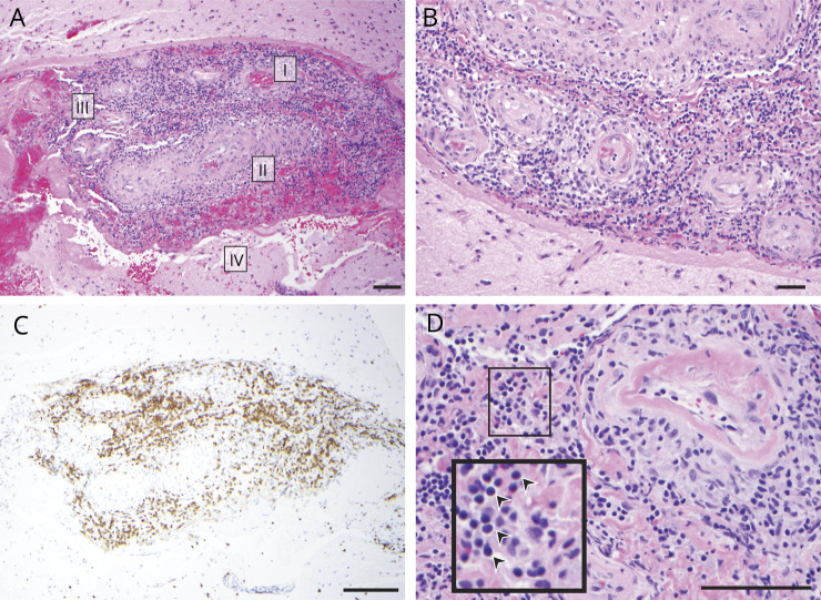
Figure 1. Histopathologic features of a diagnostic biopsy for PACNS patient cohort.
Biopsies from all patients in our primary angiitis of the CNS (PACNS) cohort (n = 8) showed inflammation of small to medium-sized CNS blood vessels. (A) Representative images from a patient biopsy (patient 3) demonstrating several hallmark features of PACNS histopathology, including (I) perivascular inflammation, (II) intramural inflammation with thickening of blood vessel wall, (III) leptomeningeal inflammation, and (IV) rupture of blood vessel wall. (B) Additional section of meningeal vessels (brain parenchyma, bottom left), showing transmural inflammation. (C) Immunohistochemistry with anti-CD3 shows enrichment of T cells among immune cell infiltrates corresponding to panel A. (D) High magnification of perivascular and intramural inflammation. Arrowheads in the inset indicate mononuclear lymphocytes. Tissue sections were stained with hematoxylin and eosin unless otherwise noted. Scale bar, 100 μm.

