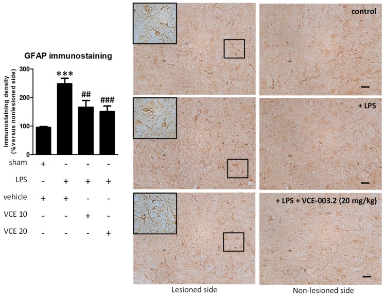
Figure 5.
Intensity of the immunostaining for GFAP measured in a selected area of the substantia nigra pars compacta of control and LPS-lesioned mice orally treated for 28 days after lesion with vehicle (corn oil) or VCE-003.2 at the doses of 10 (VCE 10) or 20 (VCE 20) mg/kg. Immunoreactivity values are measured in the lesioned side over the non-lesioned side, and correspond to means ± SEM of more than six subjects per group. Data were assessed using the one-way analysis of variance followed by the Bonferroni test (*** p < 0.005 versus vehicle-treated control (sham) mice; ## p < 0.01, ### p < 0.005 versus vehicle-treated LPS-lesioned mice). Representative immunostaining images for sham and LPS-lesioned mice treated with vehicle or VCE-003.2 at 20 mg/kg are shown at right (scale bar = 50 µm), including a specific inlet showing the morphological characteristics of GFAP-labeled cells (2x magnified).

