Abstract
Background
Vascular endothelial growth factor (VEGF) is a signal protein, implicated in various physiological and pathophysiological processes together with other common inflammatory biomarkers. However, their associations have not yet been fully elucidated. In the present study, we investigated associations between VEGF and four specific VEGF mRNA isoforms with levels of 11 inflammation molecules, derived from peripheral blood mononuclear cells (PBMCs) extracts.
Methods
Healthy participants from the STANISLAS Family Study (n = 285) were included. Levels of VEGF (four mRNA isoforms and protein levels) and inflammatory molecules (IL-1α, IL-1β, IL-2, IL-4, IL-6, IL-8, IL-10, INF-γ, TNF-α, MCP-1, EGF) were measured in PBMCs extracts. Multiple regression analyses were performed, adjusted for age and gender.
Results
The analyses revealed significant associations between VEGF protein levels and levels of IL-4 (β = 0.028, P = 0.013), MCP-1 (β = 0.015, P<0.0001) and EGF (β = 0.017, P<0.0001). Furthermore, mRNA isoform VEGF165 was associated with MCP-1 and IL-1α (P = 0.002 and P = 0.008, respectively); and mRNA isoform VEGF189 was associated with IL-4 and IL-6 (P = 0.019 and P = 0.034, respectively).
Conclusions
To our knowledge, the present study represents the first investigation that successfully demonstrates links between VEGF protein levels and inflammatory molecules levels derived from PBMCs extracts and identifies associations between specific VEGF mRNA isoforms and inflammatory molecules.
Impact
These findings provide novel insights that may assist in the development of new tissue and mRNA isoform specific measurements of VEGF levels, which may positively contribute to predicting the risk of common complex diseases and response of currently used anti-VEGF agents, and developing of novel targeted therapies for VEGF-related pathophysiology.
Introduction
Vascular endothelial growth factor A, VEGF-A (commonly referred as VEGF), is a multifunctional signal protein, which works as an important regulator of both physiological and pathological angiogenesis and has been related to a variety of pathologies, such as cancer and cardiovascular diseases (CVD) [1]. VEGF has become a perspective target for the design of anti-cancer treatments; anti-VEGF medications have already entered the clinical environment, however, the trade-off for the therapy is a common occurrence of cardiovascular side effects [2, 3].
VEGF is a prototype member of a cytokine family, which also includes placental growth factor (PLGF), VEGF-B, VEGF-C and VEGF-D, present in regulation of lymphangiogenesis, vasodilatation, chemotactic for different cells and vascular permeability [1]. Levels of VEGF represent a highly heritable phenotype (>60.5% as demonstrated in STANISLAS Family Study (SFS)) [4]. More than 50% of this variability is explained by ten single nucleotide polymorphisms identified through two genome-wide association studies (GWAS) [5, 6].
The VEGF gene produces more than 14 messenger RNA (mRNA) isoforms; the most predominant are VEGF121, VEGF145, VEGF165 and VEGF189, denoted by their length (number of amino acids). These isoforms differ in biochemical properties and receptor-binding characteristics that result in different effects on vessel growth [7]. Various tissues express different ratios of VEGF mRNA isoforms, including tumours, where growth appears to be most rapid when the isoform VEGF164 is expressed [7]. Therefore, disease susceptibility may depend on transcription of specific VEGF mRNA isoforms, rather than the currently measured VEGF protein. As a result, studies exploring specific VEGF mRNA isoforms for association with inflammatory molecules, intermediate phenotypes and diseases are warranted.
Human VEGF isoforms are classified into two main families: the VEGFxxx family and the VEGFxxxb family (xxx denoting the number of amino acids) [8]. The isoforms of these families differ only in the sequence of carboxy-terminal six amino acids, as the result of alternative splicing of exon eight of the VEGF gene [9]. This difference leads to isoforms with opposite functions; VEGFxxx isoforms (e.g. VEGF165) have pro-angiogenic properties whereas the VEGFxxxb isoforms (e.g. VEGF165b) have anti-angiogenic properties [10, 11]. VEGFxxxb isoforms present more than half of the total VEGF expressed in vitreous fluid, circulating plasma, urine, renal cortex, colonic epithelium, bladder smooth muscle, lung and pancreatic islets, whereas in tissues with physiological angiogenesis (placenta) or pathological angiogenesis (melanoma, colorectal or bladder cancer cells) VEGFxxx isoforms represent the majority of total VEGF [8].
VEGF is secreted from various cells: fibroblasts, tumour cells and inflammatory cells, such as lymphocytes [12, 13]. Lymphocytes are the most numerous components of peripheral blood cells (70–90% of PBMCs) and hold a central role in the regulation of the immune system [14]. Originating from a common lymphoid progenitor, there are three subgroups of lymphocytes, namely T-lymphocytes (70–85%), B-lymphocytes (5–20%) and natural killer cells (NK) (5–20%), each with their own function in immune response [15, 16]. Chronic inflammation often results in the infiltration of inflammatory cells at specific sites, among which the predominant are T-lymphocytes [17]. In addition to VEGF, other important signalling proteins are secreted from lymphocytes and are regulated by intracellular signalling control mechanisms or extracellular regulators [18]. Together, they are involved in complex molecular pathways that impact on physiological balance in human organism. The most important cytokine families involve hematopoietins, interferons, tumour necrosis factors-related molecules and chemokines [19]. VEGF levels have been previously related with cellular adhesion molecules (CAMs) [20], interleukin-1α (IL-1α)[21], interleukin-1β (IL-1β) [22], interleukin-4 (IL-4) [23–25], interleukin-6 (IL-6) [26, 27], nuclear factor-κB (NF-κB) transcription factor [28], endothelial growth factor (EGF) and others [29–31].
A detailed knowledge of biological pathways of VEGF is indispensable for further progress in pharmacological studies and detection of different isoforms of VEGF is of crucial importance for anticancer therapy with limited side effects. In addition, unravelling the relations between different biomarkers in physiological state of organisms can provide the basic knowledge that will help to understand these relations in pathological conditions.
The aim of this investigation is to assess the relationship between VEGF and inflammatory molecules, including cytokines and growth factors: IL-1α, IL-1β, IL-2, IL-4, IL-6, IL-8, IL-10, interferon-γ (INF-γ), tumour necrosis factor α (TNF-α), monocyte chemotactic protein 1 (MCP-1) and EGF, derived from PBMCs extracts in healthy individuals. PBMCs represent one of the most important sources of inflammatory molecules; therefore, we examined associations of cytokines and growth factors contained within PBMCs extracts utilizing a multiplex chemiluminescent biochip. Furthermore, because of the importance of VEGF isoforms, we examined the mRNA expression profiles of the four most biologically important VEGF isoforms (VEGF121, VEGF145, VEGF165 and VEGF189) and explored associations of each specific VEGF mRNA isoform with inflammatory molecules.
Materials and methods
Population
All subjects involved in this study make part of the STANISLAS Family Study (SFS). Information pertaining to this cohort has previously been described [32, 33]. Briefly, the SFS is a 10-year longitudinal survey with 3 visits at 5-year intervals, involving 1,006 families from Vandoeuvre-lès-Nancy, France, first recruited between 1993 and 1995. All subjects were of Northwest-European origin, without the presence of chronic disorders e.g. CVD or cancer, and without previous personal history of CVD. Study protocols were approved by the institutional ethics committees CCPPRB de Lorraine (Comité consultatif de protection des personnes dans la recherche biomédicale) and CNIL (Commission Nationale de l'Informatique et des Libertés). All individuals gave written informed consent for their participation in the study.
A subset of 285 participants (138 females and 147 males) from the SFS was selected for the measurement of twelve inflammatory molecules derived from PBMCs extracts.
A subset of 110 participants from the SFS was selected to quantify four specific VEGF mRNA isoforms from total RNA derived from PBMCs (i.e. VEGF121, VEGF145, VEGF165 and VEGF189), based on the sample availability.
Laboratory measurements
Isolation of PBMCs
Isolation of PBMCs was based on the method first described by Boyum in 1968 [34]. Briefly, whole blood was collected in tubes with sodium heparin and transported at room temperature. Hanks’ Balanced Salt Solution (SIGMA Aldrich, reference: H6648) was added into 15 mL tubes with blood (VHanks = Vblood) and poured gently into a 15 mL tube with Ficoll paque plus (Sigma Aldrich, reference: 17-1440-02) solution (VFicoll = VHanks+ Vblood). The contents were centrifuged for 30 min at 300 g at room temperature.
A PBMCs ring was retrieved and collected into a 15 mL tube, filled with Hanks’ Balanced Salt Solution and centrifuged for 10 min at 1000 g at room temperature. The supernatant was aspirated and 2 mL of Hanks’ Balanced Salt Solution were added. The solution was well suspended, filled up to 15 mL with Hanks’ Balanced Salt Solution and centrifuged for 10 min at 1000 g at room temperature (second washing). The PBMC ring was collected into Eppendorf tube with 1 mL of Hanks’ Balanced Salt Solution. PBMCs populations were evaluated by microscopic observation after May-Grunwald-Giemsa staining and the PBMCs concentration was normalized to 106 cells/mL in Hank’s Buffer. After final centrifugation 5 min at 1000 g at room temperature the supernatant was aspirated and the pellet of PBMCs was processed immediately or stored at -80 °C to maintain stability.
Total protein extraction
The lysis solution (lysate) composed of cell lysis buffer (CelLytic-M, SIGMA Aldrich, reference: C2978) and protease inhibitor (0.5%, Protease Inhibitor Cocktail, SIGMA Aldrich, reference: P8215) was added to the PBMC pellet, as recommended by the manufacturer (SIGMA Aldrich). This was stirred for 15 min at room temperature, and centrifuged for 15 min at 12000 g at 4 °C. The supernatant was collected and was immediately used for further analysis or stored at -80 °C to maintain stability.
Protein measurement
Quantification of inflammatory molecules (IL-1α, IL-1β, IL-2, IL-4, IL-6, IL-8, IL-10, MCP-1, TNF-α, INF-γ, EGF and VEGF (combination of VEGF121, VEGF145, VEGF165 and VEGF189 isoforms protein levels)) from PBMCs extracts was performed by Randox high sensitivity multiplex cytokine and growth factor array (Evidence Investigator Analyzer, Randox Laboratories Ltd., Crumlin, United Kingdom), which is a Randox patented 9 x 9 mm2 activated biochip with spatially discrete test regions containing antibodies specific to each of the inflammatory molecules assessed. In the present study, the combination of the four isoforms measured with the Cytokine array is referred to as “VEGF protein level”.
Gene expression analysis
Total RNA was extracted and quantified from isolated PBMCs using the MagNA Pure LC RNA HP isolation kit and RNA HP Blood External lysis protocol (Roche Diagnostics, France) as previously described [35, 36]. In short, 200 units of M-MuLV Reverse Transcriptase with 0.25 μg of oligos (dT) (Promega, France) were used to perform reverse transcription of total RNAs. Quantitative real-time PCR was performed on LightCycler instrument (Roche Diagnostics, Mannheim, Germany) with TaqMan Master Kit for all VEGF transcripts. Specific primers were used to selectively quantify the transcripts coding for the VEGF isoforms (S1 Table). All quantifications were carried out in duplicates, the starting amount of cDNA template was the same in all samples (25 ng) to remove any bias resulting from difference in the initial RNA quantity. Positive controls with known concentration were included in every run to ensure reproducibility by comparing the expression of VEGF transcripts between different PCR runs. A standard curve was generated by plotting the whole range of series dilution against the initial template quantity; this showed a linear standard curve and an efficiency of amplification near 99%. The absolute quantity of VEGF transcripts (number of copies/μL) in every sample was calculated by comparing the sample Ct value to the standard curve (Standard Curve Quantification method, The LightCycler 480 Software, Roche Diagnostic, France).
Statistical analysis
VEGF protein levels were tested for normal distribution using the Kolmogorov-Smirnov test of normality. Consequently, VEGF protein levels were log transformed (log10) to normalize a distribution of data. Data was tested for outliers (1.5×IQR interquartile range), which were removed before further analysis. Non-parametric correlation analyses (Spearman correlation) were performed. Linear regression models were applied, adjusted for age and gender, to test for possible associations between VEGF protein levels and inflammatory molecules.
Associations of specific VEGF mRNA isoforms and inflammatory molecules were assessed using identical statistical procedures. Only VEGF isoform 145 values didn’t follow the normal distribution and were log transformed before further analysis.
Mixed models adjusted for family structure were also applied to correct for possible issues of stratification due to familial resemblance.
All analyses were performed using SPSS 20.0 statistical software (SPSS, Armonk, New York: IBM Corp). Significance was determined at a two-tailed 0.05 level. Graphs for correlation and regression analyses were performed using R 3.5.2 statistical software with Jtools package.
Bioinformatics analysis
Alignment between different isoforms of VEGF was performed using Clustal-Omega program with the following parameters: default transition matrix Gonnet, gap opening penalty 6 bits and gap extension 1 bit. Clustal-Omega uses the HHalign algorithm and its default settings as its core alignment engine. The algorithm is described in details in Söding, J. [37].
Results
All twelve cytokines were detected in the studied population. All values represent the concentrations of proteins (pg/mL) measured in the cellular extracts. Concentrations are based on equal number of PBMCs (106) in all samples, counted before cell lysis. The characteristics of the study population are presented in Table 1.
Table 1. Characteristics of the study population (n = 285).
| Characteristic | Mean | SD |
| Age (years) | 39.13 | 14.57 |
| Gender (%) male | 51.58 | - |
| Interleukin 1 alpha (pg/mL) | 0.90 | 4.84 |
| Interleukin 1 beta (pg/mL) | 5.76 | 27.61 |
| Interleukin 2 (pg/mL) | 1.19 | 1.72 |
| Interleukin 4 (pg/mL) | 7.59 | 1.47 |
| Interleukin 6 (pg/mL) | 0.66 | 1.43 |
| Interleukin 8 (pg/mL) | 68.11 | 147.06 |
| Interleukin 10 (pg/mL) | 0.64 | 0.24 |
| Interferon gamma (pg/mL) | 0.55 | 3.06 |
| Tumour Necrosis Factor alpha (pg/mL) | 3.87 | 16.01 |
| Monocyte Chemoattractant Protein 1 (pg/mL) | 9.40 | 5.60 |
| Epidermal Growth Factor (pg/mL) | 6.80 | 6.50 |
| Vascular Endothelial Growth Factor (pg/mL) | 56.99 | 67.62 |
SD: Standard deviation
Associations between VEGF levels and inflammatory molecules
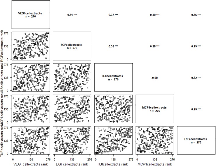
VEGF protein levels were correlated with EGF, IL-1β, IL-8, MCP1 and TNF-α (Fig 1). Results are presented in Table 2, the significant P-values are presented in bold.
Fig 1. Correlation graph between VEGF protein levels (VEGF cellular extracts) and EGF, IL-8, MCP1 and TNF-α cellular extracts levels.
‘***’ p ≤ 0.001.
Table 2. Correlation analysis between VEGF protein levels and inflammation molecules.
| VEGF | ||
|---|---|---|
| Correlation coefficient | P-value | |
| EGF | 0.5529 | < .0001 |
| IFN-γ | 0.1677 | 0.2599 |
| IL-1α | 0.001 | 0.9945 |
| IL-1β | 0.2896 | 0.0483 |
| IL-2 | -0.0911 | 0.5427 |
| IL-4 | 0.2256 | 0.1274 |
| IL-6 | -0.0077 | 0.959 |
| IL-8 | 0.3393 | 0.0197 |
| IL-10 | -0.0571 | 0.7028 |
| MCP1 | 0.3868 | 0.0072 |
| TNF-α | 0.394 | 0.0061 |
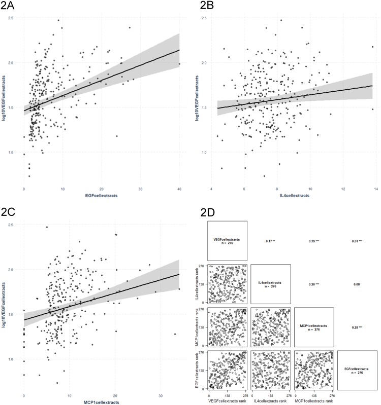
VEGF protein levels were associated with IL-4, MCP-1 and EGF levels in linear regression models adjusted for age and gender (Fig 2). Results are presented in Table 3.
Fig 2. Regression graphs for VEGF protein levels (VEGF cellular extracts) as dependent variable and EGF (2A), IL-4 (2B) and MCP1 (2C) cellular extracts levels as independent variables. The related correlation graph is presented in (2D).
‘***’ p ≤ 0.001, ‘**’ p ≤ 0.01.
Table 3. Significant determinants of VEGF protein levels extracted from PBMCs (linear regression analyses adjusted for age and gender).
| VEGF | |||
|---|---|---|---|
| Inflammatory molecule | β | SE | P |
| Interleukin 4 | 0.028 | 0.011 | 0.013 |
| Monocyte Chemoattractant Protein 1 | 0.015 | 0.003 | <0.0001 |
| Epidermal Growth Factor | 0.017 | 0.003 | <0.0001 |
SE: Standard error
Associations between VEGF mRNA isoforms and levels of inflammatory molecules extracted from PBMCs
The Randox cytokine array detects four most common VEGF isoforms, however it does not quantify them individually but displays a sum of all VEGF detected in the sample. It has been previously confirmed that various isoforms portray different roles in pathophysiological processes; this is why their quantification and association to other inflammatory molecules are of particular interest. As a result, we investigated associations between 11 inflammatory molecules derived from PBMCs extracts and the expression of specific VEGF mRNA isoforms derived and quantified from PBMCs in a subset of individuals from the SFS cohort (Table 4).
Table 4. Characteristics of the subsample of the population used for assessment of associations between inflammatory molecules levels from PBMCs extracts and VEGF mRNA isoforms (n = 110).
| Mean | SD | |
|---|---|---|
| Age (years) | 47.35 | 10.78 |
| Gender (%) male | 48.18 | - |
| Interleukin 1 alpha (pg/mL) | 0.81 | 2.75 |
| Interleukin 1 beta (pg/mL) | 4.88 | 17.42 |
| Interleukin 2 (pg/mL) | 0.90 | 1.3 |
| Interleukin 4 (pg/mL) | 7.75 | 1.55 |
| Interleukin 6 (pg/mL) | 0.614 | 1.22 |
| Interleukin 8 (pg/mL) | 67.65 | 135.91 |
| Interleukin 10 (pg/mL) | 0.65 | 0.24 |
| Interferon gamma (pg/mL) | 0.30 | 0.36 |
| Tumour Necrosis Factor alpha (pg/mL) | 3.85 | 16.55 |
| Monocyte Chemoattractant Protein 1 (pg/mL) | 9.58 | 5.59 |
| Epidermal Growth Factor (pg/mL) | 6.84 | 7.09 |
| VEGF 121 | 47.96 | 19.95 |
| VEGF 145 | 47.19 | 22.52 |
| VEGF 165 | 250.52 | 111.77 |
| VEGF 189 | 15.03 | 7.17 |
VEGF: Vascular endothelial growth factor, VEGF transcript copies number/25 ng cDNA
Studies have established that VEGF165 and VEGF121 are the most abundantly expressed VEGF isoforms [38, 39]. Our detection of mRNA isoforms of VEGF in PBMCs has confirmed these findings.
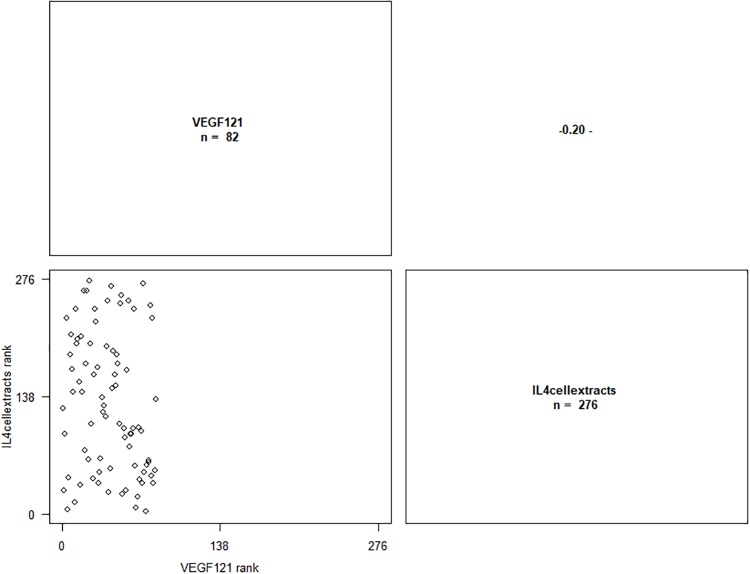
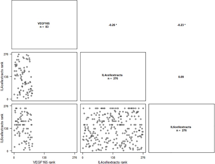
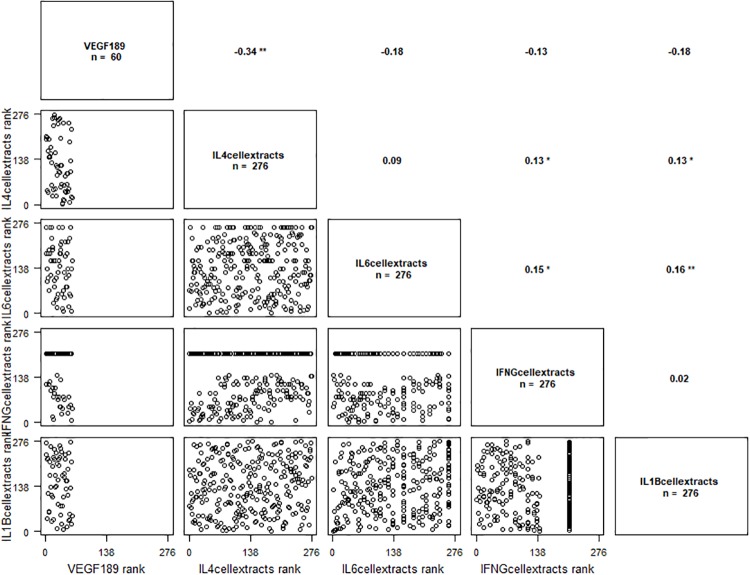
VEGF121 isoform was correlated with IL-4 (Fig 3), VEGF165 isoform was correlated with IL4 and IL6 (Fig 4) and VEGF189 isoform was correlated with IFN-γ, IL-1β, IL-4 and IL-6 (Fig 5). Results are presented in Table 5, the significant P-values are presented in bold.
Fig 3. Correlation graph between VEGF121 isoform mRNA levels and IL-4 cellular extracts levels.
‘-’ p ≤ 0.1.
Fig 4. Correlation graph between VEGF165 isoform mRNA levels and IL-4 and IL-6 cellular extracts levels.
‘*’ p ≤ 0.05.
Fig 5. Correlation graph between VEGF189 isoform mRNA levels and IL-4, IL-6, INFγ et IL-1β cellular extracts levels.
‘**’ p ≤ 0.01, ‘*’p ≤ 0.05.
Table 5. Correlation analysis between VEGF isoforms and inflammation molecules.
| VEGF121 | VEGF145 | VEGF165 | VEGF189 | |||||
|---|---|---|---|---|---|---|---|---|
| Correlation coefficient | P-value | Correlation coefficient | P-value | Correlation coefficient | P-value | Correlation coefficient | P-value | |
| EGF | -0.1626 | 0.2748 | -0.0607 | 0.6855 | -0.1426 | 0.3388 | -0.2128 | 0.1509 |
| IFN-γ | -0.2412 | 0.1025 | 0.0034 | 0.9821 | -0.2872 | 0.0503 | -0.3415 | 0.0188 |
| IL-1α | 0.1276 | 0.3928 | -0.236 | 0.1102 | 0.1256 | 0.4001 | -0.0042 | 0.9778 |
| IL-1β | -0.171 | 0.2504 | -0.1735 | 0.2435 | -0.1907 | 0.1992 | -0.3088 | 0.0347 |
| IL-2 | -0.1759 | 0.237 | 0.129 | 0.3874 | -0.1727 | 0.2457 | -0.1155 | 0.4393 |
| IL-4 | -0.4345 | 0.0023 | -0.1329 | 0.3731 | -0.3643 | 0.0118 | -0.3302 | 0.0234 |
| IL-6 | -0.2215 | 0.1346 | -0.1797 | 0.2269 | -0.3769 | 0.009 | -0.3368 | 0.0206 |
| IL-8 | -0.0797 | 0.5942 | 0.0147 | 0.9216 | -0.1354 | 0.3641 | -0.1641 | 0.2702 |
| IL-10 | -0.0535 | 0.7208 | -0.0787 | 0.5992 | -0.0619 | 0.6794 | -0.0676 | 0.6518 |
| MCP1 | -0.008 | 0.9577 | -0.1209 | 0.4184 | -0.0007 | 0.9963 | -0.2326 | 0.1156 |
| TNF-α | -0.0662 | 0.6584 | 0.0679 | 0.6503 | -0.1271 | 0.3944 | -0.2303 | 0.1194 |
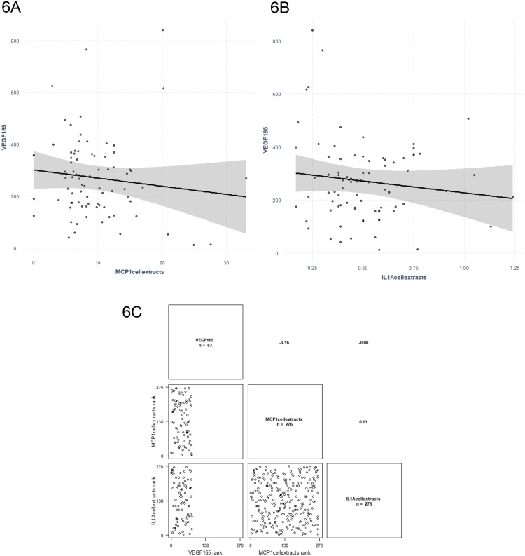
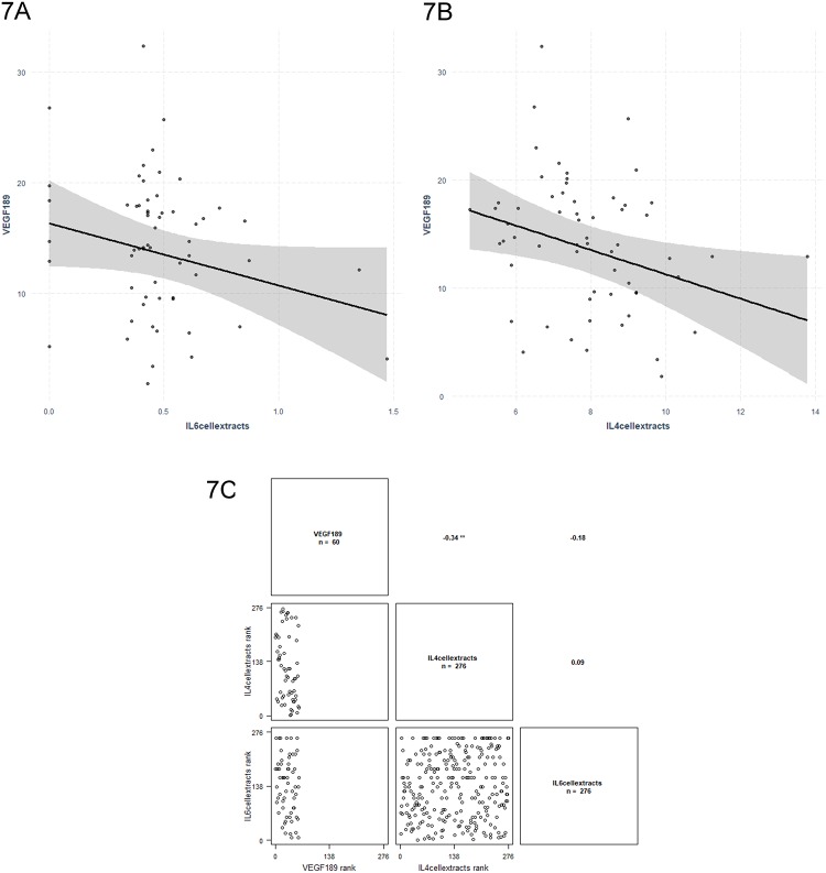
Two VEGF mRNA isoforms were significantly associated with four inflammatory molecules in linear regression analysis. VEGF165 was associated with MCP-1 (P = 0.002) and IL-1α (P = 0.008) (Fig 6), whereas VEGF189 was associated with IL-4 (P = 0.019) and IL-6 (P = 0.034) (Fig 7). Results are presented in Table 6.
Fig 6. Regression graphs for VEGF165 isoform mRNA levels as dependent variable and MCP1 (6A) and IL-1α (6B) cellular extracts levels as independent variables. The related correlation graph is presented in (6C).
‘**’ p ≤ 0.01.
Fig 7.
Regression graphs for VEGF189 isoform mRNA levels as dependent variable and IL-6 (7A) and IL-4 (7B) cellular extracts levels as independent variables. The related correlation graph is presented in (7C).
Table 6. Significant associations between the expression of VEGF mRNA isoforms and inflammatory molecules (linear regression models adjusted for age and gender).
| VEGF165 | VEGF189 | |||||
|---|---|---|---|---|---|---|
| Inflammatory molecule | β | SE | P | β | SE | P |
| Monocyte Chemoattractant Protein 1 | -0.319 | 0.006 | 0.002 | - | - | - |
| Interleukin 1 alpha | -0.269 | 0.010 | 0.008 | - | - | - |
| Interleukin 4 | - | - | - | -0.290 | 0.017 | 0.019 |
| Interleukin 6 | - | - | - | -0.260 | 0.110 | 0.034 |
VEGF: Vascular endothelial growth factor
The STANISLAS cohort used in this study is composed of related individuals, therefore the analysis have been repeated using mixed models adjusted for family structure. The mixed model gave the same significant results as the linear regression model.
Multiple alignment of VEGF isoforms
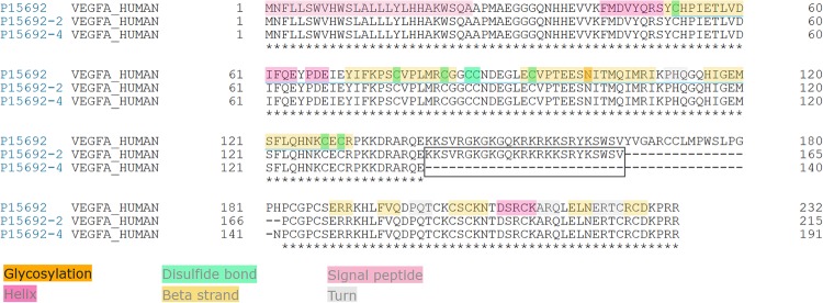
Analysis of primary and secondary structures of VEGF isoforms provides elements of understanding that may explain the commitment to different signaling pathways. VEGF189 and VEGF165 represent the same protein and differ only by 25 amino acids (Fig 8).
Fig 8. Alignment and elements of secondary structure of three VEGF isoforms (82% identity).
P15692 corresponds to VEGF206 considered as canonical sequence of VEGF. P15692-2 represents VEGF189 and P15692-4 represents VEGF165. Annotations are only depicted on the canonical sequence and stars indicate perfect alignment. The black frame shows the difference in amino acid composition (25) between VEGF189 and VEGF165.
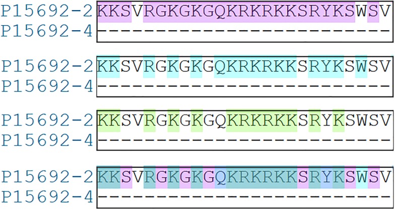
Thus, VEGF189 compared to VEGF165 owns additional residues, allowing to interact with molecules, such as IL-4 and IL-6. Without them, the signaling pathway seems to implicate others actors, such as MCP-1 and IL-1α. A detailed analysis shows that these surnumerary amino acids harbor specific properties (Fig 9).
Fig 9. Focus on the 25 specific amino acids of VEGF189 versus VEGF165.
P15692-2 corresponds to VEGF189 and P15692-4 to VEGF165. Analysis of amino acid properties revealed that VEGF189 owns 25 amino acids with three features. First, 22 are polar (top alignment highlighted in purple), second, 16 belong to the category of bulky amino acids (highlighted in blue), and third, 13 have a positive charge (highlighted in green). The last one is a superposition of the three.
These additional residues may modify polarity, steric hindering and the electrostatic potential of VEGF189 leading to change its counterparts and providing elements that could explain why these two close proteins are not associated with the same molecules.
Discussion
In the present study, we explored the relationship between VEGF protein levels and inflammatory molecules derived from PBMCs extracts from healthy individuals from the SFS cohort. In addition, specific mRNA VEGF isoforms from PBMCs were assessed for association with inflammatory molecules. To the best of our knowledge, this is the first study to investigate levels of inflammatory molecules in PBMCs extracts and their relationship with VEGF protein levels and specific VEGF mRNA isoforms.
In summary, VEGF protein levels were correlated with EGF, IL1-β, IL-8, MCP1 and TNFα levels, while linear regression model adjusted for age and gender demonstrated that IL-4 (P = 0.013), MCP-1 (P≤0.0001) and EGF (P≤0.0001) levels were significant predictors of VEGF levels. The mRNA VEGF121 isoform was correlated with IL-4 levels, while the VEGF165 was correlated with IL-4 and IL-6 levels and the VEGF189 was correlated with IFN-γ, IL-1β, IL-4 and IL6 levels. The regression models adjusted for age and sex demonstrated that for the expression of the mRNA VEGF165 isoform the MCP-1 (P = 0.002) and IL-1α (P = 0.008) levels were significant predictors, while for the expression of the mRNA VEGF189 isoform, the IL-4 (P = 0.019) and IL-6 (P = 0.034) levels were significant predictors.
PBMCs are particularly interesting cells, worthy of investigation because of their essential role in the synthesis and release of inflammatory cytokines and growth factors. Therefore, it is biologically plausible to explore associations of inflammatory molecules with VEGF protein levels and specific VEGF mRNA isoforms (i.e. VEGF121, VEGF145, VEGF165 and VEGF189) derived from PBMCs. PBMCs are a complement of cells derived from whole blood, composed of lymphocytes, monocytes, and dendritic cells. In humans, the frequencies of these cell populations vary across individuals. Lymphocytes are typically in the range of 70% to 90% of total PBMCs, monocytes range from 10% to 30% of total PBMCs, while dendritic cells are uncommon, estimated at only 1% to 2% of total PBMCs [16].
PBMCs are commonly used in immunology to examine cytokine secretion under modified condition [40, 41], in vaccine development [42, 43] and can serve as tissue for gene expression studies [44]. Gene expression of VEGF isoforms has been systematically measured in PBMCs for different purposes; to compare the expression of different isoforms in particular disease [45, 46] or to understand the control of VEGF expression [47]. Besides VEGF expression profile, PBMCs have been used to detect RNAs for characterization of cardiovascular diseases [48, 49], diabetic nephropathy [50], rheumatic diseases [51], infectious diseases [52] and many other diseases. Protein expression of inflammatory cytokines from PBMCs has also been extensively studied. Increased expression of TNF-α and IL-6 along with increased secretion of leptin was found in obese cardiovascular patients [53]. Moreover, studies showed that protein expression or plasmatic levels of cytokines released by PBMCs can be indicators of pathologic state of organism [54]. Indeed, inflammation plays a central role in development of many chronic diseases [55] and PBMCs play an important role in inflammatory cytokine production.
Our investigation exploring the association between VEGF and inflammatory molecules followed a two-staged approach. Firstly, VEGF protein levels detected by microarray were analyzed. However, this method is not sensitive to specific VEGF isoforms; therefore, we used PBMCs also for mRNA isolation and quantification of VEGF expression isoforms. This enabled quantification of the expression of four isoforms in the second stage of the study. Two of the inflammatory molecules, IL-4 and MCP-1, were significantly associated to VEGF protein levels, and were also associated with the expression of VEGF specific isoforms 189 and 165, respectively.
Firstly, IL-4 was related with VEGF levels derived from PBMCs extracts and VEGF189 mRNA levels detected from PBMCs. IL-4 is a multi-functional cytokine with anti-inflammatory and anti-tumor activity, produced by lymphocytes, basophils and mast cells [56]. Its anti-inflammatory properties are due to the ability of controlling the production of pro-inflammatory mediators by inhibiting their induction [57]. IL-4 has been previously related to VEGF as an important factor in the recruitment of tumor-associated macrophages (TAMs), which are known to promote angiogenesis, tissue remodeling and immunosuppression. As it was demonstrated in the study of Linde et. al [23, 58], VEGF is responsible for recruitment of monocytes from the peripheral circulation, whereas IL-4 induces their differentiation into tumor-promoting M2-like macrophages. The association of VEGF and IL-4 was also detected in human coronary artery endothelial cells [59] and in smooth muscle cells [25]; both studies noticed induced release of VEGF while treated with IL-4. Faffe et al. [25] also reported the increasing of VEGF-mRNA expression, caused by IL-4, but observed no alteration of VEGF promoter activity. Additionally, IL-4 has been shown to increase VEGF levels in fibroblast-like synoviocytes when applied alone, but portrayed an anti-angiogenic effect in the presence of transforming growth factor (TGF)-β, by inhibiting the VEGF production, in the study of patients with rheumatoid arthritis [56]. Thus, these results demonstrate the importance of understanding the synergy of different molecules, which can elucidate important biological pathways and thus open new possibilities for treatment of complex diseases. Previous studies have not proposed a specific VEGF isoform related to IL-4. Of note, our study is the first to propose a link between the isoform 189 and IL-4, but an additional research is warranted to confirm this finding.
Secondly, MCP-1 was another molecule statistically associated with both VEGF protein levels and VEGF mRNA. MCP-1 is an angiogenic chemokine, involved in the regulation, migration and infiltration of monocytes and macrophages [60]. Previous research found that MCP-1 induced angiogenesis is mediated by VEGF [61]. More specifically, the researchers identified that MCP-1 up-regulated hypoxia-inducible factor 1 alpha gene expression in human aortic endothelial cells, which as a result induced VEGF165 expression in the aortic wall. The importance of MCP-1 in regulation of angiogenesis and immune system was discussed in the study of breast cancer tissue, where twelve inflammatory molecules were measured and evaluated for correlations [62]. This study confirmed that the expression of MCP-1 was correlated significantly with TAM accumulation in primary breast tumours, and with VEGF. Prognostic analysis revealed that high expression of MCP-1/VEGF was an independent indicator of early relapse of cancer. A study in vascular smooth muscle cells of rats [63] demonstrated that VEGF was responsible for the mitogenic activity of MCP-1, which leads to vessel wall remodeling. Moreover, severe hypoxia, previously known to induce VEGF release, potentiated the growth-promoting effect of MCP-1 [63]. This effect on vessel growth was studied also for therapeutic purposes. The therapy with dual delivery of VEGF and MCP-1 showed beneficial effects during transplantation of endothelial cells for therapeutic vascularisation [64], where VEGF improved survival of transplanted cells and MCP-1 induced mural cell recruitment. In the current study, MCP-1 was associated with VEGF protein levels and specific mRNA VEGF165 isoform, which confirmed the results of previous studies.
Thirdly, our study identified association of VEGF isoform 165 also with protein IL-6. Several studies demonstrated IL-6 induction of VEGF expression. For example, Choen et al. demonstrated that treatment of cell lines with IL-6 results in significant increase of VEGF mRNA [65]. Moreover, IL-6 was shown to promote cervical tumorigenesis by activating VEGF-mediated angiogenesis via a STAT3 pathway [66]. Similarly, it has been shown to induce VEGF expression and as a result increase angiogenesis in gastric carcinoma patients [26] and in malignant mesothelioma [67]. These results suggest that treatment with IL-6R antibody might constitute a potential target therapy for certain cancer types. We would, again, reiterate the importance of testing for specific VEGF isoforms, which would, in combination with different targeted proteins, such as IL-6, contribute to specific and efficient therapies.
An association was observed also between IL-1α and VEGF165 isoform. IL-1α is a prototypical proinflammatory cytokine, known also as hematopoietin 1, which is involved in various immune responses, inflammatory processes, and hematopoiesis [68]. It is produced by monocytes and macrophages and released in response to cell injury [69]. It has been previously shown that IL-1α stimulates VEGF secretion by PBMCs in a dose-depended manner, via induction of VEGF mRNA synthesis. Four VEGF isoforms were encoded with de novo synthetized mRNA, VEGF121, VEGF165, VEGF189, and VEGF206. Moreover, VEGF121 and VEGF165 gave the major signals both in unstimulated and IL-1α stimulated PBMCs, which is also in favour of our results [21]. Correlation of VEGF production with IL-1α and IL-6 in cell lines suggests that the expression of VEGF is regulated by IL-1α and IL-6 in pancreatic cancer [70] and in adenoma cells [71].
Finally, an association of VEGF protein levels and EGF has been observed in the current study. EGF plays an important role in the growth, proliferation and differentiation of numerous cell types, by binding with high affinity to the cell surface receptor [72]. Major role of both related growth factors in cancer development has been researched for various types of cancers and different mechanism of their common pathways have been proposed [73–75]. Combination of VEGF/EGF signaling pathways has been proposed for new therapeutic approaches of anticancer therapy [76–79] and is showing positive predictions for future development of new medications.
VEGF isoforms are expressed in different quantities, based on the cell type that is producing them. Most cells express VEGF121, VEGF165 (the most abundant) and VEGF189. Quantification of VEGF isoforms in our study confirmed the reports from literature, as the most expressed isoform from the PBMCs population was VEGF165, followed by VEGF121. VEGF145 was also quantified in our research, although it is more commonly found in cells of placental origin [80, 81]. The notion that the identification of VEGF isoforms is important in fundamental biological and pharmacological studies has arisen the last few years. Specific VEGF isoforms were studied in different types of cancer [82], cardiovascular disease [83], kidney disease [84], autoimmune disease [85] and many others. Pro-angiogenic VEGFxxx and anti-angiogenic VEGFxxxb splicing variants are particularly interesting; it has been demonstrated that their ratio varies based on different pathology, therefore, determination of pro and anti-angiogenic isoforms could serve as diagnostic tools for such diseases [86–88]. Moreover, the quantification of pro and anti-angiogenic isoforms was shown to have a predictive value in response to a treatment with bevacizumab [89]. However, there is no available method that could separate pro and anti-angiogenic isoforms routinely, therefore, most of the studies quantify only pro-angiogenic isoforms. Likewise, in our research, only pro-angiogenic isoforms were detected and associated with specific protein levels. Still, we are aware of the importance of separation and quantification of all VEGF splice variants. We believe that in the future studies, anti-angiogenic isoforms should be routinely quantified and distinguished from pro-angiogenic isoforms for attempting diagnosis of different chronic diseases and prediction of response of anti-VEGF agents (pharmacogenomics).
With such progress, the discovery of anticancer treatments, which will specifically target VEGF isoforms and would decrease the risk of adverse effects of current anti-VEGF therapies will be only a matter of time. VEGF isoforms have already shown a good potential for significant and independent roles in the prediction of different types of cancers [90, 91] and differentiation of isoforms for therapeutic purposes is now being extensively studied [92, 93]. On the other hand, in the complex system of biological pathways, there is usually more than one factor impacting on a pathological development. Therefore, knowing the roles of various proteins related to VEGF is of special importance. It has been shown in previous studies that many cytokines and growth factors upregulate VEGF mRNA or induce VEGF release [31]. In support of this, the current investigation also identifies several significant associations.
VEGF189 and VEGF165 are two isoforms of VEGF protein sharing more than 88% sequence identity. Indeed, analysis of their multiple alignment shows that they differ by only 25 amino acids. These additional amino acids modifying polarity, steric hindering and basic properties confer a subtle difference, sufficient to significantly modify their features and involve them in different signaling pathways. Thus, according to our results, thanks to supplemental residues VEGF189 could favor molecules interacting with IL-4 and IL-6 while VEGF165 seems to be associated rather with MCP-1 and IL-1α.
One main limitation of our study is that we were unable to confirm the obtained results for VEGFmRNA/protein levels as the availability of biological materials was unfortunately limiting.
Conclusion
This investigation has revealed significant associations between VEGF levels and its specific mRNA isoforms with inflammatory molecules derived from PBMCs cellular extracts, specifically cytokines and growth factors IL-1α, IL4, IL-6, EGF, and MCP1 in a population of healthy individuals from the SFS. This confirmed that cytokines and growth factors play an important role in VEGF regulation also at lymphocytes cellular extract level. Importantly, specific isoforms interact with different inflammatory molecules. Therefore, they should be identified in future investigations of molecular and biological pathways involving VEGF, which would contribute to an in-depth and specific understanding of the process and would ultimately offer novel targeted therapies for VEGF-related pathophysiology.
Supporting information
(DOCX)
(XLSX)
Data Availability
The data set was uploaded as supporting information.
Funding Statement
Financial support was provided by the European Union within the frames of the Operational Programme FEDER-FSE Lorraine et Massif des Vosges 2014-2020, by Agence Nationale de la Recherche, programme d’Investissements d’avenir, grant number ANR-15RHU-0004 and GEENAGE project (Functional Genomic, Epigenomic and ENvironment interplay to IMPACT management of healthy and pathological AGEing)” within the frames of the LUE project (Lorraine University of Excellence). VG was partially funded by the “Région Lorraine”. The funders had no role in study design, data collection and analysis, decision to publish, or preparation of the manuscript.
References
- 1.Ferrara N, Gerber HP, LeCouter J. The biology of VEGF and its receptors. Nat Med. 2003;9(6):669–76. 10.1038/nm0603-669 [DOI] [PubMed] [Google Scholar]
- 2.Zhang W, Shen Z, Luo H, Hu X, Zheng L, Zhu X. The Benefits and Side Effects of Bevacizumab for the Treatment of Recurrent Ovarian Cancer. Curr Drug Targets. 2017;18(10):1125–31. 10.2174/1389450117666160502150237 [DOI] [PubMed] [Google Scholar]
- 3.Riondino S, Monte GD, Fratangeli F, Guadagni F, Roselli M, Ferroni P. Anti-angiogenic drugs, vascular toxicity and thromboembolism in solid cancer. Cardiovasc Hematol Agents Med Chem. 2017. [DOI] [PubMed] [Google Scholar]
- 4.Berrahmoune H, Herbeth B, Lamont JV, Masson C, Fitzgerald PS, Visvikis-Siest S. Heritability for plasma VEGF concentration in the Stanislas family study. Ann Hum Genet. 2007;71(Pt 1):54–63. 10.1111/j.1469-1809.2006.00298.x [DOI] [PubMed] [Google Scholar]
- 5.Debette S, Visvikis-Siest S, Chen MH, Ndiaye NC, Song C, Destefano A, et al. Identification of cis- and trans-acting genetic variants explaining up to half the variation in circulating vascular endothelial growth factor levels. Circ Res. 2011;109(5):554–63. 10.1161/CIRCRESAHA.111.243790 [DOI] [PMC free article] [PubMed] [Google Scholar]
- 6.Choi SH, Ruggiero D, Sorice R, Song C, Nutile T, Vernon Smith A, et al. Six Novel Loci Associated with Circulating VEGF Levels Identified by a Meta-analysis of Genome-Wide Association Studies. PLoS Genet. 2016;12(2):e1005874 10.1371/journal.pgen.1005874 [DOI] [PMC free article] [PubMed] [Google Scholar]
- 7.Vempati P, Popel AS, Mac Gabhann F. Extracellular regulation of VEGF: isoforms, proteolysis, and vascular patterning. Cytokine Growth Factor Rev. 2014;25(1):1–19. 10.1016/j.cytogfr.2013.11.002 [DOI] [PMC free article] [PubMed] [Google Scholar]
- 8.Harper SJ, Bates DO. VEGF-A splicing: the key to anti-angiogenic therapeutics? Nature reviews Cancer. 2008;8(11):880–7. 10.1038/nrc2505 [DOI] [PMC free article] [PubMed] [Google Scholar]
- 9.Woolard J, Bevan HS, Harper SJ, Bates DO. Molecular diversity of VEGF-A as a regulator of its biological activity. Microcirculation. 2009;16(7):572–92. 10.1080/10739680902997333 [DOI] [PMC free article] [PubMed] [Google Scholar]
- 10.Nowak DG, Amin EM, Rennel ES, Hoareau-Aveilla C, Gammons M, Damodoran G, et al. Regulation of vascular endothelial growth factor (VEGF) splicing from pro-angiogenic to anti-angiogenic isoforms: a novel therapeutic strategy for angiogenesis. J Biol Chem. 2010;285(8):5532–40. 10.1074/jbc.M109.074930 [DOI] [PMC free article] [PubMed] [Google Scholar]
- 11.Harper SJ, Bates DO. VEGF-A splicing: the key to anti-angiogenic therapeutics? Nat Rev Cancer. 2008;8(11):880–7. 10.1038/nrc2505 [DOI] [PMC free article] [PubMed] [Google Scholar]
- 12.Mor F, Quintana FJ, Cohen IR. Angiogenesis-inflammation cross-talk: vascular endothelial growth factor is secreted by activated T cells and induces Th1 polarization. J Immunol. 2004;172(7):4618–23. 10.4049/jimmunol.172.7.4618 [DOI] [PubMed] [Google Scholar]
- 13.Kieran MW, Kalluri R, Cho YJ. The VEGF Pathway in Cancer and Disease: Responses, Resistance, and the Path Forward. Cold Spring Harb Perspect Med. 2012;2(12). [DOI] [PMC free article] [PubMed] [Google Scholar]
- 14.Corkum CP, Ings DP, Burgess C, Karwowska S, Kroll W, Michalak TI. Immune cell subsets and their gene expression profiles from human PBMC isolated by Vacutainer Cell Preparation Tube (CPT()) and standard density gradient. BMC Immunol. 2015;16. [DOI] [PMC free article] [PubMed] [Google Scholar]
- 15.Lackie JM. The Dictionary of Cell and Molecular Biology: Elsevier Science; 2012. [Google Scholar]
- 16.Miyahira A. Sanguine Bio Researcher Blog [Internet]. PBMC Basic: Sanguine Biosciences. 2012. [cited 2018]. https://technical.sanguinebio.com/types-of-immune-cells-present-in-human-pbmc/.
- 17.Angelo LS, Kurzrock R. Vascular endothelial growth factor and its relationship to inflammatory mediators. Clin Cancer Res. 2007;13(10):2825–30. 10.1158/1078-0432.CCR-06-2416 [DOI] [PubMed] [Google Scholar]
- 18.Turner MD, Nedjai B, Hurst T, Pennington DJ. Cytokines and chemokines: At the crossroads of cell signalling and inflammatory disease. Biochim Biophys Acta. 2014;1843(11):2563–82. 10.1016/j.bbamcr.2014.05.014 [DOI] [PubMed] [Google Scholar]
- 19.Paul WE, Seder RA. Lymphocyte responses and cytokines. Cell. 1994;76(2):241–51. 10.1016/0092-8674(94)90332-8 [DOI] [PubMed] [Google Scholar]
- 20.Krieglstein CF, Granger DN. Adhesion molecules and their role in vascular disease. Am J Hypertens. 2001;14(6 Pt 2):44s–54s. 10.1016/s0895-7061(01)02069-6 [DOI] [PubMed] [Google Scholar]
- 21.Salven P, Hattori K, Heissig B, Rafii S. Interleukin-1alpha promotes angiogenesis in vivo via VEGFR-2 pathway by inducing inflammatory cell VEGF synthesis and secretion. FASEB J. 2002;16(11):1471–3. 10.1096/fj.02-0134fje [DOI] [PubMed] [Google Scholar]
- 22.Repovic P, Fears CY, Gladson CL, Benveniste EN. Oncostatin-M induction of vascular endothelial growth factor expression in astroglioma cells. Oncogene. 2003;22(50):8117–24. 10.1038/sj.onc.1206922 [DOI] [PubMed] [Google Scholar]
- 23.De Palma M. Partners in crime: VEGF and IL-4 conscript tumour-promoting macrophages. J Pathol. 2012;227(1):4–7. 10.1002/path.4008 [DOI] [PubMed] [Google Scholar]
- 24.Hong K, Cho M, Min S, Shin Y, Yoo S, Choi J, et al. Effect of interleukin-4 on vascular endothelial growth factor production in rheumatoid synovial fibroblasts. Clin Exp Immunol. 2007;147(3):573–9. 10.1111/j.1365-2249.2006.03295.x [DOI] [PMC free article] [PubMed] [Google Scholar]
- 25.Faffe DS, Flynt L, Bourgeois K, Panettieri RA, Shore SA. Interleukin-13 and Interleukin-4 Induce Vascular Endothelial Growth Factor Release from Airway Smooth Muscle Cells: Role of Vascular Endothelial Growth Factor Genotype. Am J Respir Cell Mol Biol. 2006;34(2):213–8. 10.1165/rcmb.2005-0147OC [DOI] [PMC free article] [PubMed] [Google Scholar]
- 26.Huang SP, Wu MS, Shun CT, Wang HP, Lin MT, Kuo ML, et al. Interleukin-6 increases vascular endothelial growth factor and angiogenesis in gastric carcinoma. J Biomed Sci. 2004;11(4):517–27. 10.1007/bf02256101 [DOI] [PubMed] [Google Scholar]
- 27.Loeffler S, Fayard B, Weis J, Weissenberger J. Interleukin-6 induces transcriptional activation of vascular endothelial growth factor (VEGF) in astrocytes in vivo and regulates VEGF promoter activity in glioblastoma cells via direct interaction between STAT3 and Sp1. Int J Cancer. 2005;115(2):202–13. 10.1002/ijc.20871 [DOI] [PubMed] [Google Scholar]
- 28.Huang S, Robinson JB, DeGuzman A, Bucana CD, Fidler IJ. Blockade of nuclear factor-κB signaling inhibits angiogenesis and tumorigenicity of human ovarian cancer cells by suppressing expression of vascular endothelial growth factor and interleukin 8. Cancer Res. 2000;60(19):5334–9. [PubMed] [Google Scholar]
- 29.Lau TP, Roslani AC, Lian LH, Lee PC, Hilmi I, Goh KL, et al. Association between EGF and VEGF functional polymorphisms and sporadic colorectal cancer in the Malaysian population. Genet Mol Res. 2014;13(3):5555–61. 10.4238/2014.July.25.9 [DOI] [PubMed] [Google Scholar]
- 30.Lequoy P, Murschel F, Liberelle B, Lerouge S, De Crescenzo G. Controlled co-immobilization of EGF and VEGF to optimize vascular cell survival. Acta Biomater. 2016;29:239–47. 10.1016/j.actbio.2015.10.026 [DOI] [PubMed] [Google Scholar]
- 31.Robinson CJ, Stringer SE. The splice variants of vascular endothelial growth factor (VEGF) and their receptors. J Cell Sci. 2001;114(Pt 5):853–65. [DOI] [PubMed] [Google Scholar]
- 32.Visvikis-Siest S, Siest G. The STANISLAS Cohort: a 10-year follow-up of supposed healthy families. Gene-environment interactions, reference values and evaluation of biomarkers in prevention of cardiovascular diseases. Clin Chem Lab Med. 2008;46(6):733–47. 10.1515/CCLM.2008.178 [DOI] [PubMed] [Google Scholar]
- 33.Siest G, Visvikis S, Herbeth B, Gueguen R, Vincent-Viry M, Sass C, et al. Objectives, design and recruitment of a familial and longitudinal cohort for studying gene-environment interactions in the field of cardiovascular risk: the Stanislas cohort. Clin Chem Lab Med. 1998;36(1):35–42. 10.1515/CCLM.1998.007 [DOI] [PubMed] [Google Scholar]
- 34.Boyum A. Isolation of mononuclear cells and granulocytes from human blood. Isolation of monuclear cells by one centrifugation, and of granulocytes by combining centrifugation and sedimentation at 1 g. Scand J Clin Lab Invest Suppl. 1968;97:77–89. [PubMed] [Google Scholar]
- 35.Azimi-Nezhad M, Stathopoulou MG, Bonnefond A, Rancier M, Saleh A, Lamont J, et al. Associations of vascular endothelial growth factor (VEGF) with adhesion and inflammation molecules in a healthy population. Cytokine. 2013;61(2):602–7. 10.1016/j.cyto.2012.10.024 [DOI] [PubMed] [Google Scholar]
- 36.Marteau JB, Mohr S, Pfister M, Visvikis-Siest S. Collection and storage of human blood cells for mRNA expression profiling: a 15-month stability study. Clin Chem. 2005;51(7):1250–2. 10.1373/clinchem.2005.048546 [DOI] [PubMed] [Google Scholar]
- 37.Soding J. Protein homology detection by HMM-HMM comparison. Bioinformatics. 2005;21(7):951–60. 10.1093/bioinformatics/bti125 [DOI] [PubMed] [Google Scholar]
- 38.Cai C, Bottcher MC, Werner JA, Mandic R. Differential expression of VEGF121, VEGF165 and VEGF189 in angiomas and squamous cell carcinoma cell lines of the head and neck. Anticancer Res. 2010;30(3):805–10. [PubMed] [Google Scholar]
- 39.Keyt BA, Berleau LT, Nguyen HV, Chen H, Heinsohn H, Vandlen R, et al. The carboxyl-terminal domain (111–165) of vascular endothelial growth factor is critical for its mitogenic potency. J Biol Chem. 1996;271(13):7788–95. 10.1074/jbc.271.13.7788 [DOI] [PubMed] [Google Scholar]
- 40.Goodwin JS, Webb DR. Regulation of the immune response by prostaglandins. Clin Immunol Immunopathol. 1980;15(1):106–22. [DOI] [PubMed] [Google Scholar]
- 41.Aggarwal S, Pittenger MF. Human mesenchymal stem cells modulate allogeneic immune cell responses. Blood. 2005;105(4):1815–22. 10.1182/blood-2004-04-1559 [DOI] [PubMed] [Google Scholar]
- 42.Saurwein-Teissl M, Zisterer K, Schmitt T, Glück R, Cryz S, Grubeck-Loebenstein B. Whole virus influenza vaccine activates dendritic cells (DC) and stimulates cytokine production by peripheral blood mononuclear cells (PBMC) while subunit vaccines support T cell proliferation. Clin Exp Immunol. 1998;114(2):271 10.1046/j.1365-2249.1998.00714.x [DOI] [PMC free article] [PubMed] [Google Scholar]
- 43.McElhaney JE, Xie D, Hager WD, Barry MB, Wang Y, Kleppinger A, et al. T cell responses are better correlates of vaccine protection in the elderly. The Journal of Immunology. 2006;176(10):6333–9. 10.4049/jimmunol.176.10.6333 [DOI] [PubMed] [Google Scholar]
- 44.Langjahr M, Schubert AL, Sommer C, Uceyler N. Increased pro-inflammatory cytokine gene expression in peripheral blood mononuclear cells of patients with polyneuropathies. J Neurol. 2018;265(3):618–27. 10.1007/s00415-018-8748-4 [DOI] [PubMed] [Google Scholar]
- 45.Boiardi L, Casali B, Nicoli D, Farnetti E, Chen Q, Macchioni P, et al. Vascular endothelial growth factor gene polymorphisms in giant cell arteritis. J Rheumatol. 2003;30(10):2160–4. [PubMed] [Google Scholar]
- 46.Iacobaeus E, Amoudruz P, Strom M, Khademi M, Brundin L, Hillert J, et al. The expression of VEGF-A is down regulated in peripheral blood mononuclear cells of patients with secondary progressive multiple sclerosis. PLoS One. 2011;6(5):e19138 10.1371/journal.pone.0019138 [DOI] [PMC free article] [PubMed] [Google Scholar]
- 47.Saito K, Nakaoka H, Takasaki I, Hirono K, Yamamoto S, Kinoshita K, et al. MicroRNA-93 may control vascular endothelial growth factor A in circulating peripheral blood mononuclear cells in acute Kawasaki disease. Pediatr Res. 2016;80(3):425–32. 10.1038/pr.2016.93 [DOI] [PubMed] [Google Scholar]
- 48.Hoekstra M, van der Lans CA, Halvorsen B, Gullestad L, Kuiper J, Aukrust P, et al. The peripheral blood mononuclear cell microRNA signature of coronary artery disease. Biochem Biophys Res Commun. 2010;394(3):792–7. 10.1016/j.bbrc.2010.03.075 [DOI] [PubMed] [Google Scholar]
- 49.Aziz H, Zaas A, Ginsburg GS. Peripheral blood gene expression profiling for cardiovascular disease assessment. Genomic Med. 2007;1(3):105–12. [DOI] [PMC free article] [PubMed] [Google Scholar]
- 50.Shah VO, Dorin RI, Sun Y, Braun M, Zager PG. Aldose Reductase Gene Expression Is Increased in Diabetic Nephropathy1. The Journal of Clinical Endocrinology & Metabolism. 1997;82(7):2294–8. [DOI] [PubMed] [Google Scholar]
- 51.Giannopoulou EG, Elemento O, Ivashkiv LB. Use of RNA sequencing to evaluate rheumatic disease patients. Arthritis Res Ther. 2015;17(1):167. [DOI] [PMC free article] [PubMed] [Google Scholar]
- 52.Nikolayeva I, Bost P, Casademont I, Duong V, Koeth F, Prot M, et al. A Blood RNA Signature Detecting Severe Disease in Young Dengue Patients at Hospital Arrival. J Infect Dis. 2018. [DOI] [PMC free article] [PubMed] [Google Scholar]
- 53.Erusan RR, Nalini D, Manohar G, Malathi R. Correlation between obesity and inflammation in cardiovascular diseases—evaluation of leptin and inflammatory cytokines. Open Journal of Endocrine and Metabolic Diseases. 2012;2(02):7. [Google Scholar]
- 54.Nakagomi H, Pisa P, Pisa EK, Yamamoto Y, Halapi E, Backlin K, et al. Lack of interleukin-2 (IL-2) expression and selective expression of IL-10 mRNA in human renal cell carcinoma. Int J Cancer. 1995;63(3):366–71. 10.1002/ijc.2910630311 [DOI] [PubMed] [Google Scholar]
- 55.Hotamisligil GS. Inflammation and metabolic disorders. Nature. 2006;444(7121):860 10.1038/nature05485 [DOI] [PubMed] [Google Scholar]
- 56.Hong KH, Cho ML, Min SY, Shin YJ, Yoo SA, Choi JJ, et al. Effect of interleukin-4 on vascular endothelial growth factor production in rheumatoid synovial fibroblasts. Clin Exp Immunol. 2007;147(3):573–9. 10.1111/j.1365-2249.2006.03295.x [DOI] [PMC free article] [PubMed] [Google Scholar]
- 57.Hart PH, Vitti GF, Burgess DR, Whitty GA, Piccoli DS, Hamilton JA. Potential antiinflammatory effects of interleukin 4: suppression of human monocyte tumor necrosis factor alpha, interleukin 1, and prostaglandin E2. Proc Natl Acad Sci U S A. 1989;86(10):3803–7. 10.1073/pnas.86.10.3803 [DOI] [PMC free article] [PubMed] [Google Scholar]
- 58.Linde N, Lederle W, Depner S, van Rooijen N, Gutschalk CM, Mueller MM. Vascular endothelial growth factor-induced skin carcinogenesis depends on recruitment and alternative activation of macrophages. J Pathol. 2012;227(1):17–28. 10.1002/path.3989 [DOI] [PubMed] [Google Scholar]
- 59.Skaria T, Burgener J, Bachli E, Schoedon G. IL-4 Causes Hyperpermeability of Vascular Endothelial Cells through Wnt5A Signaling. PLoS One. 2016;11(5). [DOI] [PMC free article] [PubMed] [Google Scholar]
- 60.Deshmane SL, Kremlev S, Amini S, Sawaya BE. Monocyte Chemoattractant Protein-1 (MCP-1): An Overview. J Interferon Cytokine Res. 2009;29(6):313–26. 10.1089/jir.2008.0027 [DOI] [PMC free article] [PubMed] [Google Scholar]
- 61.Hong KH, Ryu J, Han KH. Monocyte chemoattractant protein-1-induced angiogenesis is mediated by vascular endothelial growth factor-A. Blood. 2005;105(4):1405–7. 10.1182/blood-2004-08-3178 [DOI] [PubMed] [Google Scholar]
- 62.Ueno T, Toi M, Saji H, Muta M, Bando H, Kuroi K, et al. Significance of Macrophage Chemoattractant Protein-1 in Macrophage Recruitment, Angiogenesis, and Survival in Human Breast Cancer. Clin Cancer Res. 2000;6(8):3282–9. [PubMed] [Google Scholar]
- 63.Parenti A, Bellik L, Brogelli L, Filippi S, Ledda F. Endogenous VEGF-A is responsible for mitogenic effects of MCP-1 on vascular smooth muscle cells. American Journal of Physiology-Heart and Circulatory Physiology. 2004;286(5):H1978–H84. 10.1152/ajpheart.00414.2003 [DOI] [PubMed] [Google Scholar]
- 64.Jay SM, Shepherd BR, Andrejecsk JW, Kyriakides TR, Pober JS, Saltzman WM. Dual delivery of VEGF and MCP-1 to support endothelial cell transplantation for therapeutic vascularization. Biomaterials. 2010;31(11):3054–62. 10.1016/j.biomaterials.2010.01.014 [DOI] [PMC free article] [PubMed] [Google Scholar]
- 65.Cohen T, Nahari D, Cerem LW, Neufeld G, Levi BZ. Interleukin 6 induces the expression of vascular endothelial growth factor. J Biol Chem. 1996;271(2):736–41. 10.1074/jbc.271.2.736 [DOI] [PubMed] [Google Scholar]
- 66.Wei LH, Kuo ML, Chen CA, Chou CH, Lai KB, Lee CN, et al. Interleukin-6 promotes cervical tumor growth by VEGF-dependent angiogenesis via a STAT3 pathway. Oncogene. 2003;22(10):1517–27. 10.1038/sj.onc.1206226 [DOI] [PubMed] [Google Scholar]
- 67.Adachi Y, Aoki C, Yoshio-Hoshino N, Takayama K, Curiel DT, Nishimoto N. Interleukin-6 induces both cell growth and VEGF production in malignant mesotheliomas. Int J Cancer. 2006;119(6):1303–11. 10.1002/ijc.22006 [DOI] [PubMed] [Google Scholar]
- 68.Dembic Z. The Cytokines of the Immune System: The Role of Cytokines in Disease Related to Immune Response: Elsevier Science; 2015. [Google Scholar]
- 69.Dinarello CA. Biologic basis for interleukin-1 in disease. Blood. 1996;87(6):2095–147. [PubMed] [Google Scholar]
- 70.Tang RF, Wang SX, Zhang FR, Peng L, Wang SX, Xiao Y, et al. Interleukin-1alpha, 6 regulate the secretion of vascular endothelial growth factor A, C in pancreatic cancer. Hepatobiliary Pancreat Dis Int. 2005;4(3):460–3. [PubMed] [Google Scholar]
- 71.Borg SA, Kerry KE, Royds JA, Battersby RD, Jones TH. Correlation of VEGF production with IL1 alpha and IL6 secretion by human pituitary adenoma cells. Eur J Endocrinol. 2005;152(2):293–300. 10.1530/eje.1.01843 [DOI] [PubMed] [Google Scholar]
- 72.Carpenter G, Cohen S. Epidermal growth factor. J Biol Chem. 1990;265(14):7709–12. [PubMed] [Google Scholar]
- 73.Saryeddine L, Zibara K, Kassem N, Badran B, El-Zein N. EGF-Induced VEGF Exerts a PI3K-Dependent Positive Feedback on ERK and AKT through VEGFR2 in Hematological In Vitro Models. PLoS One. 2016;11(11):e0165876 10.1371/journal.pone.0165876 [DOI] [PMC free article] [PubMed] [Google Scholar]
- 74.Witsch E, Sela M, Yarden Y. Roles for growth factors in cancer progression. Physiology (Bethesda). 2010;25(2):85–101. [DOI] [PMC free article] [PubMed] [Google Scholar]
- 75.Citri A, Yarden Y. EGF–ERBB signalling: towards the systems level. Nature Reviews Molecular Cell Biology. 2006;7:505 10.1038/nrm1962 [DOI] [PubMed] [Google Scholar]
- 76.Ciardiello F, Troiani T, Bianco R, Orditura M, Morgillo F, Martinelli E, et al. Interaction between the epidermal growth factor receptor (EGFR) and the vascular endothelial growth factor (VEGF) pathways: a rational approach for multi-target anticancer therapy. Ann Oncol. 2006;17 Suppl 7:vii109–14. [DOI] [PubMed] [Google Scholar]
- 77.Tortora G, Ciardiello F, Gasparini G. Combined targeting of EGFR-dependent and VEGF-dependent pathways: rationale, preclinical studies and clinical applications. Nat Clin Pract Oncol. 2008;5(9):521–30. 10.1038/ncponc1161 [DOI] [PubMed] [Google Scholar]
- 78.Tabernero J. The role of VEGF and EGFR inhibition: implications for combining anti-VEGF and anti-EGFR agents. Mol Cancer Res. 2007;5(3):203–20. 10.1158/1541-7786.MCR-06-0404 [DOI] [PubMed] [Google Scholar]
- 79.Winder T, Lenz HJ. Vascular endothelial growth factor and epidermal growth factor signaling pathways as therapeutic targets for colorectal cancer. Gastroenterology. 2010;138(6):2163–76. 10.1053/j.gastro.2010.02.005 [DOI] [PubMed] [Google Scholar]
- 80.Cao Y, Ji WR, Qi P, Rosin A, Cao Y. Placenta growth factor: identification and characterization of a novel isoform generated by RNA alternative splicing. Biochem Biophys Res Commun. 1997;235(3):493–8. 10.1006/bbrc.1997.6813 [DOI] [PubMed] [Google Scholar]
- 81.Cressey R, Wattananupong O, Lertprasertsuke N, Vinitketkumnuen U. Alteration of protein expression pattern of vascular endothelial growth factor (VEGF) from soluble to cell-associated isoform during tumourigenesis. BMC Cancer. 2005;5:128 10.1186/1471-2407-5-128 [DOI] [PMC free article] [PubMed] [Google Scholar]
- 82.Canavese M, Ngo DT, Maddern GJ, Hardingham JE, Price TJ, Hauben E. Biology and therapeutic implications of VEGF-A splice isoforms and single-nucleotide polymorphisms in colorectal cancer. Int J Cancer. 2017;140(10):2183–91. 10.1002/ijc.30567 [DOI] [PubMed] [Google Scholar]
- 83.Kikuchi R. An anti-angiogenic isoform of VEGF-A contributes to impaired vascularization in peripheral artery disease. 2014;20(12):1464–71. [DOI] [PMC free article] [PubMed] [Google Scholar]
- 84.Stevens M, Oltean S. Modulation of VEGF-A Alternative Splicing as a Novel Treatment in Chronic Kidney Disease. Genes (Basel). 2018;9(2). [DOI] [PMC free article] [PubMed] [Google Scholar]
- 85.Manetti M, Guiducci S, Romano E, Ceccarelli C, Bellando-Randone S, Conforti ML, et al. Overexpression of VEGF165b, an inhibitory splice variant of vascular endothelial growth factor, leads to insufficient angiogenesis in patients with systemic sclerosis. Circ Res. 2011;109(3):e14–26. 10.1161/CIRCRESAHA.111.242057 [DOI] [PubMed] [Google Scholar]
- 86.Barratt SL, Blythe T, Jarrett C, Ourradi K, Shelley-Fraser G, Day MJ, et al. Differential Expression of VEGF-Axxx Isoforms Is Critical for Development of Pulmonary Fibrosis. Am J Respir Crit Care Med. 2017;196(4):479–93. 10.1164/rccm.201603-0568OC [DOI] [PMC free article] [PubMed] [Google Scholar]
- 87.Bates DO, Cui TG, Doughty JM, Winkler M, Sugiono M, Shields JD, et al. VEGF165b, an inhibitory splice variant of vascular endothelial growth factor, is down-regulated in renal cell carcinoma. Cancer Res. 2002;62(14):4123–31. [PubMed] [Google Scholar]
- 88.Bunni J, Shelley-Fraser G, Stevenson K, Oltean S, Salmon A, Harper SJ, et al. Circulating levels of anti-angiogenic VEGF-A isoform (VEGF-Axxxb) in colorectal cancer patients predicts tumour VEGF-A ratios. Am J Cancer Res. 2015;5(6):2083–9. [PMC free article] [PubMed] [Google Scholar]
- 89.Varey AH, Rennel ES, Qiu Y, Bevan HS, Perrin RM, Raffy S, et al. VEGF 165 b, an antiangiogenic VEGF-A isoform, binds and inhibits bevacizumab treatment in experimental colorectal carcinoma: balance of pro- and antiangiogenic VEGF-A isoforms has implications for therapy. Br J Cancer. 2008;98(8):1366–79. 10.1038/sj.bjc.6604308 [DOI] [PMC free article] [PubMed] [Google Scholar]
- 90.Jeng KS, Sheen IS, Wang YC, Gu SL, Chu CM, Shih SC, et al. Is the vascular endothelial growth factor messenger RNA expression in resectable hepatocellular carcinoma of prognostic value after resection? World J Gastroenterol. 2004;10(5):676–81. 10.3748/wjg.v10.i5.676 [DOI] [PMC free article] [PubMed] [Google Scholar]
- 91.Paule B, Bastien L, Deslandes E, Cussenot O, Podgorniak MP, Allory Y, et al. Soluble isoforms of vascular endothelial growth factor are predictors of response to sunitinib in metastatic renal cell carcinomas. PLoS One. 2010;5(5):e10715 10.1371/journal.pone.0010715 [DOI] [PMC free article] [PubMed] [Google Scholar]
- 92.Clegg LE, Mac Gabhann F. A computational analysis of pro-angiogenic therapies for peripheral artery disease. Integr Biol (Camb). 2018;10(1):18–33. [DOI] [PMC free article] [PubMed] [Google Scholar]
- 93.Guyot M, Hilmi C, Ambrosetti D, Merlano M, Lo Nigro C, Durivault J, et al. Targeting the pro-angiogenic forms of VEGF or inhibiting their expression as anti-cancer strategies. Oncotarget. 2017;8(6):9174–88. 10.18632/oncotarget.13942 [DOI] [PMC free article] [PubMed] [Google Scholar]
Associated Data
This section collects any data citations, data availability statements, or supplementary materials included in this article.
Supplementary Materials
(DOCX)
(XLSX)
Data Availability Statement
The data set was uploaded as supporting information.


