

Abstract
Laparoscopic cholecystectomy can be complicated by a post- operative biloma. Bile leak from the duct of Luschka is reported to be the second most frequent cause, reported in 0.15%-2% of the patients. This case report aims to underline the significance of this anatomic variation and how the management of the aforementioned complication can be facilitated by MRI- MRCP. A 78 year old male patient underwent an elective laparoscopic cholecystectomy and was found to have a post-operative biloma. An MRCP was carried out to visualize the bile tree and bile leak was identified to be originated from a duct of Luschka. The patient was referred for an ERCP, sphingterotomy and placement of biliary stent to release the pressure in the bile ducts. In the next few days the bile leak was controlled and eventually ceased. The patient was discharged free of symptoms and no sign of bile leak was to be found on his follow up imaging. In comparison with other imaging modalities picturing the bile tree, MRCP fits the ideal profile to be used as a first line choice for clinicians, as it offers detailed anatomical images with high contrast between bile and adjacent tissues, without using any contrast agent or radiation.
Keywords: Postoperative biloma, Duct of Luschka, Bile tree variations, MRCP
Abbreviations: CBD, common bile duct; DIC-CT, drip infusion cholangiography with CT; ERCP, endoscopic retrograde cholangiopancreatography; MDCT, multidetector computed tomography; MRCP, magnetic resonance cholangiopancreatography; MRI, magnetic resonance imaging; OTC, on table cholangiogram
Introduction
Laparoscopic cholecystectomy is currently regarded as the optimal treatment for cholelithiasis, having gradually replaced open surgery. However, variant biliary anatomy may occasionally lead to complications like postoperative biloma [1]. Normal anatomy of the biliary tree is reported to be found in only 58% of the population, making anatomic variations a rather frequent occurrence with clinical significance and implications for preoperative planning and surgical treatment in patients with biliary pathology [2]. There is a wide spectrum of variations related to the branching pattern of intrahepatic or extrahepatic bile ducts, with related classifications being previously published [2], [3]. The duct of Luschka represents a well-known and widely reported anatomic variation of the biliary tree which may lead to bile leakage if injured during hepatobiliary surgery. Its pre- or postoperative identification is feasible with imaging and should be sought for especially in patients with postoperative bilomas.
Endoscopic retrograde cholangiopancreatography (ERCP) has been long used as the reference method not only for evaluation of biliary tree anatomy but also, and more importantly, for treating biliary-related pathology. However, the emergence and widespread availability of noninvasive cross-sectional imaging modalities like multidetector computed tomography, especially combined with cholangiography and magnetic resonance imaging (MRI) with magnetic resonance cholangiopancreatography (MRCP) have currently replaced ERCP as an imaging method, which is now only reserved for treating complex cases [3].
The purpose of this case report is to focus on the clinical significance of a particular anatomic variation, the duct of Luschka and its potential to be successfully postoperatively with MRCP.
Case presentation
A 78-year-old gentleman was admitted from the outpatient clinic for an elective cholecystectomy and common bile duct exploration for cholelithiasis and choledocholithiasis. He had a past medical history significant for hypertension and a right hemicolectomy 2 years ago for a sizeable sessile polyp, which proved to be tubulovillous adenoma on histology report. He had an episode of pancreatitis the previous year and therefore underwent ERCP, sphincterotomy, and extraction of common bile duct stones 2 months prior to his admission. He reports no known allergies and is on antihypertensive medication.
On this admission, the patient underwent an open cholecystectomy due to multiple adhesions caused by the previous operation, an on table cholangiogram and intraoperative common bile duct exploration. No evidence of persistent choledoholithiasis was found during on table cholangiogram. A drain was left at the Morison's pouch. The patient had an uncomplicated postoperative course until day 4 post-op, when bile was noticed inside the drain tube. The next day around 800 mL of bilious fluid was drained and an MRI-MRCP was scheduled. The MRI-MRCP reports a long cystic duct, dilated intra- and extrahepatic bile ducts concluding to a 7 mm gallstone at the lower end of the common bile duct, just before the ampulla of Vater. A subhepatic collection was noted, in connection with Luschka ducts, leading to the bed of the gallbladder (Fig. 1).
Fig. 1.

Postoperative MRCP findings. Full-thickness maximum intensity projection (MIP) MRCP image (A) showing the Luschka ducts (arrow) feeding the biloma (curved arrow), while the biliary tree appears dilated and an abrupt diameter reduction is noted at the lower part of the common bile duct, due to the presence of a gallstone. Oblique thin MIP MRCP image (B) showing the Luschka ducts (arrow) feeding the biloma (curved arrow). Oblique thin volume rendering technique (VRT) MRCP image demonstrating the Luschka ducts (arrows) feeding the biloma (curved arrow). Coronal oblique thin MIP MRCP image (D) showing the gallstone as a filling defect (arrow) causing abrupt diameter reduction of the lower common biliary duct.
Decision was made to refer the patient for an urgent ERCP. The ERCP confirmed that there was no leak from the cystic duct as it appeared ligated, but it could visualize contrast leakage from Luschka ducts at the bed of the gallbladder. A further sphincterotomy was performed and a 10 fr/10 cm biliary stent was placed. In the next few days the content of the drain was minimized and the drain was removed. The patient was discharged free of symptoms on post-op day 12.
Discussion
The term “ducts of Luschka” refers to biliary ducts measuring 1-2 mm in diameter which are typically situated within the gallbladder fossa in the lower aspect of the right hepatic lobe, being either a solitary duct or a network of multiple interconnecting ductules. These ducts may drain either to the extra- or the intrahepatic biliary tree, being connected with the right or common hepatic duct in the former case or subsegmental ducts in the latter. Proximally, ducts of Luschka may be blind-ending and not always connected with the gallbladder as it is commonly perceived. What differentiates these ducts from normal intrahepatic bile ducts is the fact that they are not accompanied by arteries or veins and thus do not form portal triads. Different terms have been used in the literature to describe the same entity including accessory biliary ducts, vasa aberrantia, subvesical, or subvesicular ducts. From a terminology point of view, the ducts of Luschka should be considered accessory and not aberrant as they coexist with normal segmental bile ducts, while the second term describes ectopic bile ducts exclusively draining a hepatic segment [2], [4], [5]. An example of an aberrant bile duct is the hepatocystic duct, which drains bile from 1 or more hepatic segments directly into the gallbladder or the cystic duct [6].
The reported prevalence of duct of Luschka is 4% based on published series, although this may be under-estimated as many of these ducts remain undetected as no routine screening is done with any imaging modality [5]. As mentioned earlier, the term “subvesical ducts” is commonly used interchangeably with the term “duct of Luschka”. However, this is not entirely true. Namely, the former term is actually an umbrella-term encompassing 4 different entities. In our case, the leak was caused by an aberrant subvesical bile duct draining its hepatic segment and situated within the connective tissue or capsule of the gallbladder fossa [5] (Fig. 2).
Fig. 2.
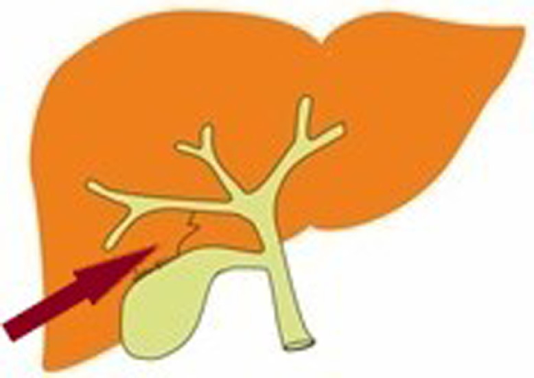
Marked by the arrow is an aberrant subvesical bile duct draining its hepatic segment. It is situated within the connective tissue or capsule of the gallbladder fossa.
The clinical significance of ducts of Luschka lies on their risk for injury during open or laparoscopic cholecystectomy, a very common surgical procedure. Moreover, these ducts can also be injured during liver resection and interventional radiological procedures. Injury of these ducts becomes clinically evident with leak of bile in the postoperative period, being its second most common cause after injury of the cystic duct [2], [4], [7]. Liver transplantation is another procedure where bile leakage has been reported due to injury of these ducts [8]. Bile leakage due to injury of a duct of Luschka is considered a very rare complication, being reported in only 0.15%-2% of patient undergoing laparoscopic cholecystectomy [1], [4], [5]. Clinical presentation of patient with postoperative bile leakages varies from asymptomatic to biloma formation and biliary peritonitis with sepsis, depending on the volume of bile leaked and its sterile or infected state. In general though, the majority of patients will complain of symptoms disproportionate to those expected for the postoperative course, usually within the first week after the operation [1], [4].
Based on their clinical significance, imaging of ducts of Luschka becomes important and can be done in the preoperative, intraoperative, and postoperative period, as this was the case in our patient. A study by Kitami et al reported the preoperative investigation of patients scheduled for cholecystectomy using a form of compute tomographic cholangiography. Subvesical ducts were found in approximately 10% of patients and all cholecystectomies were performed successfully and with no injury [9]. Albeit these promising results, computed tomographic cholangiography has been gradually replaced by MRCP, as it will be discussed later. Intraoperative detection of subvesical ducts can be done with direct visualization if an open surgical procedure is performed or with an intraoperative cholangiogram [4].
HIDA scintigraphy can be used to dynamically assess active bile leakage from an injured subvesical duct, although with lower spatial resolution and thus suboptimal anatomic detail, usually necessitating further imaging studies. ERCP was considered the reference method for evaluation of the biliary tree and treatment of biliary abnormalities. Nevertheless, with the widespread availability of multidetector computed tomography and MRI, especially with the emergence of MRCP, diagnosis of leaking ducts of Luschka has been made feasible and highly accurate and the need for an interventional technique like ERCP has been negated [3], [4]. MRI-MRCP is advantageous when it comes to the detection of variant biliary anatomy thanks to its potential for high-resolution 2D or 3D acquisitions in virtually every plane, providing detailed anatomy of the biliary tree, equivalent to ERCP [2]. MRCP is usually used as a complementary technique to standard upper abdomen MRI examination for investigation of biliary anatomy and pathology. It is typically acquired as a 3-dimensional isotropic sequence in axial or oblique coronal planes, optionally with respiratory-triggering for minimizing respiratory artifacts and fat-suppression for elimination of signal from adjacent fat. This type of MRCP typically provides T2-weighted images, visualizing the biliary tree with high signal intensity on a low signal-intensity background. Alternatively, MRCP can be performed after the intravenous administration of hepatocyte-specific MR contrast agents such as gadobenate dimeglumine which are excreted both via the renal and biliary tract. Optimal cholangiographic images can be acquired 20-40 minutes and up to 2 hours postinjection using sequences with T1-weighting [2], [3], [7], [10]. MRCP can be performed using contrast agents affecting T1-weighed sequences primarily excreted by the biliary tract, resulting in 86% sensitivity and 83% specificity for the detection of a bile leak, offering the possibility to detect leaks not communicating with the central biliary tree and to differentiate bile from free fluid of different origin [3], [10], [11], [12], [13].
Conclusions
Perioperative imaging evaluation of subvesical bile ducts and biliary anatomy in general can be performed both with drip infusion cholangiography with CT (DIC-CT) and with MRCP. Each of these 2 methods has its strengths and limitations. For instance, DIC-CT may be able to provide more accurate information regarding the number and location of stones in comparison with MRCP due to the pseudolesion artifacts encountered with the latter. Moreover, DIC-CT has the potential to assess patency of bile ducts as their opacification by definition reflects biliary flow; something not applicable for MRCP where signal intensity is generated both by flowing and static fluids. In summary, DIC-CT is characterized by shorter scanning time, high spatial resolution, and ability to evaluate biliary flow but is limited by the use of ionizing radiation and suboptimal opacification of extremely dilated bile ducts. On the other hand, MRCP uses no contrast agent or radiation and offers high contrast between bile and adjacent tissues but may suffer from artifacts, pseudolesions, and may not visualize aberrant bile ducts due to projection of other structures. A reasonable approach to investigation of biliary anatomy and pathology would be the use of MRCP as a first-line modality with DIC-CT or ERCP being reserved for patients with contraindication for MRCP or inconclusive findings [3], [14].
Footnotes
Declaration of Competing Interest: The authors certify that there is no conflict of interest with any financial organization regarding the material discussed in the manuscript. The manuscript has not been published previously (partly or in full), has not been submitted to more than 1 journal for simultaneous consideration, and no data have been fabricated or manipulated. No funding has been received in the process of making this article.
Supplementary material associated with this article can be found, in the online version, at doi:10.1016/j.radcr.2019.07.008.
Appendix. Supplementary materials
References
- 1.Ramia J.M., Muffak K., Mansilla A., Villar J., Garrote D., Ferron J.A. Postlaparoscopic cholecystectomy bile leak secondary to an accessory duct of Luschka. JSLS. 2005;9(2):216–217. [PMC free article] [PubMed] [Google Scholar]
- 2.Sureka B., Bansal K., Patidar Y., Arora A. Magnetic resonance cholangiographic evaluation of intrahepatic and extrahepatic bile duct variations. Indian J Radiol Imaging. 2016;26(1):22–32. doi: 10.4103/0971-3026.178283. [DOI] [PMC free article] [PubMed] [Google Scholar]
- 3.Hyodo T., Kumano S., Kushihata F., Okada M., Hirata M., Tsuda T. CT and MR cholangiography: advantages and pitfalls in perioperative evaluation of biliary tree. Br J Radiol. 2012;85(1015):887–896. doi: 10.1259/bjr/21209407. [DOI] [PMC free article] [PubMed] [Google Scholar]
- 4.Spanos C.P., Syrakos T. Bile leaks from the duct of Luschka (subvesical duct): a review. Langenbecks Arch Surg. 2006;391(5):441–447. doi: 10.1007/s00423-006-0078-9. [DOI] [PubMed] [Google Scholar]
- 5.Schnelldorfer T., Sarr M.G., Adams D.B. What is the duct of Luschka?—a systematic review. J Gastrointest Surg. 2012;16(3):656–662. doi: 10.1007/s11605-011-1802-5. [DOI] [PubMed] [Google Scholar]
- 6.Champetier J., Létoublon C., Alnaasan I., Charvin B. The cystohepatic ducts: surgical implications. Surg Radiol Anat. 1991;13(3):203–211. doi: 10.1007/BF01627988. [DOI] [PubMed] [Google Scholar]
- 7.Mortele K.J., Ros P.R. Anatomic variants of the biliary tree: MR cholangiographic findings and clinical applications. Am J Roentgenol. 2001;177(2):389–394. doi: 10.2214/ajr.177.2.1770389. [DOI] [PubMed] [Google Scholar]
- 8.Albishri S.H., Issa S., Kneteman N.M., Shapiro A.M.J. Bile leak from duct of Luschka after liver transplantation. Transplantation. 2001;72(2):338–340. doi: 10.1097/00007890-200107270-00031. [DOI] [PubMed] [Google Scholar]
- 9.Kitami M., Murakami G., Suzuki D., Takase K., Tsuboi M., Saito H. Heterogeneity of subvesical ducts or the ducts of Luschka: a study using drip-infusion cholangiography-computed tomography in patients and cadaver specimens. World J Surg. 2005;29(2):217–223. doi: 10.1007/s00268-004-7652-5. [DOI] [PubMed] [Google Scholar]
- 10.Catalano O.A., Singh A.H., Uppot R.N., Hahn P.F., Ferrone C.R., Sahani D.V. Vascular and biliary variants in the liver: implications for liver surgery. Radiographics. 2008;28(2):359–378. doi: 10.1148/rg.282075099. [DOI] [PubMed] [Google Scholar]
- 11.Vitellas K.M., El-Dieb A., Vaswani K.K., Bennett W.F., Fromkes J., Ellison C. Using contrast-enhanced MR cholangiography with IV mangafodipir trisodium (teslascan) to evaluate bile duct leaks after cholecystectomy: a prospective study of 11 patients. Am J Roentgenol. 2002;179(2):409–416. doi: 10.2214/ajr.179.2.1790409. [DOI] [PubMed] [Google Scholar]
- 12.Aduna M., Larena J.A., Martín D., Martínez-Guereñu B., Aguirre I., Astigarraga E. Bile duct leaks after laparoscopic cholecystectomy: value of contrast-enhanced MRCP. Abdom Imaging. 2005;30(4):480–487. doi: 10.1007/s00261-004-0276-2. [DOI] [PubMed] [Google Scholar]
- 13.Lee N.K., Kim S., Lee J.W., Lee S.H., Kang D.H., Kim G.H. Biliary MR imaging with Gd-EOB-DTPA and its clinical applications. Radiographics. 2009;29(6):1707–1724. doi: 10.1148/rg.296095501. [DOI] [PubMed] [Google Scholar]
- 14.Hirao K., Miyazaki A., Fujimoto T., Isomoto I., Hayashi K. Evaluation of aberrant bile ducts before laparoscopic cholecystectomy: helical CT cholangiography versus MR cholangiography. Am J Roentgenol. 2000;175(3):713–720. doi: 10.2214/ajr.175.3.1750713. [DOI] [PubMed] [Google Scholar]
Associated Data
This section collects any data citations, data availability statements, or supplementary materials included in this article.