Abstract
Acrometastasis caused by malignancy is a very rare phenomenon, and gastric malignancy metastasising to the hands is an even rarer entity. It accounts for only 0.1% of all metastatic osseous involvement, and may be a late manifestation of malignancy or may even be a presenting symptom. It is generally seen with lung primary, followed by kidney and breast, and less frequently with colon, liver, prostate, rectum and stomach primaries. The terminal phalanges are the most common sites of metastases, followed by the metacarpals and the proximal phalanges. We present a case of stomach carcinoma with metastases to the liver and adrenals which was managed with three lines of chemotherapy. He was lost to follow-up and reported after 1 year with swelling over his left hand, which was managed with palliative radiation to the hand in view of severe pain, followed by chemotherapy.
Keywords: radiotherapy, gastric cancer
Background
The incidence of primary gastric carcinoma varies widely in different regions of the world, and the highest incidence is seen in Japan, South-East Asia, South America and Eastern Europe. It can spread locally to adjacent structures or by lymphatic spread to distant organs. The most common sites of metastases are the lymphatics, the peritoneum and the liver. However, bony metastasis especially to the small bones of the hand is extremely rare. Acrometastasis to the hand is not common and accounts for approximately 0.1% of all metastatic osseous involvement.1 It is generally a late manifestation of disseminated disease and generally heralds a poor prognosis.2 3 In such cases, pain palliation is of prime importance.4 The terminal phalanges are the most frequent sites of metastases, followed by the metacarpals and the proximal phalanges.5 Since metastatic involvement of the small bones of the hand is rare in stomach carcinoma, we thought it prudent to bring this case report so that a database can be built for the future.
Case presentation/investigations/treatment/outcome and follow-up
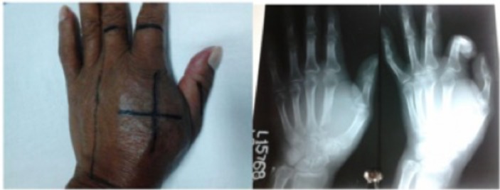
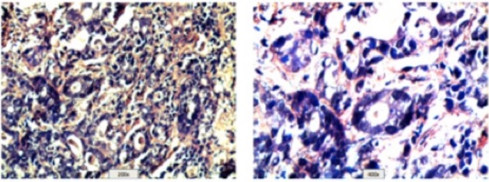
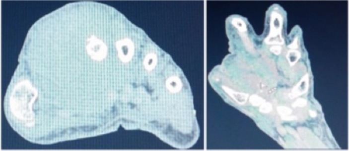
A 58-year-old man with diabetes and on regular medication presented with features of gastric outlet obstruction. On evaluation, he was found to have an advanced gastric carcinoma with metastases to the liver and adrenals. Postevaluation he was started on palliative chemotherapy with EOX (epirubicin, oxaliplatin, and capecitabine) schedule (8 cycles), to which he showed partial response and was then stable for the next 7 months. During follow-up evaluation, the disease showed signs of progression, for which he was placed on second-line chemotherapy with four cycles of oral capecitabine. He continued to have progression and was placed on third line of chemotherapy with cisplatin and docetaxel. Postchemotherapy he presented with swelling over the dorsum of the left hand, 5×4 cm in size, hard and tender, between the thumb and index finger in the interdigital space (figure 1), restricting the movement of his fingers. There was no radiological evidence of pathological fracture. Biopsy and histopathology of the lesion were done which suggested a diagnosis of deposits from adenocarcinoma (figure 2). CT of the left hand and wrist revealed an ill-defined heterogeneous soft tissue mass of 49×41×57 mm on the dorsal aspect of the hand in relation to the second metacarpal, with destruction of the base and shaft. Destruction of the trapezium and trapezoid was also seen, and the lesion was seen involving the extensor and abductor pollicis tendon. There was loss of fat planes with interossei and lumbricals (figure 3). The patient was taken up for palliative radiation to the left hand to a dose of 20 Gy in five fractions with conventional simulator and treated on Theratron Elite 100 Cobalt 60 machine. Postradiation there was immediate relief of pain and reduction of swelling. He regained complete movements of his thumb and index finger. At the end of 1 month of radiation, there was no recurrence of swelling or pain, and 6 months later the patient was alive with no recurrence of swelling on the left hand and was pain-free.
Figure 1.
Clinical and radiological images.
Figure 2.
Histopathology.
Figure 3.
CT scan images.
Discussion
Metastatic disease to the bone is commonly from the breast, prostate and lung malignancies, which account for 70% of patients with metastatic disease.4 5 Other primary sites such as the thyroid, melanoma and kidney account for a lower percentage of bony metastases.6 Some haematological malignancies such as myeloma and lymphoma can also cause bony metastases, causing severe pain and destruction. Gastrointestinal sites of primary malignancies account for only 3%–15% of cases of bony metastases, and stomach carcinoma rarely causes bony metastases especially to the smaller bones of the appendicular skeleton. The prognosis of such patients is poor and the median survival is only 6 months.2 3 Overall survival depends on the primary site and visceral metastases. However, it varies from 6 months to 5 years depending on the site of primary malignancy. These patients present with pain, difficulty in ambulation, immobility, pathological fractures, neurological deficits, anxiety, depression, nerve root compression, fatigue, insomnia, sleep disturbances and deterioration of quality of life.
In general, the axial skeleton is more frequently a site of metastases than the appendicular skeleton. In the axial skeleton spine, the pelvis and the ribs are the most common sites of metastases, and in the appendicular skeleton the proximal femurs and humerus are the commonly involved sites. The acral sites, that is, the hands and the feet, are very rare sites of metastases.
The case in discussion was of locally advanced metastatic stomach carcinoma with visceral metastases and acral metastases. The reason to present this case was that stomach carcinoma rarely causes bony metastases, and having acral metastases makes it an even rarer finding.
Metastases to the bones most often occur in the red marrow, which is found in highest concentration within the axial skeleton,1 and commonly the spread is by haematogenous route. The predilection of tumours to metastasise to the bones are due to transforming growth factor beta (TGFb), insulin-like growth factor I and II, fibroblast growth factor (FGF), platelet-derived growth factor (PDGF), and chemotactic attraction from the bone cells by osteocalcin and type I collagen. Bony metastases may be osteolytic or osteoblastic. Breast and lung primary causes osteolytic lesion, whereas prostate and thyroid primaries present with osteoblastic lesions. Clinical examination is the cornerstone of evaluation of bony lesions. These patients present with swelling, tenderness and restriction in movements. Serum alkaline phosphatase (Sr ALP) is raised in most of the cases.7 X-ray examination reveals destructive lesions and swelling. Tc-99M bone scintigraphy is the best modality for screening of such patients and is an indicator of osteoblastic activity.8 9 However, false positive rates can be seen in arthritis, trauma and Paget’s disease. CT scan also evaluates the bony metastases. It localises the lesion within the bone better, is useful in defining the extent of the cortical destruction, and helps in assessing the risk of pathological fracture and in guiding needle biopsy. T1-weighted and short TI inversion recovery (STIR) images of MRI are a better diagnostic tool to evaluate involvement of the neurovascular structure. Positron emission tomography-CT evaluation shows increased metabolic activity and is useful in detecting osteolytic bone metastases. Pain management of bony metastases can be done by pharmacological methods using the WHO step ladder pattern, surgical intervention or by radiotherapy. The WHO analgesic approach starts with the use of non-opioid analgesics, then weak opioids, followed by strong opioids such as morphine. Adjuvant medications such as gabapentin, pregabalin and amitriptyline can be added for neuropathic relief of pain. Antianxiety and antidepressant measures are also useful for pain relief. Surgical options for management are done for the prevention or treatment of pathological fracture. Radiation therapy is very effective for palliation of bony metastases, with partial pain relief in up to 80%–90% of patients and complete pain relief in up to 50% of patients.
Since acrometastasis to the hand is a rare occurrence and is seen in advanced malignancies, the survival of these patients is very poor. Extensive search of the literature also does not show any survival data per se for such presentation, but generally these patients do not survive for more than 6–8 months. The most common sites of metastatic deposits are the distal phalanges, although involvement of all bones has been documented in the literature. As far as incidence is concerned, metacarpals are involved in 17% of cases, phalanges in 66% of cases and carpal bones in 17% of cases. The most common primary site is the lung (40%–50%), followed by the breast and the kidneys (11%–15%). Rarely colon, prostate, thyroid, oesophagus and bony malignancy can metastasise to the hands. These metastases usually involve the osseous structures, but also the soft tissues by direct involvement. How the tumours metastasise to the hands is still unclear, and many factors have been postulated for its mechanism: history of trauma, hormonal influence, haemodynamics, host immune response, lymphatic spread, prostaglandins, increased blood flow, cigarette smoking, involvement of the dominant hand as metastatic site and so on. Generally these patients present with a swollen, painful, erythematous and warm hand. Presentation may be confused with gout, pulp space infection, fracture, osteomyelitis, septic arthritis, rheumatoid arthritis and tenosynovitis.
Review of literature by Flynn et al analysed 257 cases of acrometastases from various malignancies. Based on their observation, the following inferences were made:
Male preponderance.
Lung carcinoma as the most common primary site, followed by the kidney and the breast.
Association with cigarette smoking.
Elderly age.
Involvement of the right hand more than the left hand.
Third finger most commonly involved, followed by the thumb, fourth finger, second finger and fifth finger.
Distal phalanx most commonly involved, followed by the metacarpal bones, proximal phalanges and middle phalanges. The postulated reason behind involvement of these sites was that these regions of the body had reduced circulatory speed and are preferential for secondary tumour growth, which may explain the greater prevalence of lesions within the phalanges of the hand.
Lesions are mostly osteolytic, sclerotic and mixed.
Amputation was the most common treatment modality, followed by radiation, excision and systemic therapy.
Radiation therapy reduced the pain and associated symptoms and improved the quality of life.
The mean overall survival was poor and the average length of survival was 6–8 months.
Patient’s perspective.
I was a distraught person with so much suffering after being diagnosed as a case of carcinoma stomach. On top of that I had this lesion on my left hand with swelling and severe pain. Since I had already received 3 cycles of chemotherapy and this lesion developed suddenly I was advised radiation for symptom relief. After radiation I had a marked improvement in the pain and was nearly pain free.
Learning points.
Acrometastasis to the hand is a rare occurrence, and few cases have been reported in the literature with gastric carcinoma as the primary malignancy.
The prognosis of such patients is very poor and may be worsened due to delayed diagnosis.
A very high level of suspicion and detailed clinical inspection not to ignore the minor symptoms are the cornerstone for diagnosis and further management.
As far as possible, non-surgical methods should be applied, and achieving the best quality of life of patients should be kept in mind.
Pain palliation by radiation therapy is very effective, along with other appropriate treatments.
Acknowledgments
I am grateful to my colleagues Dr Manish Kumar, Dr Richa Ranjan, Dr Kishore Kumar and the patient who provided me support to write about this article. I am grateful to all for above.
Footnotes
Contributors: AK is the sole author of this article.
Funding: The authors have not declared a specific grant for this research from any funding agency in the public, commercial or not-for-profit sectors.
Competing interests: None declared.
Provenance and peer review: Not commissioned; externally peer reviewed.
Patient consent for publication: Obtained.
References
- 1. Kerin R. Metastaitc tumors of the hand. J Bone Joint Surg Am 1983;65:1331–5. [PubMed] [Google Scholar]
- 2. Shannon FJ, Antonescu CR, Athanasian EA, et al. Metastatic thymic carcinoma in a digit: a case report. J Hand Surg Am 2000;25:1169–72. 10.1053/jhsu.2000.17863 [DOI] [PubMed] [Google Scholar]
- 3. Hsu CS, Hentz VR, Yao J, et al. Tumors of hand. Lancet Ocnol 2007;8:157–66. [DOI] [PubMed] [Google Scholar]
- 4. Amadio PC, Lombardi RM. Metastatic tumors of the hand. J Hand Surg Am 1987;12:311–6. 10.1016/S0363-5023(87)80299-X [DOI] [PubMed] [Google Scholar]
- 5. Basora J, Fery A, et al. Metastatic malignancy of the hand. Clin Orthop Relat Res 1975;108:182–6. 10.1097/00003086-197505000-00029 [DOI] [PubMed] [Google Scholar]
- 6. Elhassan B, Fakhouri A. Metastases of squamous cell carcinoma of lung to the first web space of the hand. J Bone Joint Surg Br;200:1243–6. [DOI] [PubMed] [Google Scholar]
- 7. Kusumoto H, Haraguchi M, Nozuka Y, et al. Characteristic features of disseminated carcinomatosis of the bone marrow due to gastric cancer: the pathogenesis of bone destruction. Oncol Rep 2006;16:735–40. 10.3892/or.16.4.735 [DOI] [PubMed] [Google Scholar]
- 8. Wilner D. Cancer metastases to bone. Radiology of bone tumor and allied disorders. Vol I. Philadelphia: W B saunders, 1982:3641–908. [Google Scholar]
- 9. Gold RI, Seeger LL, Bassett LW, et al. An integrated approach to the evaluation of metastatic bone disease. Radiol Clin North Am 1990;28:471–83. [PubMed] [Google Scholar]


