Abstract
Introduction:
We present one of the largest case series of Macrodystrophia lipomatosa, a rare congenital disorder of localized gigantism characterized by overgrowth of all the mesenchymal elements, predominantly involving the fibroadipose tissue.
Aims:
To detail the radiological features, pattern of distribution, associated conditions and to suggest an appropriate terminology to describe the condition.
Methods and Material:
It is a retrospective study. Data from PACS server dating from 2000 and 2018 was used. The cases with isolated enlarged limb or digit/digits with or without nerve involvement were included in the study.
Statistical Analysis Used:
Frequency and percentage were used for analysis of categorical variables.
Results:
A total of 31 cases was included for the final analysis, out of which 19 were males and 12 were females. Unilateral limb involvement was seen in 30 cases. The most common pattern identified was the ’nerve territory oriented’ type in 28 cases confined to the hand or foot, ’diffuse or pure lipomatous’ type in one case and mixed type was seen in two cases. The most common nerve territory involved was along the median nerve in the upper limb and along the medial plantar nerve in the lower limb. Neural involvement was seen in 16 cases of the upper limb and 10 cases of the lower limb. Syndactyly was seen in two cases, polydactyly in one case and symphalangism in one case.
Conclusions:
A diagnosis of macrodystrophia lipomatosa can be confidently made in cases with congenital isolated limb or digit/digits enlargement with or without fibrolipohamartoma of nerve. Radiographs and ultrasound are sufficient along with clinical examination to make accurate diagnosis. MRI is useful for assessing the extent and for planning surgery.
Keywords: Digital gigantism, fibrolipohamartoma nerve, lipomatous overgrowth, local gigantism, macrodactyly, macrodystrophia lipomatosa, megalodactyly
INTRODUCTION
Macrodystrophia lipomatosa is a rare disorder of localized gigantism characterized by overgrowth of all the mesenchymal elements, predominantly involving the fibroadipose tissue. It is a congenital and nonhereditary condition. The term “macrodystrophia lipomatosa”was originally used to describe localized gigantism involving the lower limb by Feriz in 1925.[1] Golding later extended the term to describe localized gigantism affecting the upper limb.[2] The term macrodactyly is often interchangeably used to describe macrodystrophia lipomatosa.[3] The etiology of macrodystrophia lipomatosa is poorly understood, and several hypotheses exist. Recent studies have shown an association with PIK3CA gene mutation.[4] Pathologically, there is an increase in adipose tissue in the subcutaneous plane with a fine lattice of fibrous tissue and can occasionally involve bone marrow, periosteum, muscles, and nerve sheath in a sclerotomal distribution.[2,5] The disease is usually present since birth and the affected region/digit increases in length and girth until puberty when growth ceases. The affected individuals are usually asymptomatic except for the cosmetic disfigurement. Some of them manifest with hindered function, premature degenerative changes or very rarely due to symptoms of neurovascular compression.
Macrodystrophia lipomatosa is often associated with fibrolipomatous hamartoma (FLH) of the median nerve.[6,7] The other associated conditions are clinodactyly, syndactyly, polydactyly, and symphalangism. The rarity of this condition and the variety of terms that have been used to describe it has resulted in confusion. The purpose of this study was to report one of the largest case series, elaborate its associated findings, radiographic features and to suggest the use of the term macrodystrophia lipomatosa exclusively for isolated congenital localized gigantism with or without nerve involvement.
MATERIALS AND METHODS
It is a retrospective study. After obtaining Institutional Review Board approval, a case search was conducted in our PACS server from April 2000 to April 2018 to identify the cases of macrodystrophia lipomatosa. The search terms used were “macrodactyly,”“fibrolipohamartoma,”“macrodystrophia lipomatosa,”and “localized gigantism.”The inclusion criteria were cases with an isolated enlarged limb or digit/digits, with or without nerve involvement. Exclusion criteria were cases with a diagnosis of localized gigantism due to other causes such as neurofibromatosis, vascular malformations, cutaneous hemangiomas, and overgrowth syndromes such as Proteus syndrome and Klippel-Trenaunay syndrome.
The plain radiographs in anteroposterior and lateral views were assessed. Ultrasound was performed on Philips EPIQ 5G with L18-5 high-resolution linear probe and Siemens Acusons 2000 with 14 L5 high-resolution linear probe.
Magnetic resonance imaging
Magnetic resonance imaging (MRI) was done with either a 1.5 Tesla Magnetom Avanto, Siemens machine or a 3 Tesla Intera Achieva, Philips machine. The imaging protocol used was T2 spectral attenuated inversion recovery (SPAIR) axial, coronal and sagittal, PD axial and coronal and T1 sagittal. The parameters for acquisition of hand MRI were repetition time (TR)/echo time (TE) (in milliseconds) of 5100/65 for SPAIR sequence, TR/TE of 360/20 for T1 and 2270/30 for PD sequence; 384 × 320 matrix; 3 mm slice thickness and 16–18 cm field of view. The parameters for acquisition of foot MRI were TR/TE (in milliseconds) of 3900/65 for SPAIR sequence, TR/TE of 700/20 for T1 and 1420/30 for PD sequence; 384 × 320 matrix; 3 mm slice thickness; and 14–20 cm field of view. The parameters for acquisition of whole extremity MRI were TR/TE (in milliseconds) of 5100/44 for SPAIR sequence, TR/TE of 480/20 for T1 and 2270/30 for PD sequence; 320 × 224 matrix; 3 mm slice thickness and 23–25 cm field of view.
Image analysis
Images were reviewed by two radiologists having at least 2 years of experience in musculoskeletal radiology using Centricity PACS-RA1000 Workstation. The obtained data and the images were evaluated for distribution in sex, age at presentation, involvement of upper or lower extremity, whether unilateral or bilateral, part of the limb involved, type of macrodystrophia lipomatosa, nerve territory involved, the presence of nerve involvement, and presence of associated anomalies.
Based on our observation type of macrodystrophia lipomatosa was classified into three types. “Nerve territory-oriented type”which involved only the hand or foot in a nerve territory/sclerotomal distribution with or without nerve enlargement. “Diffuse or pure lipomatous type”which involved the entire extremity with all the digits involved in the hand/foot without affecting of the nerves. “Mixed pattern”which showed nerve territory distribution in the hand/foot, along with diffuse lipomatous enlargement of rest of the limb. The presence of nerve involvement was confirmed when there was enlargement of the nerve with interfascicular adipose tissue proliferation splaying the fascicles giving a coaxial cable appearance. Nerve territory involved was either along a single nerve or mixed nerve territory. For example, involvement along the median nerve/median plantar nerve distribution only or along the median and ulnar nerve/medial and lateral plantar nerves.
Statistical analysis
Frequency and percentage analysis were used for analysis of categorical variables. The analysis was performed using SPSS (version 16.0, Chicago, IL USA).
RESULTS
Initial search fetched a total of 38 cases, of which seven cases were excluded. Three cases were incompletely evaluated, and the available radiological features were not specific for macrodystrophia. One case had discolored skin with a small arteriovenous malformation at the distal phalanx level (which was confirmed by digital subtraction angiography). Two cases had slow flow venous malformation which was confirmed with ultrasound. One case had isolated fibrolipohamartoma of the median nerve without localized gigantism. A total of 31 cases was included for the final analysis. Twenty-five patients had radiographs of the affected region, out of which, 20 patients were further investigated with MRI, four patients had both MRI plus ultrasound and one patient had only ultrasound. A total of six patients had only MRI.
Demographics
Our study group comprised of 19 (61.3%) males and 12 (38.7%) females. The age at presentation ranged between 6 months and 56 years. The most common clinical presentation was localized overgrowth and cosmetic deformity. Unilateral limb involvement was the most common type in our study population seen in 30 (96.7%) cases. We had only one case of bilateral involvement (3.3%). There was a slight predilection for the upper limb with 16 (51.6%) cases, and 15 cases involved the lower limb (49.4%).
Type of macrodystrophia lipomatosa
The most common type identified was the “nerve territory oriented”type in 28 (90.3%) cases confined to the hand or foot, “diffuse or pure lipomatous”type was seen in 1 (3.22%) case, and mixed type was seen in 2 (6.45%) cases [Table 1].
Table 1.
Distribution according to the pattern of macrodystrophia lipomatosa in our study population
| Type of macrodystrophia lipomatosa | Number of cases |
|---|---|
| Nerve territory oriented | 28 |
| Diffuse lipomatous type | 1 |
| Mixed pattern | 2 |
Nerve territory involvement
The most common nerve territory involved was along the median nerve in the upper limb (11 cases) and along the medial plantar nerve in the lower limb (10 cases). One case was along the ulnar nerve territory. Mixed nerve territory distribution was seen in a total of 9 cases, four cases in the upper limb and five cases in the lower limb. The 2nd and 3rd digit were more commonly involved in the hand and feet which corresponds to the median and the medial plantar nerve territories, respectively. Findings are tabulated in Table 2.
Table 2.
Distribution according nerve territory involvement in our study
| Upper limb | Lower limb | |
|---|---|---|
| Median | 11 | |
| Ulnar | 1 | |
| Median and ulnar | 4 | |
| Medial plantar nerve | 10 | |
| Lateral plantar nerve | - | |
| Medial and lateral plantar nerves | 5 |
Presence of fibrolipomatous hamartoma/neural involvement
Neural involvement in the form of enlarged nerve with inter-fascicular adipose tissue proliferation giving a coaxial cable appearance (either on ultrasound or MRI) was seen in 16 (100%) cases of the upper limb and 10 (66%) cases of the lower limb. Four cases in the lower limb and one case of “pure lipomatous type”did not show any nerve involvement.
Presence of associated anomalies
The incidence of associated anomalies was less which accounted only for 12.9% (Four cases). Syndactyly was seen in two cases, polydactyly in one case and symphalangism in one case. None of our cases had clinodactyly.
DISCUSSION
Macrodystrophia lipomatosa was first described by Feriz[1] in 1925 and is a disorder of localized gigantism characterized by abnormal proliferation of the fibroadipose tissue. It is a congenital and nonhereditary disorder. There is overgrowth that usually occurs in a specific sclerotomal distribution.[2] Barsky classified macrodystrophia lipomatosa as static or progressive based on the growth pattern and former being the most common type.[3] In static type, the growth of the enlarged digit is at the same rate as the other digits. In the progressive type, the growth of the affected digit is more rapid than the rest of the extremity. The etiology of macrodystrophia lipomatosa is poorly understood, and several hypotheses exist, none of which are conclusive. Recent studies have shown an association with PIK3CA gene mutation.[4]
In our study, males were more commonly affected than females. Unilateral involvement is the norm and was the most common pattern in our study group. Bilateral involvement is rare, and as noted in previous studies,[8] we also had only one case. There were case reports of abdominal wall involvement.[9] None of our cases had abdominal wall lipomatosis.
Clinically, macrodystrophia is present since birth with swelling of the affected digit or limb leading to cosmetic disfigurement [Figure 1a and b]. Some may present with hindered/loss of function, pain due to neurovascular compression or degenerative changes.
Figure 1.

A 2-year-old child with localized gigantism. (a and b) Clinical images show gross enlargement of the third and fourth digits of the left hand with syndactyly. Abnormal soft-tissue hypertrophy predominantly involving the volar aspect. (c and d) Anteroposterior and lateral radiographs show grossly enlarged left middle and index fingers, abnormal soft tissue along the volar aspect with resultant dorsal bending of the digits, syndactyly, and symphalangism.
Figure 2.
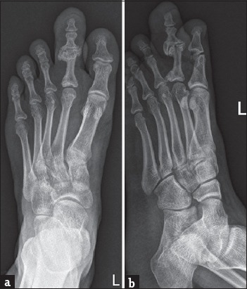
(a and b) Radiograph of the foot shows isolated enlargement of the 2nd digit with soft tissue and osseous hypertrophy of the phalanges along with bony excrescences of the proximal interphalangeal joint.
According to the pattern of distribution, the macrodystrophia lipomatosa was divided into “nerve territory oriented type”and “diffuse or pure lipomatous type.” [10] In the “nerve territory oriented type,”there are hypertrophied soft tissues especially that of fat, with or without fatty infiltration of the nerves. In the pure lipomatous type, there was a marked hypertrophy of adipose tissue without the involvement of the nerves. We also observed a “mixed pattern type”where there was an overlap of “nerve territory oriented type”and “diffuse lipomatous type”in two cases. In our study, the most common pattern identified was the “nerve territory-oriented type.”The most common nerve territory distribution was along the median nerve in the upper limb and along the medial plantar nerve in the lower limb. In the hands and feet, the involvement of the 2nd and 3rd digit is common corresponds to the median and the medial plantar nerve supply respectively. Our results are in concordance with the literature.[11-14] Prasetyono et al., and Gupta et al., had similar observations in their study.
The presence of nerve involvement was confirmed on either ultrasound or MRI when there was enlargement of the nerve with interfascicular adipose tissue proliferation splaying the nerve fascicles giving a coaxial cable appearance. The term “fibrolipohamartoma”or “lipomatosis of nerve”is most commonly used to describe the proliferation of mature adipocytes within peripheral nerves.[14] The circumferential proliferation of fat around the epineurium of the nerve has also been reported.[15] Earlier studies have suggested FLH of the nerve and macrodystrophia lipomatosa as separate entities.[5,16] However, neural involvement in macrodystrophia and FLH of the nerve are interchangeably used as they define the same pathological and radiological finding. In our study, there was 100% of upper limb cases, and 66% of lower limb cases had neural involvement.
Imaging in cases with localized gigantism, using radiographs and ultrasonography (USG), is required for confirming the diagnosis and assessing the accurate distribution of the disease for further management.
Plain radiographs are first and most commonly used imaging modality.[17] The common findings are soft tissue overgrowth with osseous hypertrophy of the affected digit or limb. The osseous changes are increased length, width, and cortical thickening. In digits, these changes are seen along the volar aspect leading to dorsal bending [Figure 1c and d]. Bony outgrowths with or without degenerative changes in the adjacent joints can be ascertained [Figure 2]. In long-standing cases, there may be ankylosis. Other associated findings such as syndactyly and polydactyly [Figure 3] and symphalangism[18] [Figure 1c and d] can be readily appreciated on the plain radiographs.
Figure 3.
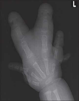
Anteroposterior radiograph of the hand shows, postaxial hexadactyly, proximal phalanx of the 6th digit articulates with the 5th metacarpal phalangeal joint. Syndactyly of left 3rd and 4th digits.
High-resolution ultrasound reveals soft-tissue hypertrophy and extremely helpful in assessing the involvement of the nerves. Fat hypertrophy can be subcutaneous, inter- or intra-muscular in distribution. Fibrolipohamartoma of the nerve appears as an enlarged nerve with prominent fascicles and interfascicular fat hypertrophy [Figure 4]. Focused USG is also very helpful in excluding the clinical mimics of the localized overgrowth such as vascular malformation which can be missed on plain radiographs.
Figure 4.
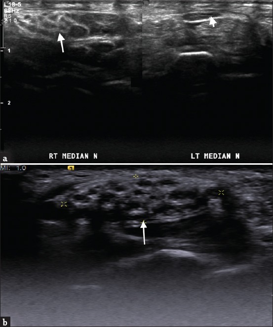
(a and b) Ultrasound images of two different patients, showing an enlarged median nerve with prominent hypoechoic nerve fascicles and hyperechoic interfascicular fat (large arrows). Figure on the right in 4a shows normal left median nerve (small arrow).
Computed tomography is not indicated, as it does not provide additional information. MRI is helpful, especially in assessing “diffuse lipomatous”and “mixed pattern”types.[19,20] MRI demonstrates the nonencapsulated fibrofatty proliferation of the soft tissues which follows the subcutaneous fat in all sequences and the osseous hypertrophy. MRI is useful in assessing the FLH of the nerve [Figures 5 and 6] and also in assessing the neurovascular bundle in other associations such as syndactyly [Figure 7]. It is the investigation of choice in cases of diffuse lipomatous and mixed type, where ultrasound is suboptimal [Figure 8].
Figure 5.
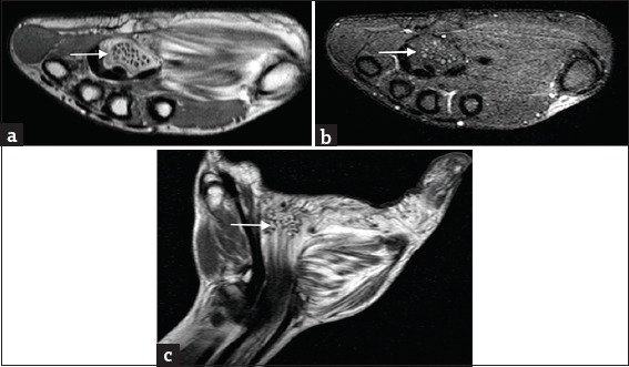
Typical appearance of the fibrolipohamartoma of median nerve on magnetic resonance imaging. (a) Coaxial cable appearance (arrow) in the axial images with interfascicular fat, (b) suppressing on fat suppressed sequence. (c) Spaghetti like appearance in the coronal images (arrow). There is associated intermuscular fat hypertrophy in the thenar muscles.
Figure 6.
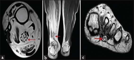
Fibrolipohamartoma of tibial nerve on magnetic resonance imaging, non fat suppressed images. (a) Coaxial cable appearance (red arrow) in the axial images with interfascicular fat. (b) Spaghetti-like appearance in the coronal images (red arrow). (c) Fibrolipomatous hamartoma of the medial plantar nerve. There is associated intramuscular fat hypertrophy of the gastrocnemius and soleus muscle and in the subcutaneous plane.
Figure 7.
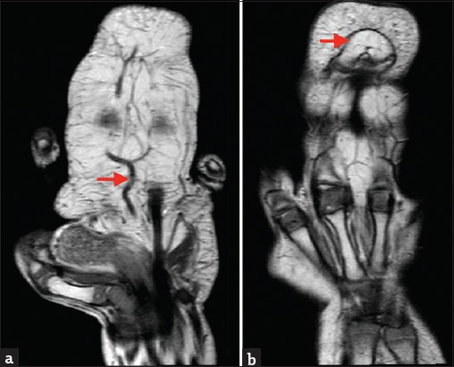
(a) Magnetic resonance imaging of the hand, non fat suppressed images showing syndactyly, spaghetti-like median nerve and the common artery which is bifurcating into the digital arteries (red arrow). (b) Shows fusion of the distal phalanges - symphalangism (red arrow).
Figure 8.
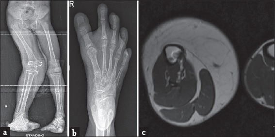
(a) Radiograph of the lower limbs shows an elongated right lower limb with limb length discrepancy, hypertrophied soft tissue, and postepiphysiodesis fixation screws. (b) Radiograph of the foot anteroposterior view in the same patient, shows elongated 1–4 digits with associated soft tissue hypertrophy. (c) T2 axial image at the level of the mid-third of leg shows hypertrophy of fat in the subcutaneous and intermuscular fascial planes.
Macrodystrophia lipomatosa can be associated commonly with syndactyly, symphalangism, polydactyly, brachydactyly, or clinodactyly and rarely with lipomatous growths in subcutaneous plane and intestines, pulmonary cysts, and calvarial abnormalities.[2,5] In our study, we had three cases with syndactyly and one case with symphalangism and one case with polydactyly.
Localized gigantism associated with cutaneous hemangiomas/varicose veins or cutaneous discoloration or palmar and plantar cerebroid thickening or cafe au lait spots should indicate an alternate diagnosis.
Treatment options include serial follow-up or surgical intervention in the form of debulking with staged reconstruction or epiphysiodesis or ray amputation. The choice of treatment offered and the treatment taken by our study group is beyond the scope of this study.
Limitation
As it was a retrospective study, the natural course of the disease could not be assessed. Furthermore, imaging was limited to the clinically affected region(s), hence accurate evaluation of associated anomalies in other areas was not possible. Another limitation was that we had only a few cases with “diffuse lipomatous”and “mixed pattern”of the macrodystrophia lipomatosa, which precluded analysis of statistical significance of different types and their associations.
CONCLUSION
The most commonly used terminologies for congenital isolated limb or digit enlargement are macrodystrophia lipomatosa and macrodactyly. However, the usage of the term “macrodactyly”for the diffuse involvement of an entire limb with or without digital enlargement as seen in three of our cases, may not be appropriate. In conclusion, we have reserved the term “macrodystrophia lipomatosa”for congenital isolated limb or digit/digits enlargement with or without fibrolipohamartoma of nerve. The presence of cutaneous hemangiomas/varicose veins, cutaneous discoloration, palmar, and plantar cerebroid thickening or cafe au lait spots should indicate an alternate diagnosis. The term “isolated fibrolipohamartoma of nerve”can be used in cases when there is only nerve enlargement without subcutaneous, inter-muscular and intra-muscular fibrofatty hypertrophy, as seen in one of our cases. Plain radiographs and high-resolution ultrasound are sufficient for the diagnosis of macrodystrophia lipomatosa with or without fibrolipohamartoma. MRI is indicated to assess the status of the neurovascular bundle in case of complex cases with syndactyly for operative planning.
Financial support and sponsorship
Nil.
Conflicts of interest
There are no conflicts of interest.
REFERENCES
- 1.Feriz H. Macrodystrophia lipomatosa progressiva. Virchows Arch. 1925;260:308–68. [Google Scholar]
- 2.Goldman AB, Kaye JJ. Macrodystrophia lipomatosa: Radiographic diagnosis. AJR Am J Roentgenol. 1977;128:101–5. doi: 10.2214/ajr.128.1.101. [DOI] [PubMed] [Google Scholar]
- 3.Barsky AJ. Macrodactyly. J Bone Joint Surg Am. 1967;49:1255–66. [PubMed] [Google Scholar]
- 4.Rios JJ, Paria N, Burns DK, Israel BA, Cornelia R, Wise CA, et al. Somatic gain-of-function mutations in PIK3CA in patients with macrodactyly. Hum Mol Genet. 2013;22:444–51. doi: 10.1093/hmg/dds440. [DOI] [PMC free article] [PubMed] [Google Scholar]
- 5.Majumdar B, Jain A, Sen D, Bala S, Mishra P, Sen S, et al. Macrodystrophia lipomatosa: Review of clinico-radio-histopathological features. Indian Dermatol Online J. 2016;7:293–6. doi: 10.4103/2229-5178.185465. [DOI] [PMC free article] [PubMed] [Google Scholar]
- 6.Brodwater BK, Major NM, Goldner RD, Layfield LJ. Macrodystrophia lipomatosa with associated fibrolipomatous hamartoma of the median nerve. Pediatr Surg Int. 2000;16:216–8. doi: 10.1007/s003830050728. [DOI] [PubMed] [Google Scholar]
- 7.Silverman TA, Enzinger FM. Fibrolipomatous hamartoma of nerve. A clinicopathologic analysis of 26 cases. Am J Surg Pathol. 1985;9:7–14. doi: 10.1097/00000478-198501000-00004. [DOI] [PubMed] [Google Scholar]
- 8.Aydos SE, Fitoz S, Bökesoy I. Macrodystrophia lipomatosa of the feet and subcutaneous lipomas. Am J Med Genet A. 2003;119A:63–5. doi: 10.1002/ajmg.a.10179. [DOI] [PubMed] [Google Scholar]
- 9.Khan RA, Wahab S, Ahmad I, Chana RS. Macrodystrophia lipomatosa: Four case reports. Ital J Pediatr. 2010;36:69. doi: 10.1186/1824-7288-36-69. [DOI] [PMC free article] [PubMed] [Google Scholar]
- 10.Cerrato F, Eberlin KR, Waters P, Upton J, Taghinia A, Labow BI, et al. Presentation and treatment of macrodactyly in children. J Hand Surg Am. 2013;38:2112–23. doi: 10.1016/j.jhsa.2013.08.095. [DOI] [PubMed] [Google Scholar]
- 11.Fritz TR, Swischuk LE. Macrodystrophia lipomatosa extending into the upper abdomen. Pediatr Radiol. 2007;37:1275–7. doi: 10.1007/s00247-007-0606-y. [DOI] [PubMed] [Google Scholar]
- 12.Ranawat CS, Arora MM, Singh RG. Macrodystrophia lipomatosa with carpal-tunnel syndrome. A case report. J Bone Joint Surg Am. 1968;50:1242–4. [PubMed] [Google Scholar]
- 13.Upadhyay D, Parashari UC, Khanduri S, Bhadury S. Macrodystrophia lipomatosa: Radiologic-pathologic correlation. J Clin Imaging Sci. 2011;1:18. doi: 10.4103/2156-7514.78264. [DOI] [PMC free article] [PubMed] [Google Scholar]
- 14.Prasetyono TO, Hanafi E, Astriana W. A review of macrodystrophia lipomatosa: Revisitation. Arch Plast Surg. 2015;42:391–406. doi: 10.5999/aps.2015.42.4.391. [DOI] [PMC free article] [PubMed] [Google Scholar]
- 15.Toms AP, Anastakis D, Bleakney RR, Marshall TJ. Lipofibromatous hamartoma of the upper extremity: A review of the radiologic findings for 15 patients. AJR Am J Roentgenol. 2006;186:805–11. doi: 10.2214/AJR.04.1717. [DOI] [PubMed] [Google Scholar]
- 16.Prasad NK, Mahan MA, Howe BM, Amrami KK, Spinner RJ. A new pattern of lipomatosis of nerve: Case report. J Neurosurg. 2017;126:933–7. doi: 10.3171/2016.2.JNS151051. [DOI] [PubMed] [Google Scholar]
- 17.Blacksin M, Barnes FJ, Lyons MM. MR diagnosis of macrodystrophia lipomatosa. AJR Am J Roentgenol. 1992;158:1295–7. doi: 10.2214/ajr.158.6.1590127. [DOI] [PubMed] [Google Scholar]
- 18.Gupta SK, Sharma OP, Sharma SV, Sood B, Gupta S. Macrodystrophia lipomatosa: Radiographic observations. Br J Radiol. 1992;65:769–73. doi: 10.1259/0007-1285-65-777-769. [DOI] [PubMed] [Google Scholar]
- 19.Turkington JR, Grey AC. MR imaging of macrodystrophia lipomatosa. Ulster Med J. 2005;74:47–50. [PMC free article] [PubMed] [Google Scholar]
- 20.Sone M, Ehara S, Tamakawa Y, Nishida J, Honjoh S. Macrodystrophia lipomatosa: CT and MR findings. Radiat Med. 2000;18:129–32. [PubMed] [Google Scholar]


