Abstract
Study Design:
This is prospective study.
Purpose:
The purpose of this study is to assess the functional, neurological, and radiological outcomes of the patients of subaxial cervical spine injuries treated by anterior corpectomy and stabilization with anterior cervical locking plate and cage filled with bone.
Overview of the Literature:
The principles in the treatment of unstable cervical spine injuries are reduction and stabilization of the injured segment, maintenance of cervical lordosis and decompression where indicated and ranges from nonoperative to combined anterior and posterior surgical fusion. There is, however, debate on the indications for anterior, posterior, or combined surgery.
Materials and Methods:
The present study of 99 patients includes prospective patients of subaxial cervical spine injuries between February 2014 and February 2016 admitted and operated to Indira Gandhi Medical College, Shimla. Bony fusion, neurological recovery, Neck Disability Index and complication were studied in all patients. The mean follow-up period was 27 months (range 12–42 months).
Results:
Of the 99 procedures, 77 (77.8%) involved a single vertebral level, 19 (19.2%) involved two levels, and 3 (3%) involved three levels corpectomy. The mean Neck Disability Index was 7.57 ± 5.42. Definitive Bridwell Grade 1 fusion was seen in 64.6% of the cases. No deterioration of neurological symptoms was seen. Dysphagia was the most common complication in 79 (79.8%) patients. One patient had minimal screw back out.
Conclusions:
Anterior cervical corpectomy and stabilization with cage filled with bone and cervical reflex locking plate are good method for subaxial cervical spine injuries with good fusion rates and probably procedure of choice for posttraumatic multiple disc prolapse with reduced hazards of multiple grafts.
Keywords: Corpectomy, fusion, neck disability index, subaxial cervical spine injuries
Introduction
The cervical spine is injured in 2.4% of blunt trauma injury patients.[1] It is more common in elderly, male, and European American ethnicity. The annual incidence rate is 64/lakhs with two peaks, one in the second and third decade of the male population and another in elderly females.[1] The most common mechanism of injury is noted to be accidental falls followed by motor vehicle accident. The most common site of injury is the atlantoaxial region, with the most commonly injured levels in the subaxial cervical spine being C6 and C7. The C2 vertebra is the most common level of injury (24%) and lower two cervical vertebrae C6 and C7 constitutes the second-most common level of injury C6 (20.25%) and C7 (19.08%). C3 is the least likely injured structure, i.e., 4.27%. The subaxial cervical spine (C3–C7) is particularly vulnerable to traumatic injury due to its considerable mobility and its proximity to the more rigid thoracic spine.[1,2]
The principles in the treatment of unstable cervical spine injuries are reduction and stabilization of the injured segment, maintenance of cervical lordosis, and decompression where indicated. Methods of treatment range from nonoperative to combined anterior and posterior surgical fusion. There is, however, debate on the indications for anterior, posterior, or combined surgery. The anterior approach is a less traumatic and provides the ability for decompression, reduction of dislocated facet joints, interbody grafting with reconstruction and maintenance of lordosis. Although fusion rates are high with the use of autograft, it is associated with significant graft site morbidity while the use of allograft, which is devoid of any donor site problems, is associated with high rates of pseudoarthrosis. To overcome these problems associated with both allograft and autograft, titanium mesh cages (filled with local bone saved from corpectomy) are used with advantages of immediate anterior column stability, shorter operation time, maintenance of intervertebral disc height and lordotic angle, avoidance of morbidity associated with autologous bone graft (iliac crest) harvesting, good biocompatibility, and obtain comparable fusion rate to autogenous tricortical iliac bone.[3,4]
Although multiple discectomies are an alternative means of decompression in cases associated with posttraumatic prolapsed intervertebral discs, we preferred corpectomy to address spinal stenosis caused by age-related degenerative spondylotic changes (osteophytes), and cervical corpectomy should result in higher fusion rates because there are only two fusion surfaces.[5,6]
In contrast, posterior approaches may be injurious to adjacent levels; this has been postulated to cause late deformity and with concerns regarding the rate of wound infection, the inability to address a disrupted disc before reduction.[7]
We stabilize the subaxial cervical spine injuries anteriorly with anterior cervical locking plate and cage filled with bone graft after corpectomy, and the aim of our study is to directly decompress the cord, to facilitate early ambulation and to evaluate the clinical improvement, neurological outcome, and radiographic fusion rates.
Materials and Methods
The present study includes prospective patients of subaxial cervical spine injuries admitted and operated to Indira Gandhi Medical College, Shimla, between February 2014 and February 2016 and these patients were assessed radiologically for fusion using Bridwell criteria, neurologically using the American Spinal Injury Association (ASIA) chart, and for functional outcome as per Neck Disability Index and clinical neck movements pictures were taken. Ethical clearance was taken from the Institutional Ethics Committee and informed written consent was taken from all the patients.
Inclusion criteria
Subaxial Cervical Spine Injury Classification (SLIC) score ≥4
Relative sagittal plane translation >3.5 mm
Relative sagittal plane rotation >11°
Three columns injury and two columns injury with neurological deficit.
Exclusion criteria
Patients medically unfit for surgery
Patients operated through posterior approach
SLIC scores <3
Single column injury and two columns injury without neurological deficit.
In our institution, Philadelphia hard cervical collar or cervical traction (either head halter or Crutchfield tongs) is applied till the fracture is reduced. We excluded the single level subluxation from our study treated with discectomy and not corpectomy. Patients with neglected/irreducible subluxation, vertebral body fracture, and multiple level disc prolapses are included in this study as they needed corpectomy.
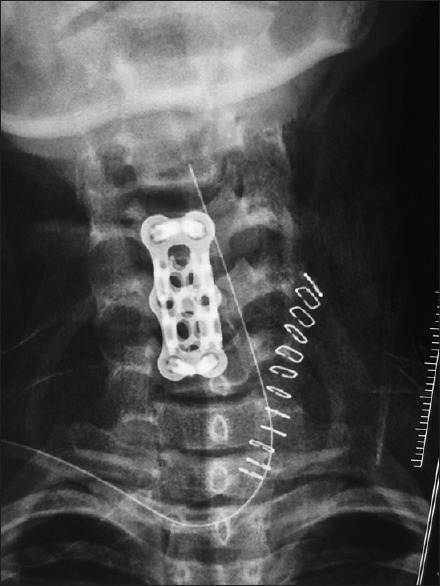
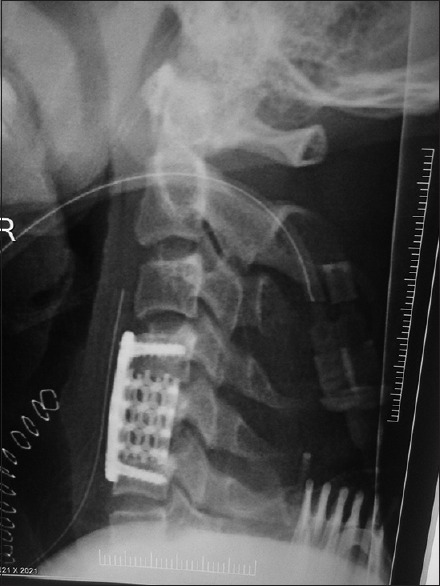
On admission of the patient history, clinical examination, routine and specific blood investigations were done which includes complete hemogram (red blood cell count, hemoglobin, hematocrit, mean corpuscular volume, mean corpuscular hemoglobin, platelet count, white blood cell count, and white blood cell differential count) renal function test, serum electrolytes, fasting blood sugar, electrocardiogram, viral markers for Hepatitis B, Hepatitis C, HIV I, and HIV II. The radiological examination includes cervical spine radiograph anteroposterior view, lateral view, and chest radiograph posteroanterior view. Computerized tomography cervical spine with three-dimensional reconstruction was done in all patients, and the relevant findings were documented [Figure 1]. Intravenous antibiotic (3rd generation cephalosporin) and injection hydrocortisone 1000 mg intravenous are given at the time of induction for general anesthesia. All patients were given intravenous antibiotics (3rd generation cephalosporin) for 5 days’ postsurgery. All patients were mobilized with Philadelphia collar as soon as possible by 1st–2nd day. Postoperatively, routine anteroposterior and lateral view of cervical spine was obtained [Figures 2 and 3] to assess the placement of cervical locking plate. The negative suction drain was removed 2nd postoperative day after the wound inspection. The patient was discharged on the 5th postoperative day. Sutures were removed on the 14th postoperative day in the outpatient department.
Figure 1.

Preoperative computed tomography scan showing neglected irreducible subluxation C4 over C5 vertebra
Figure 2.

Immediate postoperative anteroposterior view after corpectomy C5 vertebra
Figure 3.

Immediate postoperative lateral view after corpectomy C5 vertebra
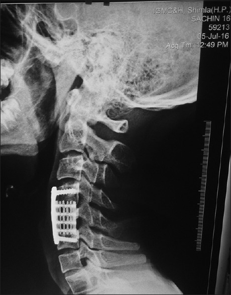
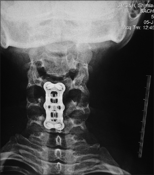
Patients were advised to follow-up after 6 weeks then after postoperative 3 months, then every 6 months [Figures 4 and 5]. Static cervical exercises advised after the surgery. Cervical movements allowed at 6 weeks. At follow-up, a detailed clinical examination was done, and radiological assessment was done. Fusion of bone graft was assessed using Birdwell fusion Grade 1. Neck-specific disability was measured as per the Neck Disability Index.
Figure 4.

1-year followup X-ray lateral view showing Birdwell Fusion Grade 1
Figure 5.

1-year follow-up X-ray anteroposterior view showing Birdwell Fusion Grade 1
For statistical analysis, Chi-squared test and t-tests were used, and P < 0.05 was considered statistically significant.
Results
All cases were followed up for 12–42 months (mean 27 ± 8 months). The mean age was 41.8 ± 15.25 years (range, 15–88 years). The study group consisted of 82 male and 17 female patients. The most common mode of trauma was fall from the height, i.e., 66.7% followed by roadside accident 33.3%. Of 99 patients, preoperatively, three patients had ASIA B neurology, 21 patients had ASIA C neurology, 41 had ASIA D neurology, 34 patients had ASIA E neurology, and postoperatively, one patient had ASIA B neurology, four patients had ASIA C neurology, 36 patients had ASIA D neurology, and 58 patients had ASIA E neurology. No patient showed any deterioration of the neurological symptoms after surgery [Table 1]. The study group consisted of majority of 43 patients with vertebral body fracture, 11 patients with fracture dislocation, nine patients with pure subluxation, and remaining 36 were categorized into posttraumatic disc prolapsed without significant vertebral body injury. According to AO (Arbeitsgemeinschaft für Osteosynthesefragen) classification, 60.6% were type B, 33.3% were type C and 6.1% were type A.
Table 1.
Neurological charting preoperative and postoperative
| Number of patients | ASIA grading | ||||
|---|---|---|---|---|---|
| ASIA A | ASIA B | ASIA C | ASIA D | ASIA E | |
| Preoperative | 0 | 3 | 21 | 41 | 34 |
| Postoperative | 0 | 1 | 4 | 36 | 58 |
ASIA – American Spinal Injury Association
The most common level of vertebral fracture was C5 in 21 (21.2%) followed by C6 in 14 (14.1%). Of the 99 procedures, 77 (77.8%) involved a single vertebral level, 19 (19.2%) involved two levels, 3 (3%) involved three levels corpectomy, and most common vertebra to be corpectomized was C5 in 28 (28.3%) followed by C6 in 22 (22.2%). The incidence of neurological deficit was more in patients in which two or three vertebra corpectomy was done (20 out of 22, i.e., 90.90%) compared with patients, in which single-level corpectomy was done (47 out of 77, i.e., 61.03%) and it is significant (P = 0.029) [Table 2]. In our study, 22 (22.22%) patients had multiple level corpectomy and of that 17 (17.17%) patients were in age group >45 years and is statistically significant (P = 0.006) [Table 3].
Table 2.
Number of corpectomy vertebra versus neurological deficit
| Number of corpectomy vertebra | Neurological deficit | |
|---|---|---|
| Present | Absent | |
| One | 47 | 30 |
| Two | 17 | 2 |
| Three | 3 | 0 |
Table 3.
Number of corpectomy vertebra and age groups
| Number of vertebra corpectomy | Age (years) | |||||
|---|---|---|---|---|---|---|
| 15-30 | 31-45 | 46-60 | 61-75 | >75 | Total | |
| One | 27 | 28 | 16 | 6 | 0 | 77 |
| Two | 2 | 3 | 8 | 5 | 1 | 19 |
| Three | 0 | 0 | 2 | 1 | 0 | 3 |
Functional outcome was assessed using Neck Disability Index. About 62.6% of patients had mild disability and 27.3% had no disability, 6.1% had moderate disability and 4.0% had severe disability according to the Neck Disability Index. Mean Neck Disability Index is 7.57 ± 5.42.
Fusion was assessed using the Bridwell criteria.[8] Of the 99 patients, 64 (64.6%) patients showed Grade 1 fusion, 31 (31.3%) patients showed Grade 2 fusion, and 4 (4.0%) patients showed Grade 3 fusion.
Of 99 patients, 79 (79.8%) patients had postoperative transient dysphagia which gradually resolved in all patients and only one patient has minimal screw back out.
Discussion
The subaxial cervical spine is having considerable mobility and proximity to the more rigid thoracic region, hence prone to traumatic disruption. Many surgical series recommend early treatment with aggressive canal decompression, improving neurological outcomes and also offering immediate stabilization.[9,10,11,12,13] In subaxial cervical spine injuries, SLIC score is used to determine the threshold for surgical intervention. In our study, for these patients with SLIC score 4 preferentially early surgery was indicated in view of:
Refinements of spinal instrumentation and early mobilization
Most of the patients in our study with SLIC score 4 were in young age group (55% in 15–30 years and 80% in 15–45 years)
Severe radiculopathy involving motor and sensory impairment
Radiological parameters associated with failure in conservative management of these injuries, such as ≥40% of height compression, kyphotic angulation higher than 15° or 20% of subluxation of one vertebra on another are not addressed by the SLIC score.[14,15]
Surgical stabilization has been described using both anterior and posterior approach or combined approach.[8,16] In our study, subaxial cervical spine injuries treated operatively with anterior corpectomy and stabilization with cage filled with autologous bone graft of vertebral body and cervical locking plates are studied. The advantages of using interbody cages for reconstruction after anterior cervical corpectomy fusion include, avoidance of morbidity associated with autologous bone graft (iliac crest) harvesting, compared with multilevel anterior cervical discectomy and fusion should result in lesser fusion rates because of increased graft-host interfaces where fusion needs to occur.[17]
The purpose of this prospective cohort study was to analyze the clinical and radiographic results of cages in the surgical treatment of patients with traumatic cervical spine instability.
Most of the patients with Grade 1 fusion have no disability (P = 0.021). This comparison is significant. Most patients with Grade 1 fusion had less incidence of neurological deficit (P = 0.043) and is significant. Most of the patients with Grade 1 fusion had ASIA E neurology (P = 0.001) and is highly significant.
The Neck Disability Index score used in this study was highly correlated with the neurological outcome in the form of ASIA score, and the radiological outcome in the form of Bridwell fusion criteria.
The incidence of neurological deficit was more in patients which required multiple level corpectomies, and most of the patients which required multiple level corpectomies were in elder age groups.
Of 99 patients, 79 (79.8%) patients had postoperative transient dysphagia which gradually resolved in all patients.
Only one patient had minimal screw back out and is without any deficit and is currently under the sequential follow-up.
Conclusions
Anterior cervical corpectomy and stabilization with cage filled with bone and cervical reflex locking plate is good and safe method in treating subaxial cervical spine injuries. This procedure has benefits of high primary stability, anatomical reduction, and direct decompression of spinal cord with minimal complications.
Financial support and sponsorship
Nil.
Conflicts of interest
There are no conflicts of interest.
References
- 1.Torretti JA, Sengupta DK. Cervical spine trauma. Indian J Orthop. 2007;41:255–67. doi: 10.4103/0019-5413.36985. [DOI] [PMC free article] [PubMed] [Google Scholar]
- 2.Goldberg W, Mueller C, Panacek E, Tigges S, Hoffman JR, Mower WR, et al. Distribution and patterns of blunt traumatic cervical spine injury. Ann Emerg Med. 2001;38:17–21. doi: 10.1067/mem.2001.116150. [DOI] [PubMed] [Google Scholar]
- 3.Park HW, Lee JK, Moon SJ, Seo SK, Lee JH, Kim SH, et al. The efficacy of the synthetic interbody cage and Grafton for anterior cervical fusion. Spine (Phila Pa 1976) 2009;34:E591–5. doi: 10.1097/BRS.0b013e3181ab8b9a. [DOI] [PubMed] [Google Scholar]
- 4.Wang JC, McDonough PW, Endow KK, Delamarter RB. Increased fusion rates with cervical plating for two-level anterior cervical discectomy and fusion. Spine (Phila Pa 1976) 2000;25:41–5. doi: 10.1097/00007632-200001010-00009. [DOI] [PubMed] [Google Scholar]
- 5.Bohlman HH, Emery SE, Goodfellow DB, Jones PK. Robinson anterior cervical discectomy and arthrodesis for cervical radiculopathy.Long-term follow-up of one hundred and twenty-two patients. J Bone Joint Surg Am. 1993;75:1298–307. doi: 10.2106/00004623-199309000-00005. [DOI] [PubMed] [Google Scholar]
- 6.Cauthen JC, Kinard RE, Vogler JB, Jackson DE, DePaz OB, Hunter OL, et al. Outcome analysis of noninstrumented anterior cervical discectomy and interbody fusion in 348 patients. Spine (Phila Pa 1976) 1998;23:188–92. doi: 10.1097/00007632-199801150-00008. [DOI] [PubMed] [Google Scholar]
- 7.Koller H, Reynolds J, Zenner J, Forstner R, Hempfing A, Maislinger I, et al. Mid- to long-term outcome of instrumented anterior cervical fusion for subaxial injuries. Eur Spine J. 2009;18:630–53. doi: 10.1007/s00586-008-0879-3. [DOI] [PMC free article] [PubMed] [Google Scholar]
- 8.Gelb DE, Aarabi B, Dhall SS, Hurlbert RJ, Rozzelle CJ, Ryken TC, et al. Treatment of subaxial cervical spinal injuries. Neurosurgery. 2013;72(Suppl 2):187–94. doi: 10.1227/NEU.0b013e318276f637. [DOI] [PubMed] [Google Scholar]
- 9.Aebi M, Zuber K, Marchesi D. Treatment of cervical spine injuries with anterior plating.Indications, techniques, and results. Spine (Phila Pa 1976) 1991;16:S38–45. doi: 10.1097/00007632-199103001-00008. [DOI] [PubMed] [Google Scholar]
- 10.Dai L, Jia L. Central cord injury complicating acute cervical disc herniation in trauma. Spine (Phila Pa 1976) 2000;25:331–5. doi: 10.1097/00007632-200002010-00012. [DOI] [PubMed] [Google Scholar]
- 11.Goffin J, Plets C, Van den Bergh R. Anterior cervical fusion and osteosynthetic stabilization according to Caspar: A prospective study of 41 patients with fractures and/or dislocations of the cervical spine. Neurosurgery. 1989;25:865–71. [PubMed] [Google Scholar]
- 12.Hirsh LF, Duarte LE, Wolfson EH, Gerhard W. Isolated symptomatic cervical spinous process fracture requiring surgery. Case report. J Neurosurg. 1991;75:131–3. doi: 10.3171/jns.1991.75.1.0131. [DOI] [PubMed] [Google Scholar]
- 13.Joaquim AF, Patel AA, Vaccaro AR. Cervical injuries scored according to the subaxial injury classification system: An analysis of the literature. J Craniovertebr Junction Spine. 2014;5:65–70. doi: 10.4103/0974-8237.139200. [DOI] [PMC free article] [PubMed] [Google Scholar]
- 14.Hadley MN, Walters BC, Grabb PA, Oyesiku NM, Przybylski GJ, Resnick DK, et al. Guidelines for the management of acute cervical spine and spinal cord injuries. Clin Neurosurg. 2002;49:407–98. [PubMed] [Google Scholar]
- 15.Lemons VR, Wagner FC., Jr Stabilization of subaxial cervical spinal injuries. Surg Neurol. 1993;39:511–8. doi: 10.1016/0090-3019(93)90038-3. [DOI] [PubMed] [Google Scholar]
- 16.Vasan T. Circumferential fusion for sub-axial cervical spine fracture – Subluxations. The Indian J Neurotrauma. 2007;4:65–70. [Google Scholar]
- 17.Uribe JS, Sangala JR, Duckworth EA, Vale FL. Comparison between anterior cervical discectomy fusion and cervical corpectomy fusion using titanium cages for reconstruction: Analysis of outcome and long-term follow-up. Eur Spine J. 2009;18:654–62. doi: 10.1007/s00586-009-0897-9. [DOI] [PMC free article] [PubMed] [Google Scholar]


