Abstract
Background:
Supraorbital craniotomy (SOC) has brought a paradigm shift in approaching anterior skull base lesions. With better understanding of relevant anatomy, the indications are being stretched from highly selected, small-to-moderate-sized tumors to large and complex anterior skull base lesions.
Objective:
We share our experience and discuss the nuances of surgery for large anterior skull base meningiomas using the SOC.
Methods:
This is a single institute study using prospectively collected retrospective data from seven cases of large anterior skull base meningiomas (>3 cm) using the SOC. We reviewed the indications, safety, and procedural complications in these cases.
Results:
Simpson's Grade 2 excision was achieved in all these seven cases, with faster postoperative recovery. Follow-up clinical outcome and cosmesis were satisfactory.
Conclusion:
SOC is a safe alternative for the standard skull base approaches in treating large anterior skull base meningiomas. The SOC can be effectively used to treat selected large anterior skull base meningiomas.
Keywords: Brow craniotomy, meningiomas, minimally-invasive, skull base, supraorbital
Introduction
Meningiomas are the most common benign intracranial tumors. Meningiomas constitute 30% of all intracranial tumors, of which anterior skull base meningiomas amount to 8.8%.[1] The most common midline anterior skull base locations are tuberculum sellae (3.6%) followed by olfactory groove (3.1%).[2] The traditional approaches to these lesions would require a large pterional/frontal craniotomy or one of their variations. The disadvantage of these large exposures is unnecessary exposure of the brain, which can advertently be injured during retraction or instrument passes. A tailored approach for anterior skull base pathology using supraorbital craniotomy (SOC-brow craniotomy) was first described by Krause in the early 1900s and then popularized by Axel Perneczky in the 1990s. Since then, this has gained confidence of the operating surgeons worldwide and SOC is being utilized more frequently in surgeries of the anterior skull base.[3,4] More recently, Mahmoud et al. have described variants of SOC in the approaches to orbital tumors.[5] Selection of lesions for resection using SOC depends upon the relation and position of the lesion in the anterior cranial fossa, which is determined using preoperative imaging. The goal of this approach would be to have an adequate access to the skull base and be able to excise the lesion, with minimal retraction of the brain. The counterargument for this approach lies in the adequacy of the surgical exposure when compared with conventional approaches to the anterior cranial fossa. The main limitation of this approach is that it has a steep learning curve as compared to traditional approaches, made especially difficult as it needs frequent adjustments of the operating table, microscope, and microinstruments needing coaxial control through the narrow surgical window.[6] In this article, we have attempted to describe the advantages, disadvantages, and technical nuances in approaching large anterior skull base meningiomas using SOC.
Methods
This is a single institute study from a retrospective review of prospectively collected data. Seven cases of large anterior skull base meningiomas were operated through SOC at our institute over a 5-year period from 2013 to 2018. The patients were selected for SOC after a preoperative imaging review by the institutional tumor board. In general, we included meningiomas of the anterior skull base, of any size, except when they had extensive involvement of the frontal or ethmoidal sinus, or have gross optic nerve involvement. We also avoid in patients where there is radiological evidence of involvement of the circle of Willis or one of the major branches. All patients underwent standard positioning, same operative techniques in exposure, and closure of SOC to maintain uniformity. Any adverse events or surgical complications were also noted. Patients had immediate postoperative imaging with either contrast-enhanced computed tomography or magnetic resonance imaging. All patients had regular follow-up examinations postoperatively.
Operative procedure
All procedures were performed under general endotracheal anesthesia. Preprocedural imaging data were acquired with skin fiducials and registered on SonoWand navigation system (SONOWAND, Trondheim, Norway).
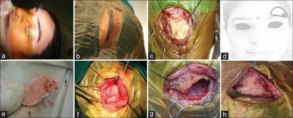
Next, the patient was positioned supine with head fixed on a Sugita frame. Head was elevated 10° and extended 15°–20° as it allowed the frontal lobe to fall back. Degree of head rotation toward the opposite side was based on the location of the tumor [Figure 1]. We chose nondominant side approach for midline tumors and ipsilateral side for laterally placed tumors. The incision was made within the eyebrow (ciliary incision) extending from 0.5 cm lateral to the supraorbital notch to the lateral edge of the brow. Initial incision was carried across the skin and dermis. A pericranial flap was elevated, with the base directed inferiorly over the orbital rim to exteriorize the frontal sinus if needed. Monopolar Bovie is generally avoided during exposure of skin and subcutaneous tissue. The attachment of periorbita to the rim of the supraorbital ridge is better left undisturbed to prevent postoperative periorbital edema. Blunt dissection of a small portion of temporalis muscle and fascia at the superior temporal line was performed, and a 5-mm burr hole (or just adequate to pass the foot plate of the B1/2 craniotome) was made on the lateral aspect of the exposure below the temporalis muscle for a better cosmetic result as shown in Figure 2. Frontalis branch of the facial nerve was avoided by staying subfacial and not exposing beyond the first 10 mm of the temporalis muscle. A craniotome was then used to make two cuts: the first cut was made from the burr hole just flush with the supraorbital ridge and the second cut was made in the shape of an inverted “U” and connecting the burr hole with the medial edge of the first cut. Navigation was used to mark this border, and all effort was made to avoid exposing the frontal sinus. In case of inadvertent exposure of the sinus, the mucosa was stripped off and gel foam-soaked povidone-iodine was used to pack the sinus, which was later exteriorized using the harvested pericranium and fibrin glue. Next, a 4-mm cutting burr was used to drill the bony irregularities on the supraorbital ridge and the orbital roof to maximize the surgical exposure.
Figure 1.
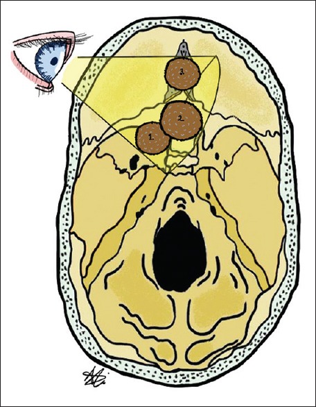
We can approach various targets in supraorbital craniotomy with the help of rotation of head, patient positioning, maneuvering the microscope, and adjusting the Operating Table. Lesions include olfactory groove (1), planum sphenoidale (2) and clinoidal (3) Meningiomas
Figure 2.
(a) Position of the patient with neck extended and rotation toward contralateral side; (b) surgical drapes before surgery; (c) subcutaneous and muscle flap turned upward; (d) illustrative picture of size of craniotomy; (e) bone flap elevated after craniotomy; (f) dural exposure and visualization of frontal lobe; (g) water tight dural closure after tumor excision; (h) replacement of bone flap with miniplates and screws
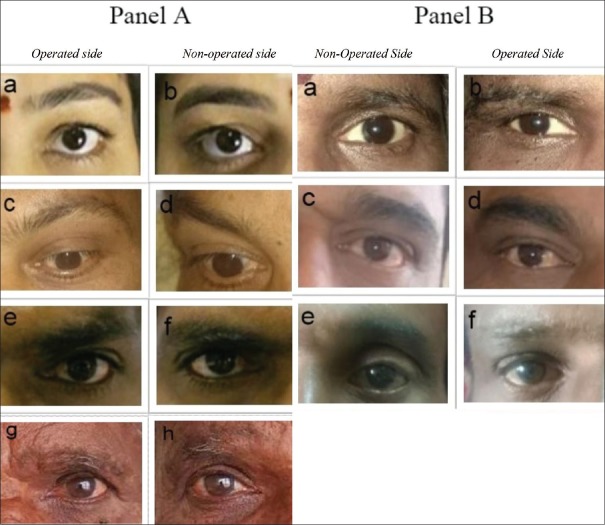
Dura was opened in semilunar fashion with the base at orbital rim. Cerebrospinal fluid (CSF) cisterns were opened to aid brain relaxation, and the arachnoid plane was dissected. The tumor was devascularized and debulked in piecemeal fashion, using a combination of ultrasonic aspirator and the bipolar coagulation, followed by dissection of the tumor from the surrounding brain tissue. Intraoperative navigation guidance was used for better anatomical orientation during surgery. Once the excision was achieved, the operative cavity was thoroughly inspected for residue with angled endoscopes in all cases. The dural attachment was thoroughly coagulated and hemostasis achieved. In cases where preoperative imaging revealed extension in the optic canal, the falciform ligament was cut and deroofing of the optic canal was carefully done using a 2-mm Diamond drill. The dura was closed in a watertight manner, and the cranial flap was fixed with miniplates and screws. Bone cement may be occasionally used to cover the gap at the edges of craniotomy and the replaced bone flap for better cosmesis. Multilayered meticulous closure of muscle and subcutaneous tissue was performed. The skin was closed in subcuticular fashion using nonabsorbable ethylon (nylon), which was subsequently removed on day 7–10. Absorbable sutures were avoided as there is a higher chance of thickened scar at this location secondary to the occasional inflammation caused by the absorbable suture material. The various steps are illustrated in Figure 2. Photographs were taken preoperatively and at different time points during the follow-up to keep a record of cosmetic outcomes [Figure 3].
Figure 3.
(A) Postoperative scars (a, c, e, and g) and nonoperative contralateral side (b, d, f, and h) in patients who underwent left supraorbital brow craniotomy. (B) Nonoperative side (a, c, and e) and postoperative scars (b, d, and f) in patients who underwent right supraorbital brow craniotomy (sides intentionally swapped to preserve patient privacy)
Results
We operated seven cases of large anterior skull base meningiomas (defined as maximum diameter >3 cm).[6] Of seven meningiomas, four were located in the planum sphenoidale, two were in the orbital roof at the fronto-orbital junction, and one was located at tuberculum sellae. Headache was the most common presentation and was seen in 5/7 cases. One patient presented with progressive mono-ocular loss of vision and another patient had right hemiparesis. Simpson's Grade 2 excision was achieved in all cases. One patient had postoperative CSF leak which was managed conservatively with a lumbar drainage. One patient had transient frontal branch of facial nerve weakness, which recovered over 6 weeks. Tube shaft instruments and endoscope were useful adjuncts in all our cases. Frontal sinus was inadvertently opened in one case, which was packed with gel foam and exteriorized with harvested pericranial flap. Case illustrations from 2 cases are shown in Figures 4 and 5.
Figure 4.
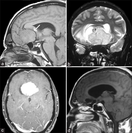
(a) T1-weighted sagittal section magnetic resonance imaging brain with isointense lesion in the planum sphenoidale, white arrow indicates anterior cerebral artery being pushed upward and posteriorly by the lesion. (b) T2-weighted coronal section magnetic resonance imaging brain with heterointense lesion the planum sphenoidale. (c) T1-weighted postcontrast axial section magnetic resonance imaging brain with contrast enhancing lesion in the planum sphenoidale. (d) T1-weighted postcontrast sagittal section magnetic resonance imaging brain postoperative image with complete excision of the lesion
Figure 5.
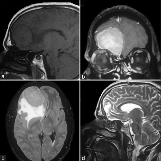
(a) T1-weighted sagittal section magnetic resonance imaging brain with isointense lesion in the right orbital roof; (b) T1-weighted coronal section magnetic resonance imaging brain with contrast enhancing lesion in the right orbital roof; (c) T2-weighted axial section magnetic resonance imaging brain FLAIR sequence with lesion in the right frontal region demonstrating the severe perilesional edema; (d) T2-weighted sagittal section magnetic resonance imaging brain with complete excision of the right frontal lesion
Discussion
Initial efforts on surgical management of large anterior skull base meningiomas were limited to partial frontal lobectomy followed by excision of the lesion or a subtemporal bony decompression.[7] Dandy was the first to describe bifrontal craniotomy with transbasal approach though it still necessitated resection of part of the frontal lobe to excise meningioma. With the advent of microscopes, many surgeons continued bifrontal approach for anterior skull base lesions without resecting normal frontal lobes. Unilateral approaches were popularized by Yasargil et al. when the familiarity with pterional craniotomy and trans-sylvian approach increased. These procedures were further modified into fronto-orbito-zygomatic approach which provided more basal exposure, necessitating lesser brain retraction.
The ideal surgical approach should provide adequate exposure of tumor, the surrounding structures, and the dural attachments. It should aid to minimize brain retraction and avoid manipulation of vital neurovascular structures. A tailored supraorbital approach can be used based on the location of meningioma in the anterior cranial fossa. With adequate patient positioning, trajectory adjustment, and brain relaxation, most of the large anterior skull base meningiomas can be excised. SOC has been used to address various other pathologies such as craniopharyngiomas and aneurysms.[8,9] It has also been sparingly used in the treatment of arteriovenous malformations, gliomas, pituitary lesions, and cavernous sinus lesions.[10] Gazzeri et al. reported a series of 41 cases of meningioma of anterior skull base treated with the SOC approach, of which complete excision of tumors was achieved in 84.5%.[11] In our series of seven cases of large meningiomas of anterior skull base [Table 1], Simpson's Grade 2 resection was achieved in all the cases. Resection of the meningioma was made possible by the use of surgical adjuncts, such as the tube shaft microinstruments and an endoscope. Tube shaft instruments have an extremely thin shaft design and thereby provide an almost completely unobstructed view. These are ideal attributes for surgeries performed in the coaxial plane where the regular microinstruments obstruct the surgical field. Endoscopes help in visualizing the extent of tumor resection in depth and obscure bleeding from a tumor in the surgical field. We do not consider large frontal sinus as a contraindication for supraorbital brow craniotomy.
Table 1.
Clinical symptomatology and radiological characteristics
| Case number | Age | Sex | Symptom | Duration | Location of Tumor | Size of Tumor (in cms) | T1 | T2 | Contrast | Simpson extent of excision |
|---|---|---|---|---|---|---|---|---|---|---|
| 1 | 45 | F | Dural Stretch Headache | 8 Months | Planum sphenoidale | 3.9×3.3×2.7 | Hypo | Hetero | Enhancing | Grade 1 |
| 2 | 44 | M | Headache and seizure | 2 Months | Planum sphenoidale extending to tuberculum sellae | 3.6×3.9×3.6 | Iso | Hetero | Enhancing | Grade 1 |
| 3 | 50 | M | Headache | 6 Months | Planum sphenoidale | 3.5×3.9×2.9 | Iso | Hetero | Enhancing | Grade 2 |
| 4 | 86 | M | Right hemiparesis | 2 Months | Planum sphenoidale | 4.1×3.7×2.8 | Iso | Hetero | Enhancing | Grade 2 |
| 5 | 55 | F | Progressive loss of vision | 1 Month | Tuberculum sellae | 2.3×2.5×1.8 | Iso | Hyper | Enhancing | Grade 2 |
| 6 | 42 | M | Headache | 1 month | Right frontal pole | 4.2×6.x 5.1 | Hypo | Hetero | Enhancing | Grade 2 |
| 7 | 48 | F | Headache | 24 Months | Left frontal pole | 3.5×2.7×3.2 | Hyper | Hyper | Enhancing | Grade 2 |
A similar case series of 23 patients operated using the SOC was reported by Iacoangeli et al. They suggested using conventional approaches if lateral extent of a tumor lies beyond the anterior circulation vessels and optic nerve, in case of complete encasement of carotid arteries, invasion of ethmoidal sinuses, and presence of severe bifrontal edema.[12]
Complications encountered with this approach are usually in relation to craniotomy such as cosmetic deformities in the frontotemporal area, and this can be overcome by filling the defect around the bone flap with bone cement and fixing the bone flap with miniplates and screws. Zumofen et al. performed a PubMed/Medline database search for publications on supraorbital craniotomy performed for either aneurysm repair or tumor resection; overall, 2783 patients with 3085 lesions were found in various case series or case cohort-type studies. Approach-related complications included 3.3% CSF collection or leak, 4.3% permanent, and 1.6% temporary supraorbital hypesthesia, 2.9% permanent and 1% temporary facial nerve palsy, and 1% wound-healing disturbance or infection.[13] The esthetic outcome was typically reported as highly acceptable. Opening of frontal sinus can cause CSF leak which can be avoided with neuronavigation. Inadvertently opened frontal sinus should be sealed with bone wax. Thermal injuries to the eyebrow due to microscopic light on 100% intensity can be avoided by protecting the surrounding area with constant irrigation and cushioning with wet gauze.[9] Our series had one patient with CSF leak which was managed conservatively with 3 days of lumbar drain and strict bed rest. Two patients had transient ptosis which improved completely within duration of 2 weeks. Ptosis encountered in brow craniotomies is more often due to a muscular cause and is transient.
Potential errors while performing a supraorbital brow craniotomy may arise due to inadequate preoperative planning, inadequate positioning of the patient, and inadequate placement of craniotomy. All these may result in the suboptimal outcomes. With neuronavigation and a meticulous preoperative planning, these pitfalls can be avoided. Drilling the inner edge of the craniotomy provides additional working angle when the lesions are deep-seated. Damage to the neurovascular bundle in the surgical field is likely due to lack of orientation. Dural tears while performing craniotomies and inappropriate closure of the dural reflection can cause postoperative CSF leaks and should be avoided. Watertight dural closure with nonabsorbable sutures and tissue glues help in preventing CSF leaks. Other anticipated local tissue-related problems would be an improper replacement of bone flap and improper wound closure techniques which would hamper the cosmetic outcome of the patient.
Conclusion
Supraorbital keyhole approach should be a part of every neurosurgeons armamentarium in the management of skull base lesions. As shown by our case series, large anterior skull base meningiomas can be safely operated with SOC with good cosmetic outcomes. The utilization of endoscope, navigation, tube shaft instruments, hemostatic agents, and tissue sealants makes the surgery safer and more effective. With careful selection of appropriate lesions and adequate experience, excellent outcomes can be achieved with minimal complications and near natural cosmesis.
Declaration of patient consent
The authors certify that they have obtained all appropriate patient consent forms. In the form the patient(s) has/have given his/her/their consent for his/her/their images and other clinical information to be reported in the journal. The patients understand that their names and initials will not be published and due efforts will be made to conceal their identity, but anonymity cannot be guaranteed.
Financial support and sponsorship
Nil.
Conflicts of interest
There are no conflicts of interest.
Acknowledgment
We acknowledge Asad Ali, research fellow at Neuro-Oncology Institute, Cleveland Clinic, for helping with the Figure art.
References
- 1.Abbassy M, Woodard TD, Sindwani R, Recinos PF. An overview of anterior skull base meningiomas and the endoscopic endonasal approach. Otolaryngol Clin North Am. 2016;49:141–52. doi: 10.1016/j.otc.2015.08.002. [DOI] [PubMed] [Google Scholar]
- 2.Raizer J, Sherman Sojka WJ. 2012. Neuro-Oncology; pp. 115–24. [Google Scholar]
- 3.Perneczky A, Müller-Forell W, Van Lindert E, Fries G. Stuttgart, Germany: Thieme Medical Publishers; 1999. Keyhole Concept in Neurosurgery. [Google Scholar]
- 4.Reisch R, Perneczky A. Ten-year experience with the supraorbital subfrontal approach through an eyebrow skin incision. Neurosurgery. 2005;57:242–55. doi: 10.1227/01.neu.0000178353.42777.2c. [DOI] [PubMed] [Google Scholar]
- 5.Mahmoud M, Nader R, Al-Mefty O. Optic canal involvement in tuberculum sellae meningiomas: Influence on approach, recurrence, and visual recovery. Neurosurgery. 2010;67:ons108–18. doi: 10.1227/01.NEU.0000383153.75695.24. [DOI] [PubMed] [Google Scholar]
- 6.da Silva CE, de Freitas PE. Large and giant skull base meningiomas: The role of radical surgical removal. Surg Neurol Int. 2015;6:113. doi: 10.4103/2152-7806.159489. [DOI] [PMC free article] [PubMed] [Google Scholar]
- 7.Morales-Valero SF, Van Gompel JJ, Loumiotis I, Lanzino G. Craniotomy for anterior cranial fossa meningiomas: Historical overview. Neurosurg Focus. 2014;36:E14. doi: 10.3171/2014.1.FOCUS13569. [DOI] [PubMed] [Google Scholar]
- 8.van Lindert E, Perneczky A, Fries G, Pierangeli E. The supraorbital keyhole approach to supratentorial aneurysms: Concept and technique. Surg Neurol. 1998;49:481–9. doi: 10.1016/s0090-3019(96)00539-3. [DOI] [PubMed] [Google Scholar]
- 9.Fatemi N, Dusick JR, de Paiva Neto MA, Malkasian D, Kelly DF. Endonasal versus supraorbital keyhole removal of craniopharyngiomas and tuberculum sellae meningiomas. Operative Neurosurg. 2009;64:ons269–87. doi: 10.1227/01.NEU.0000327857.22221.53. [DOI] [PubMed] [Google Scholar]
- 10.Ormond DR, Hadjipanayis CG. The supraorbital keyhole craniotomy through an eyebrow incision: Its origins and evolution. Minim Invasive Surg. 2013;2013:296469. doi: 10.1155/2013/296469. [DOI] [PMC free article] [PubMed] [Google Scholar]
- 11.Gazzeri R, Nishiyama Y, Teo C. Endoscopic supraorbital eyebrow approach for the surgical treatment of extraaxialand intraaxial tumors. Neurosurg Focus. 2014;37:E20. doi: 10.3171/2014.7.FOCUS14203. [DOI] [PubMed] [Google Scholar]
- 12.Iacoangeli M, Nocchi N, Nasi D, DI Rienzo A, Dobran M, Gladi M, et al. Minimally invasive supraorbital key-hole approach for the treatment of anterior cranial fossa meningiomas. Neurol Med Chir (Tokyo) 2016;56:180–5. doi: 10.2176/nmc.oa.2015-0242. [DOI] [PMC free article] [PubMed] [Google Scholar]
- 13.Zumofen DW, Rychen J, Roethlisberger M, Taub E, Kalbermatten D, Nossek E, et al. A review of the literature on the transciliary supraorbital keyhole approach. World Neurosurg. 2017;98:614–24. doi: 10.1016/j.wneu.2016.10.110. [DOI] [PubMed] [Google Scholar]


