Abstract
Incidental sonographic findings in thyroid and estrogen-responsive organs have been described in children and adults, but no publications describe incidental findings of these organs in infancy. We describe ultrasound features in thyroid, breast buds, testes, uterus, and ovaries in infants up to 32 weeks old that vary from the expected tissue architecture. Infants described in this paper were enrolled as healthy term neonates in a longitudinal study of normal feeding practices. Radiology reports for ultrasound exams in these infants described a range of findings that are similar to those reported in older populations. Knowledge of these asymptomatic variants occurring in infancy may guide radiologists in interpretation of these findings during clinical exams.
Keywords: Organ size, Newborn, Thyroid gland, Breast, Testis, Ovary, Uterus
Introduction
Ultrasound (US) is employed during infancy to evaluate palpable lumps, endocrine anomalies, and other abdominal and pelvic concerns. During examinations, anatomical variations may be identified, and the radiologist must ascertain whether these findings are significant. They may be normal variants or may be pathological abnormalities that have not yet produced symptoms. Incidental findings can have a significant impact on clinical management, as they may generate follow-up imaging or other diagnostic evaluations [1], leading to increased costs for health care systems and psychological strain for patients and families. The burden of incidental findings imposed by screening for lung cancer with chest imaging was considered so high as to outweigh the benefits of screening; consequently, programs for population-wide screening for lung cancer in the United States were stopped [2]. Since such findings occur without symptomatic presentation, they may be most readily identified in healthy individuals undergoing screening examinations. Screening imaging is not typically performed in healthy children, so clinical knowledge of normal variants and management decisions regarding potentially worrisome findings in children is incomplete. We present a pictorial review of incidental findings identified in a cohort of healthy term infants enrolled in a prospective study of infant feeding and early development (IFED) that included interval sonographic assessments of thyroid, testes, breast buds, uterus, and ovaries over the first 32 weeks of life [3]. Since feeding protocols in IFED were within the range of normal infant feeding practices, the subjects offered a unique opportunity to observe incidental US findings of these organs in asymptomatic neonates. Although variations in these organs have been described in older individuals, changing hormonal influences and the potential for ongoing regression of fetal structures warrant special attention in neonates and young infants.
Thyroid
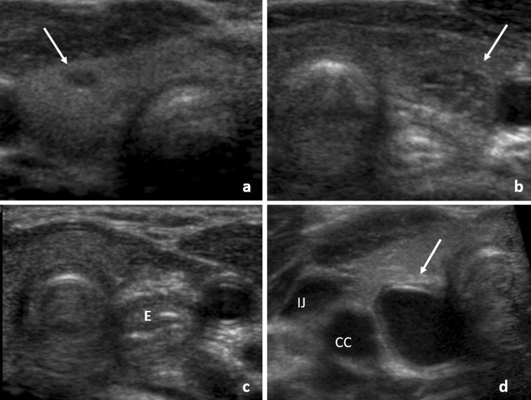
In children, 18% of thyroids imaged by US show some anomalies, most commonly cysts and ectopic thymus [4]. These were also the most common incidental findings among infants enrolled in IFED, in which sonographers performed US of the thyroid in both boys and girls at ages 4, 8, 16, and 24 weeks and in girls also at age 32 weeks (Table 1). Incidental thyroid findings during US increase with age [4], so the appearance of these less common features in infants is of interest to imagers. Cysts were the most common finding in our sample, identified in 16 children ranging from 4 to 24 weeks old (Fig. 1a). At times, radiologists observed echogenic foci within a cyst, indicating the presence of a colloid cyst. Radiologists detected an ectopic thymus in 14 children ranging from 4 to 16 weeks old (Fig. 1b). Thymic tissue was characterized by stippled echogenicities on a hypoechoic background. Due to aberrant migration during embryogenesis, thymic tissue can be found in the thyroid gland and is likely to be more conspicuous in children in whom thymic tissue has not yet involuted. An anatomic survey of pediatric thyroids suggested the presence of ectopic intrathyroidal thymic tissue, and ectopic parathyroid glands are common enough to be considered normal in pediatric individuals [5]. The neonatal thyroid may be affected by maternal thyroid disorders, but expectant mothers were excluded from IFED if they reported a history of thyroid abnormalities [3]. Congenital agenesis of the left thyroid lobe was seen at the first thyroid US at 4 weeks (Fig. 1c). This is a rare anomaly and is defined by the congenital absence of one of the thyroid lobes. It is an asymptomatic condition that is often discovered incidentally when the remaining lobe becomes symptomatic. However, it may be associated with ectopic thyroids, thyroglossal duct cysts, and parathyroid adenomas, so follow-up is advised [6]. Another 4-week-old infant exhibited a circumscribed, anechoic cyst posterior to the right thyroid lobe (Fig. 1d). The cyst appeared to have a trilaminar wall and was thought to be a foregut duplication cyst. These developmental remnants can lead to a compression of the airway via a mass effect or can become infected; resection is then recommended [4].
Table 1.
Incidental findings during thyroid ultrasound (US) in children < 8 months old
| Finding | N (%)a | First noted on US at weeks of life:modeb (range), weeks |
|---|---|---|
| Cyst or colloid cyst | 16 (4.7%) | 4 (4–24) |
| Ectopic thymus | 14 (4.1%) | 4 (4–16) |
| Hypoechoic nodule | 7 (2.0%) | 4 (4–8) |
| Absent left thyroid lobe | 1 (0.3%) | 4 (no range) |
| Congenital foregut cyst | 1 (0.3%) | 4 (no range) |
aNumber (N) is the number of infants with the given feature mentioned at any of the study visits. % is given with reference to the 342 infants who completed the 4-week study visit, which was the first visit during which thyroid was imaged.
bMode represents the central tendency among visits at 4 weeks, 8 weeks, 16 weeks, 24 weeks, and 32 weeks
Fig. 1.
Thyroid findings. Thyroid cysts were found in 16 infants, as in the right lobe of this 8-week–old male (a). An ectopic thymus, characterized by stippled echogenicities, was present in 14 children, as in the left thyroid lobe of this 8-week-old female (b). In one 4-week-old female, the left hemithyroid was absent (c), with the esophagus (E) in the thyroid bed. One 4-week-old male had a cystic lesion (arrow) posterior to the right thyroid lobe and medial to the internal jugular vein (IJ) and the common carotid artery (CC) (d)
Breast bud
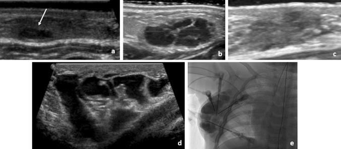
Palpable breast lesions in children may be alarming, but they are nearly all benign [7]. In the IFED study, sonographers examined breast buds in both boys and girls within 72 h of birth (0.4 weeks) and at 4, 8, 16, and 24 weeks and in girls also at 32 weeks (Table 2). Breast cysts were the most common incidental finding, occurring in seven subjects ranging from 0.4 to 8 weeks old (Fig. 2a). Breast cysts are thought to arise from an imbalance between glandular secretion and resorption, while cysts are thought to be secondary to the obstruction of Montgomery’s glands. These cysts may be symptomatic, but they are benign and require no follow-up [7]. Ductal ectasia was described in four IFED subjects, one female (age 24 weeks) and three males (ages 4, 16, and 24 weeks) (Fig. 2b, c). In adults, ductal ectasia may occur secondary to an obstructing papillary neoplasm, but in children it is benign and requires no follow-up (24). A 5:2 male predominance of ductal ectasia in young children has been reported [8]. Accessory breast tissue was also identified in the axilla in one infant at its first visit within 72 h of life. Accessory axillary breast tissue is a common finding in women, and its clinical significance lies in being able to recognize it as breast tissue rather than a mass. One subject displayed anechoic multilocular tubular and cystic features in the soft tissues lateral to the breast bud, suggestive of a lymphatic malformation (Fig. 2d). Lymphatic malformations are present from birth and grow with the child, and they are usually diagnosed in the first few years of life. Lymphatic malformations are treated percutaneously with an injection of doxycycline, an irritant that causes the lymphatic channels to collapse and sclerose [9]. The infant in the IFED study with an incidentally identified lymphatic malformation went on to treatment with sclerotherapy (Fig. 2e).
Table 2.
Incidental findings during breast ultrasound (US) in infants < 6 months old
| Finding | N (%)a | First noted on US at weeks of life: modeb (range), weeks |
|---|---|---|
| Breast cysts | 7 (1.8%) | 0.4, 4c (0.4–8) |
| Ducts ectatic or prominent | 4 (1.0%) | 4 (4–24) |
| Chest wall cyst | 1 (0.3%) | 16 (no range) |
| Accessory breast tissue | 1 (0.3%) | 0.4 (no range) |
aNumber (N) is the number of infants with the given feature mentioned at any of the study visits. % is given with reference to the 398 children who completed the first study visit at 72 h of life (0.4 weeks)
bMode represents the central tendency among visits at 0.4 weeks (72 h), 4 weeks, 8 weeks, 16 weeks, and 24 weeks
Fig. 2.
Breast findings. Seven children had breast cysts, as in this 24-week-old female (a). Ductal ectasia was identified in four children, including this 4-week-old boy (b). The ectasia had resolved by the age of 8 weeks (c). One 16-week-old boy was observed to have a multiloculated chest wall cyst adjacent to the breast tissue (d). This lesion was subsequently diagnosed as a lymphatic malformation and was treated with sclerotherapy (e)
Testes
In adults, incidentally discovered anomalies in the testes and paratesticular tissues include calcifications, cysts, and nonpalpable masses [10, 11]. These findings are typically identified during workup of infertility, which calls into question whether they are truly asymptomatic and whether the descriptions of them are relevant to children. In the IFED study, boys were examined with US within 72 h of birth (0.4 weeks) and at 4, 8, 16, and 24 weeks (Table 3). The most common incidental finding was an epididymal cyst, observed in 34 out of 204 subjects (Fig. 3a). A prevalence of 14.4% has been reported in the pediatric population, with the prevalence increasing with age to 35.5% in boys over 15 years old [12]. Epididymal cysts contain fluid similar to that in the rete testis, and they are treated clinically as benign cysts that do not warrant further evaluation. Other benign findings in IFED infants included five cases of scrotal pearls (Fig. 3b). Scrotal pearls are benign extratesticular calcifications that are typically mobile between the folds of the tunica vaginalis [10]. They are thought to result from fibrinous deposits or sequelae of torsed appendage testis or epididymis. The young age of the infants in which the scrotal pearls were found suggests that the process of scrotal pearl formation may in some cases occur in utero. As with epididymal cysts, no further follow-up is recommended for this anomaly. Three infants in IFED were discovered to have an inguinal hernia, a common developmental anomaly. One infant had scrotal edema that had resolved by the following visit. Microlithiasis was identified in six IFED subjects ranging in age from 0.4 to 24 weeks (Fig. 3c). Microlithiasis typically has a US appearance of diffuse, intratesticular, nonshadowing echogenic foci. The prevalence of pediatric microlithiasis is approximately 2.9% in North America, with regional differences [13]. The management and significance of testicular microlithiasis is a subject of debate. Some evidence suggests an association between microlithiasis and cancer risk in children [13].
Table 3.
Incidental findings during scrotal ultrasound (US) in boys < 6 months old
| Finding | N (%)a | First noted on US at weeks of life: modeb (range), weeks |
|---|---|---|
| Epididymal cyst | 34 (16.2%) | 4 (0.4–24) |
| Microlithiasis | 6 (3.0%) | 24 (0.4–24) |
| Scrotal pearl or calcification | 5 (2.5%) | 4 (0.4–4) |
| Inguinal hernia | 3 (1.5%) | 0.4 (no range) |
| Scrotal wall edema | 1 (0.5%) | 0.4 (no range) |
aNumber (N) is the number of infants with the given feature mentioned at any of the study visits. % is given with reference to the 204 boys who completed the first study visit at 72 h of life (0.4 weeks)
bMode represents the central tendency among visits at 0.4 weeks (72 h), 4 weeks, 8 weeks, 16 weeks, and 24 weeks
Fig. 3.
Testicular and paratesticular findings. Epididymal head cysts were present in 16.2% of boys, as in this 32-week-old boy (a). Five boys had scrotal pearl (arrow), like this 0.4-week-old boy (b). Microlithiasis (c) was present in six boys, as in this 8-week-old boy
Uterus
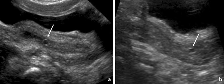
Fewer anomalies were incidentally discovered in the uterus, which was imaged with a curved transducer at greater depth and lower spatial resolution. At 4 weeks old, an endometrial cyst together with endometrial fluid was identified in one girl, and a cervical cyst in another girl (Fig. 4); both anomalies had resolved by the girls’ next visits at 8 weeks. Endometrial fluid is a common finding in peripubertal and postpubertal females and may occur also in neonates due to the influence of maternal hormones [14]. Cervical (nabothian) cysts are a common anomaly in postpubertal females, arising from transient obstruction of mucous-producing ducts in the cervix. Both anomalies have been described in neonates and are self-limited, insignificant conditions [14]. Clinically uterine anomalies, such as Müllerian fusion abnormalities, can incidentally be detected or may go unnoticed in prepubertal girls because of the smaller size of the uterus.
Fig. 4.
Uterus findings. Endometrial fluid and an endometrial cyst (arrow) were present in one 4-week–old girl (a). A cervical cyst was present in another 4-week-old girl (b)
Ovaries
In the IFED study, ovarian follicles and cysts were among the primary observations, so are not reported here as incidental findings. No other variants in ovarian appearance were discovered during the IFED study. Paraovarian anomalies, such as venolymphatic malformations or mesenteric duplication cysts, may be obscured by bowel gas and are rarely reported.
Conclusions
As in older individuals, US examinations of organs in infants can incidentally uncover abnormalities. Incidental findings are more common in superficial structures, which can easily be examined with high-frequency US. Knowledge of the frequency of detection and the clinical prognosis of such anomalies may be useful in speaking with families and in avoiding unnecessary diagnostic tests.
Funding
This research was supported in part by the Intramural Research Program of the National Institutes of Health (NIH), National Institute of Environmental Health Sciences (project number Z01-ES044006). Data collection at the Children’s Hospital of Philadelphia (CHOP) was supported through subcontract PHR-UPS2-S-09–00196 under contract HHSN291200555546C between the National Institute of Environmental Health Sciences and Social & Scientific Systems Inc (NIEHS). This project was supported by the Nutrition Center at the Children’s Hospital of Philadelphia and by the National Center for Research Resources, Grant UL1TR000003. The findings and conclusions in this report are those of the author(s) and do not necessarily represent the views of the Centers for Disease Control and Prevention and the National Institute of Health.
Compliance with ethical standards
Conflict of interest
The authors have no conflicts of interest to declare.
Ethical approval
All the procedures performed in studies involving human participants were in accordance with the ethical standards of the institutional and/or national research committee and with the 1964 Helsinki Declaration and its later amendments or comparable ethical Title Page standards. All applicable international, national, and/or institutional guidelines for the care and use of animals were followed. This article does not contain any studies with human participants or animals performed by any of the authors.
Informed consent
Informed consent was obtained from all individual participants included in the study.
Footnotes
Publisher's Note
Springer Nature remains neutral with regard to jurisdictional claims in published maps and institutional affiliations.
References
- 1.Berland LL, et al. Managing incidental findings on abdominal ct: white paper of the ACR incidental findings committee. J Am Coll Radiol. 2010;7(10):754–773. doi: 10.1016/j.jacr.2010.06.013. [DOI] [PubMed] [Google Scholar]
- 2.de Koning HJ, et al. Benefits and harms of computed tomography lung cancer screening strategies: a comparative modeling study for the US preventive services task force. Ann Intern Med Article. 2014;160(5):311. doi: 10.7326/M13-2316. [DOI] [PMC free article] [PubMed] [Google Scholar]
- 3.Adgent MA, et al. A longitudinal study of estrogen-responsive tissues and hormone concentrations in infants fed soy formula. J Clin Endocrinol Metab. 2018;103(5):1899–1909. doi: 10.1210/jc.2017-02249. [DOI] [PMC free article] [PubMed] [Google Scholar]
- 4.Avula S, Daneman A, Navarro OM, Moineddin R, Urbach S, Daneman D. Incidental thyroid abnormalities identified on neck US for non-thyroid disorders. Pediatr Radiol. 2010;40(11):1774–1780. doi: 10.1007/s00247-010-1684-9. [DOI] [PubMed] [Google Scholar]
- 5.Carpenter GR, Emery JL. Inclusions in the human thyroid. J Anat. 1976;122(Pt 1):77–89. [PMC free article] [PubMed] [Google Scholar]
- 6.De Sanctis V, Soliman AT, Di Maio S, Elsedfy H, Soliman NA, Elalaily R. Thyroid hemiagenesis from childhood to adulthood: review of literature and personal experience. Pediatric Endocrinol Rev Per. 2016;13(3):612–619. [PubMed] [Google Scholar]
- 7.Valeur NS, Rahbar H, Chapman T. Ultrasound of pediatric breast masses: what to do with lumps and bumps. Pediatr Radiol. 2016;45(11):1585–1599. doi: 10.1007/s00247-015-3402-0. [DOI] [PubMed] [Google Scholar]
- 8.McHoney M, Munro F, Mackinlay G. Mammary duct ectasia in children: report of a short series and review of the literature. Early Hum Dev. 2011;87(8):527–530. doi: 10.1016/j.earlhumdev.2011.04.005. [DOI] [PubMed] [Google Scholar]
- 9.Cahill AM, et al. Percutaneous sclerotherapy in neonatal and infant head and neck lymphatic malformations: a single center experience. J Pediatr Surg. 2011;46(11):2083–2095. doi: 10.1016/j.jpedsurg.2011.07.004. [DOI] [PubMed] [Google Scholar]
- 10.Bushby LH, Miller F, Rosairo S, Clarke JL, Sidhu PS. Scrotal calcification: ultrasound appearances, distribution and aetiology. Br J Radiol. 2002;75(891):283–288. doi: 10.1259/bjr.75.891.750283. [DOI] [PubMed] [Google Scholar]
- 11.Eifler JB, King P, Schlegel PN. Incidental testicular lesions found during infertility evaluation are usually benign and may be managed conservatively. J Urol. 2008;180(1):261–264. doi: 10.1016/j.juro.2008.03.021. [DOI] [PubMed] [Google Scholar]
- 12.Posey ZQ, Ahn HJ, Junewick J, Chen JJ, Steinhardt GF. Rate and associations of epididymal cysts on pediatric scrotal ultrasound. J Urol. 2010;184(4):1739–1741. doi: 10.1016/j.juro.2010.03.118. [DOI] [PubMed] [Google Scholar]
- 13.Trout AT. MAssociation between testicular microlithiasis and testicular neoplasia: large multicenter study in a pediatric population. Radiology. 2017;28(2):576–583. doi: 10.1148/radiol.2017162625. [DOI] [PubMed] [Google Scholar]
- 14.Paltiel HJ, Phelps A. US of the pediatric female pelvis. Radiology. 2014;270(3):644–657. doi: 10.1148/radiol.13121724. [DOI] [PubMed] [Google Scholar]


