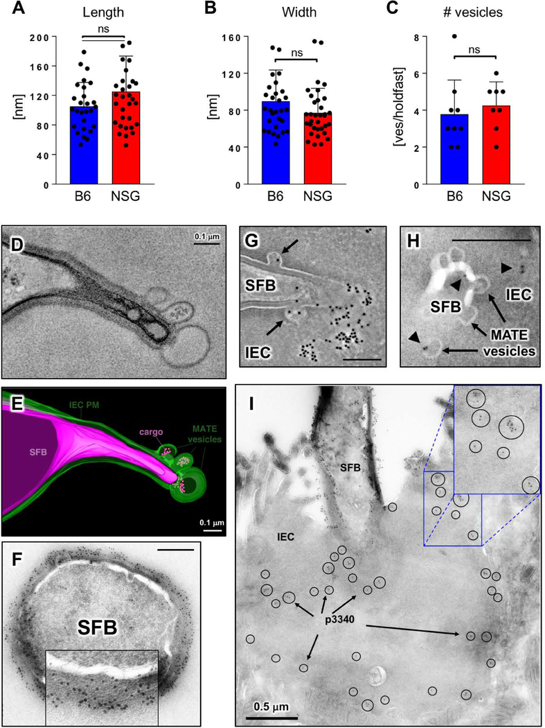
Fig. 2. MATE vesicles transfer an immunodominant SFB antigen inside IECs.
(A-C) Quantification of MATE vesicle size (A, B) and numbers per holdfast (C) in the terminal ileum of C57BL/6 (B6) and NOD.Scid.Il2rgnull (NSG) mice. Error bars, standard deviation. Statistics, unpaired two-tailed t test. (D, E) Single section and 3D reconstruction of an SFB-IEC synapse featuring MATE vesicles that contain electron dense cargo. Green, IEC PM; magenta, SFB PM. (F) Immuno-EM for P3340 on SFB in mouse intestine. Cross-section of an SFB cell. P3340 is present exclusively on the cell-wall of the bacterium. (G, H, I) Immuno-EM for P3340 on intestinal sections from terminal ileum. (G) An SFB cell interacting with a single IEC transfers P3340 into the IEC cytosol via MATE. Arrows, MATE vesicles containing P3340 labeling. (H) A close-up of the distal end of an SFB holdfast showing P3340 presence (black arrowheads) on the microbe, in a MATE vesicle, and inside the IEC cytosol. (I) Immuno-EM of P3340 in terminal ileum of C57BL/6 mice colonized with SFB. An IEC with an attached SFB is shown. P3340 immunogold labeling is present on the cell wall of the bacteria, as well as inside the IEC cytosol in the vicinity of the SFB-IEC synapse (black circles and arrows). All scale bars are 200 nm unless otherwise noted.

